. 2001 Nov;21(21):7243-55.
doi: 10.1128/MCB.21.21.7243-7255.2001.

Targeted disruption of the transition protein 2 gene affects sperm chromatin structure and reduces fertility in mice

Affiliations

Targeted disruption of the transition protein 2 gene affects sperm chromatin structure and reduces fertility in mice

M Zhao et al. Mol Cell Biol. 2001 Nov.

Abstract

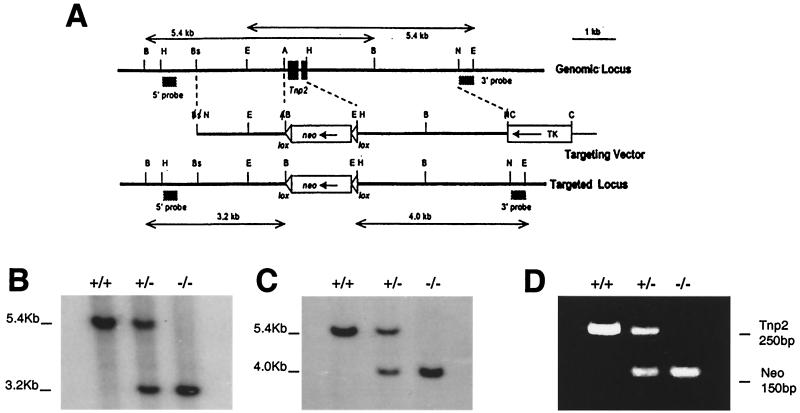
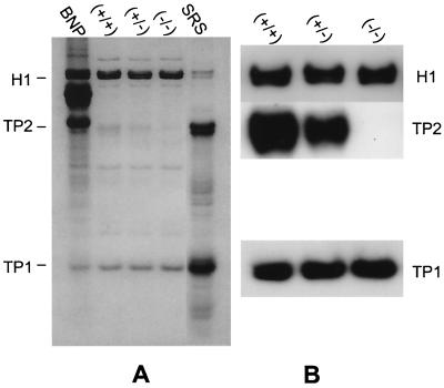
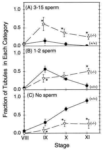
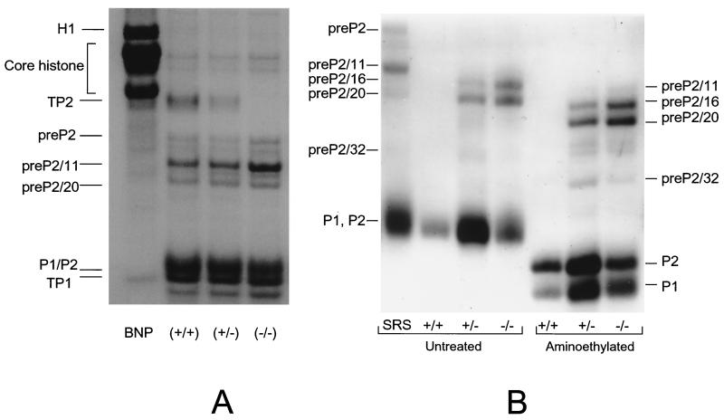
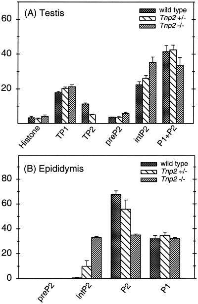
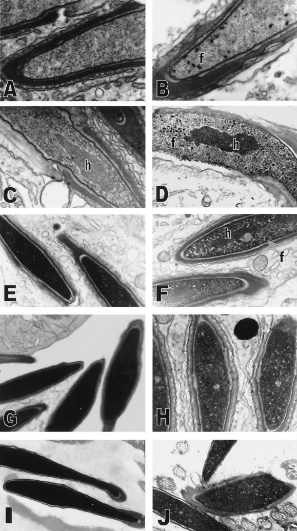
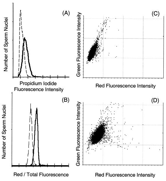
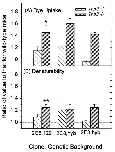
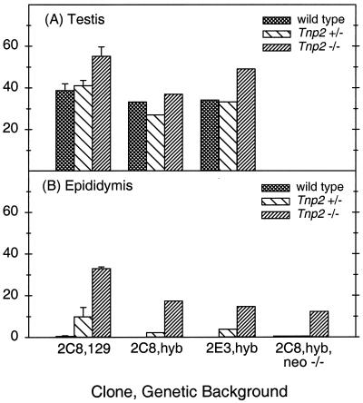
During mammalian spermiogenesis, major restructuring of chromatin takes place. In the mouse, the histones are replaced by the transition proteins, TP1 and TP2, which are in turn replaced by the protamines, P1 and P2. To investigate the role of TP2, we generated mice with a targeted deletion of its gene, Tnp2. Spermatogenesis in Tnp2 null mice was almost normal, with testis weights and epididymal sperm counts being unaffected. The only abnormality in testicular histology was a slight increase of sperm retention in stage IX to XI tubules. Epididymal sperm from Tnp2-null mice showed an increase in abnormal tail, but not head, morphology. The mice were fertile but produced small litters. In step 12 to 16 spermatid nuclei from Tnp2-null mice, there was normal displacement of histones, a compensatory translationally regulated increase in TP1 levels, and elevated levels of precursor and partially processed forms of P2. Electron microscopy revealed abnormal focal condensations of chromatin in step 11 to 13 spermatids and progressive chromatin condensation in later spermatids, but condensation was still incomplete in epididymal sperm. Compared to that of the wild type, the sperm chromatin of these mutants was more accessible to intercalating dyes and more susceptible to acid denaturation, which is believed to indicate DNA strand breaks. We conclude that TP2 is not a critical factor for shaping of the sperm nucleus, histone displacement, initiation of chromatin condensation, binding of protamines to DNA, or fertility but that it is necessary for maintaining the normal processing of P2 and, consequently, the completion of chromatin condensation.


Figures

FIG. 1
FIG. 1
Targeted deletion of the Tnp2 gene. (A) Genomic Tnp2 locus, the replacement vector used, and the targeted locus. Restriction sites: A, AvaI; B, BamHI; Bs, BsmI; C, ClaI; E, EcoRI; H, HindIII; N, NotI. The positions of the probes used to distinguish the targeted locus from the endogenous locus are indicated. (B and C) Southern blot genotyping performed by probing BamHI digests with the 5′ probe (B) or probing EcoRI digests with the 3′ probe (C). (D) PCR genotyping using primers for Tnp2 and neo.
FIG. 2
FIG. 2
Bivariate histograms of green fluorescence peak height versus peak area for acid-denatured, AO-stained preparations from wild-type mice. Regions containing intact sperm (S), free sperm heads (H), and free sperm tails (T) are marked. (A) Intact sperm. (B) Trypsin-treated sperm. (C) Sonicated sperm. (D) Histograms of green fluorescence peak height for the indicated gated areas from intact sperm (thin solid lines), trypsin-treated sperm (dashed lines), and sonicated sperm (thick solid line).
FIG. 3
FIG. 3
Analysis of 3.5% TCA-soluble proteins from testes of wild-type, heterozygous, and Tnp2 mutant mice by acid-urea gel electrophoresis. (A) Coomassie blue-stained gel. A total basic nuclear protein extract (BNP) from unfractionated wild-type mouse testis nuclei was used as a marker. (B) Immunoblots using antibodies to TP1, TP2, and H1.
FIG. 4
FIG. 4
Retention of mature spermatids in tubules at stages IX to XI in testes of wild-type (circles) and Tnp2 mutant (triangles) mice. The fractions of tubules with 3 to 15 sperm (A), 1 to 2 sperm (B), and no sperm (C) per cross-section are shown. Asterisks indicate values that are significantly different from those for the wild type (P < 0.05). Error bars indicate standard errors.
FIG. 5
FIG. 5
Basic nuclear proteins from sonication-resistant (step 12 to 16) spermatids (A) and epididymal sperm (B) from wild-type, Tnp2+/−, and Tnp2−/− mice. A total basic nuclear protein extract (BNP) from unfractionated wild-type mouse testis nuclei was used as a marker for the histone bands and TP1. The SRS marker represents sonication-resistant step 12 to 16 spermatids prepared from the testes of wild-type mice. In panel B, the heterozygote samples were overloaded on this gel so that the low level of P2 precursors would be clearly visible. Parts of the epididymal preparations were aminoethylated to separate P1 and the mature form of P2. pre-P2 is the primary translation product of the Prm2 gene; pre-P2/11, pre-P2/16, pre-P2/20, and pre-P2/32 result from proteolytic cleavage before the 11th, 16th, 20th, and 32nd amino acids, respectively.
FIG. 6
FIG. 6
Basic nuclear proteins in sonication-resistant spermatids (A) and epididymal sperm (B) as a percentage of total basic protein. Error bars indicate range (n = 2).
FIG. 7
FIG. 7
Abnormalities in chromatin condensation in spermatids from Tnp2 mutant mice. (A and B) Step 11 spermatids from wild-type (A) and Tnp2−/− (B) mice. (C and D) Step 13 spermatids from wild-type (C) and Tnp2−/− (D) mice. (E and F) Step 14 spermatids in stage II from wild-type mice (E) and in stage III from Tnp2−/− mice (F). (G and H) Step 16 spermatids in stage VIII from wild-type mice (G) and in stage VII from Tnp2−/− mice (H). (I and J) Cauda epididymal sperm nuclei from wild-type mice (I) and sperm nuclei from the vas deferens from Tnp2−/− mice (J). f, abnormal focal condensations; h, differentially condensed core of pericentromeric heterochromatin. Magnifications: A, ×25,000; B, ×16,000; C, ×31,000; D, ×10,000; E, ×12,000; F, ×13,000; G, ×11,000; H, ×16,000; I, ×12,000; J, ×12,000.
FIG. 8
FIG. 8
Flow cytometric analysis of sperm chromatin defects. (A) Distributions of propidium iodide fluorescence intensities for sperm heads prepared by sonication from Tnp2-null mice (solid line) and from wild-type mice (dashed line). (B) Distributions of the relative amount of red fluorescence (red/total fluorescence) of acid-denatured, AO-stained sperm heads prepared with trypsin from Tnp2-null mice (solid line) and from wild-type mice (dashed line). (C and D) Bivariate histograms of green versus red fluorescence for acid-denatured, AO-stained sperm heads prepared by sonication from wild-type (C) and Tnp2-null (D) mice.
FIG. 9
FIG. 9
Dye uptake and susceptibility to acid denaturation of DNA in cauda epididymal sperm nuclei from Tnp2 mutant mice on different genetic backgrounds and derived from different ES cell clones. All values were normalized to those for wild-type mice derived from the same matings as were the mutants. (A) Propidium iodide fluorescence intensity. (B) Denaturability as defined by the ratio of red fluorescence to total (red plus green) fluorescence of AO-stained sperm nuclei. For clone 2C8 on the 129 background, n = 3 to 5; for clone 2C8 and clone 2E3 on the hybrid (hyb) background, n = 2. When n = 2, the error bars represent the range of values; otherwise the error bars indicate standard errors. ∗ and ∗∗, significantly different from 1 at P values of <0.05 and <0.005, respectively.
FIG. 10
FIG. 10
Protamine 2 precursor levels (pre-P2 plus int-P2) expressed as percentages of total protamine in Tnp2 mutant mice produced from different ES cell clones, on different genetic backgrounds, and after the excision of the neo gene from the Tnp2 locus. (A) SRS nuclei (step 12 to 16 spermatids) from testes. (B) Epididymal sperm nuclei. Note that pre-P2 was not present in epididymal sperm of any genotype studied here and that there was <1% int-P2 in epididymal sperm from all wild-type mice. hyb, hybrid. Error bars indicate range (n = 2).

Similar articles

Cited by

References

    1. Alfonso P J, Kistler W S. Immunohistochemical localization of spermatid nuclear transition protein 2 in the testes of rats and mice. Biol Reprod. 1993;48:522–529. - PubMed
    1. Arango N A, Lovell-Badge R, Behringer R R. Targeted mutagenesis of the endogenous mouse Mis gene promoter: in vivo definition of genetic pathways of vertebrate sexual development. Cell. 1999;99:409–419. - PubMed
    1. Aravindan G R, Bjordahl J, Jost L K, Evenson D P. Susceptibility of human sperm to in situ DNA denaturation is strongly correlated with DNA strand breaks identified by single-cell electrophoresis. Exp Cell Res. 1997;236:231–237. - PubMed
    1. Balhorn R, Gledhill B L, Wyrobek A J. Mouse sperm in chromatin proteins: quantitative isolation and partial characterization. Biochemistry. 1977;16:4074–4080. - PubMed
    1. Balhorn R, Reed S, Tanphaichitr N. Aberrant protamine 1/protamine 2 ratios in sperm of infertile human males. Experientia. 1988;44:52–55. - PubMed

Publication types

MeSH terms

LinkOut - more resources