. 2001 Nov;21(21):7268-76.
doi: 10.1128/MCB.21.21.7268-7276.2001.

Bid, a widely expressed proapoptotic protein of the Bcl-2 family, displays lipid transfer activity

Affiliations

Bid, a widely expressed proapoptotic protein of the Bcl-2 family, displays lipid transfer activity

M D Esposti et al. Mol Cell Biol. 2001 Nov.

Abstract

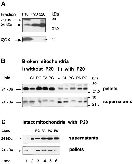
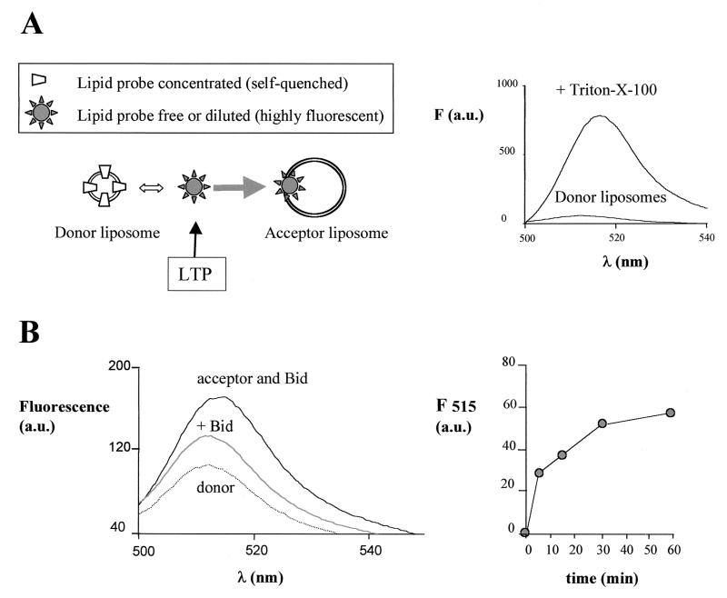
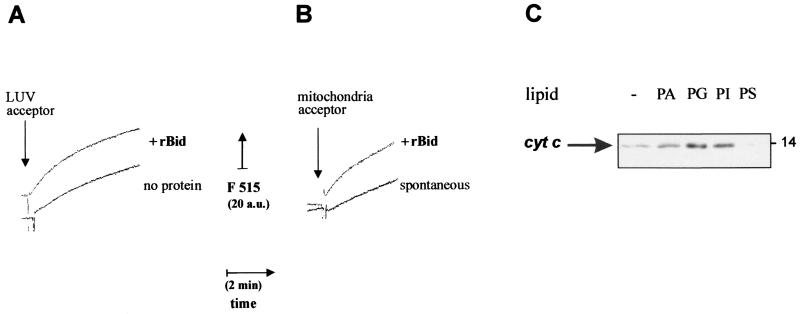
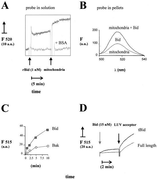
Bid is an abundant proapoptotic protein of the Bcl-2 family that is crucial for the induction of death receptor-mediated apoptosis in primary tissues such as liver. Bid action has been proposed to involve the relocation of its truncated form, tBid, to mitochondria to facilitate the release of apoptogenic cytochrome c. The mechanism of Bid relocation to mitochondria was unclear. We report here novel biochemical evidence indicating that Bid has lipid transfer activity between mitochondria and other intracellular membranes, thereby explaining its dynamic relocation to mitochondria. First, physiological concentrations of phospholipids such as phosphatidic acid and phosphatidylglycerol induced an accumulation of full-length Bid in mitochondria when incubated with light membranes enriched in endoplasmic reticulum. Secondly, native and recombinant Bid, as well as tBid, displayed lipid transfer activity under the same conditions and at the same nanomolar concentrations leading to mitochondrial relocation and release of cytochrome c. Thus, Bid is likely to be involved in the transport and recycling of mitochondrial phospholipids. We discuss how this new role of Bid may relate to its proapoptotic action.


Figures

FIG. 1
FIG. 1
Phospholipids change the mitochondrial distribution of Bid. (A) Different forms of Bid-reactive bands were detected in three subcellular fractions obtained with healthy mouse liver. P10 was the pellet of crude mitochondria obtained by the first centrifugation at 10,000 × g, while P20 was the pellet containing mainly ER-associated light membranes that was obtained by centrifugation of the S10 supernatant at 20,000 × g. S20 was the cytosolic extract remaining in the supernatants after the latter centrifugation and showed a pattern of Bid immunoblots similar to that of fraction S10. Exactly equal protein concentrations (24 μg) of each fraction were immunoblotted with a mixture of the C-20 and the R&D antibodies to detect all forms of Bid. Blots for cytochrome c (bottom) and subunit IV of cytochrome oxidase (not shown) were used as mitochondrial markers. The presence of a 28-kDa band reacting with Bid antibodies is noted by the dashed arrow. (B) Exogenous phospholipids do not affect Bid distribution in purified mitochondria alone (panels i) but change Bid distribution when the same mitochondria are coincubated with the ER-rich fraction P20 (panels ii). Mitochondria and light membranes were initially coprecipitated by centrifugation of a postnuclear supernatant of mouse liver at 22,000 × g. After the frozen pellets were thawed, mitochondria were separated from fraction P20 by centrifugation (see Materials and Methods) and further purified by multiple washes at 9,000 × g. Purified mitochondria were resuspended in the caspase assay buffer (20 mM K-HEPES, 0.25 M sucrose, 2 mM dithiothreitol, 1 mM EDTA [pH 7.4], supplemented with 0.1% [vol/vol] of protease inhibitors) at 2 mg/ml and then incubated at 30°C for 30 min with 0.2 mg of each phospholipid (all synthetic dioleyl analogues dissolved in chloroform except for cardiolipin) per ml in either the absence (panels i) or presence (panels ii) of 1 mg of fraction P20 per ml. Control (−) samples contained equivalent amounts of solvent (less than 1% of final volume). At the end of the incubation, the samples were centrifuged at 10,000 × g under cold conditions, and equal volumes of pellets and supernatants were analyzed by Western blotting with the R&D antibody. The two panels are taken from duplicate blots with slightly different exposures. (C) Intact mouse liver mitochondria were diluted to 1 mg/ml in assay buffer (20 mM K-HEPES, 0.12 M mannitol, 0.08 M KCl, 1 mM EDTA [pH 7.4], supplemented with protease inhibitors) and incubated at 30°C for 30 min with preformed liposomes (0.1 mg/ml, obtained after drying the chloroform solutions of different phospholipids). A total of 0.9 mg of fraction P20 per ml obtained separately was also added to the incubation mixture, and the mitochondria were subsequently separated by centrifugation as described in the legend to panel B. Equivalent amounts of the supernatants (top) and pellets (bottom) were probed with C-20 together with the R&D antibody as described in the legend to panel A. For reference, lanes 1 and 2 in contained mitochondria without lipids and P20 alone, respectively. Note that exogenous lipids, and especially PG, increased the proportion of the 24-kDa band of Bid that associated with the mitochondrial pellets.
FIG. 2
FIG. 2
Bid displays lipid transfer activity with liposomes. (A) The scheme in the left panel illustrates the principles of the lipid transfer assay between donor and acceptor liposomes using fluorescent lipid probes (cf. references and 27). The arrow indicates the action of an LTP, which fundamentally shifts the equilibrium dynamics of the whole process to the right. The right panel illustrates these principles experimentally. Donor liposomes containing 100 nM BODIPY FL C5-HPA show a quenched fluorescence due to the high molar fraction (see below) of the probe with respect to the other lipids in the membrane, but high fluorescence after solubilization and dilution of the probe with the detergent Triton X-100. (B) Lipid transfer activity was measured with a final concentration of 100 nM BODIPY FL C5-HPA under conditions equivalent to those in OM permeability and cell-free assays (see Materials and Methods). The donor liposomes were prepared by rapid injection of an ethanolic solution containing 1:3 (wt/wt) of the lipid probe with a mixture of purified phospholipids (44% PC, 31% PI, and 25% PS) in assay buffer. The acceptor liposomes were prepared by ethanol injection of a lipid mixture containing (wt/wt) 20% CL, 35% PC, 25% PI, and 18% PS and added at a final concentration of 3.5 μg/ml (i.e., 15-fold in excess of the concentration of donor liposomes). The results were obtained by recording time-resolved emission spectra with excitation at 485 nm. The dotted spectrum represents the fluorescence emission of donor liposomes alone, which was stable with time. Upon addition of native Bid isolated from mouse kidney (equivalent to a final concentration of approximately 10 nM Bid), a rapid increase of fluorescence occurred that stabilized to the middle spectrum shown. The subsequent addition of acceptor liposomes induced a large increase in fluorescence with time: the top spectrum was recorded after 15 min of incubation. The entire time course of the peak emission at 515 nm is shown in the right panel.
FIG. 3
FIG. 3
Correlation between lipid transfer and release of cytochrome c by recombinant Bid. (A) Lipid transfer was measured as time courses with sonicated donor liposomes prepared with a 1:3 (wt/wt) mixture of BODIPY FL C5-HPA with phospholipids (50% PC, 25% PI, and 25% PS) and acceptor liposomes consisting of LUV. These LUV were prepared by filter extrusion (38) with a phospholipid mixture closely resembling that of the OM (8, 25), namely, 50% PC, 30% PE, 12.5% PI, 5% PG, and 2.5% CL. Before the assay, both donor and LUV liposomes were purified by gel filtration with a Sigma PD10 column. Donor liposomes were equilibrated in assay buffer at a final concentration of 30 nM, and then 2 μg of LUV acceptors per ml were added in either the absence (bottom trace) or the presence (top, trace) of 250 ng of recombinant mouse Bid (rBid) per ml (i.e., 10 nM). Fluorescence emission at 515 nm was continuously monitored with excitation at 490 nm. (B) Lipid transfer from sonicated donor liposomes was measured as shown in panel A using 2.5 μg of mouse liver mitochondria per ml as acceptor membranes. The spontaneous rate of transfer (bottom trace) was accelerated when 30 nM recombinant mouse Bid (rBid) was coincubated with donor liposomes prior to the addition of mitochondria (top trace) (C) Mouse liver mitochondria were incubated for 15 min with 10 nM recombinant Bid in assay buffer. Except for the control sample (−), mitochondria were also incubated with 0.1 mg/ml liposomes formed by different negatively charged phospholipids and then separated by centrifugation as in the experiment shown in Fig. 1C. Equal volumes of the supernatants were immunoblotted for cytochrome c as in routine cell-free assays (22).
FIG. 4
FIG. 4
Specificity of the LTP-like activity displayed by recombinant Bid. (A) Donor and acceptor liposomes were mixed first for over 10 min under the conditions of the experiment shown in Fig. 2B to reach equilibrium in the spontaneous lipid transfer. A total of 1 nM (25 ng/ml) recombinant human Bid was then added as indicated, and the fluorescence changes were recorded for about 10 min. The top trace was obtained in the absence of other additions, while the bottom trace was obtained in the presence of 25 μg of BSA per ml which strongly inhibits lipid transfer (5, 33). Subsequently, 50 μg of freeze-thawed pig heart mitochondria per ml (equivalent to approximately 20 μg of membrane lipids per ml) was added to further stimulate lipid transfer. The final concentration of the lipid probe was 50 nM. (B) The increase in fluorescence of the PA analog probe was measured at 1 h after the addition of pig heart mitochondria in experiments similar to those shown in panel A. Subsequently, the reaction mixtures were centrifuged at 10,000 × g for 15 min to separate the mitochondria, and the pellets were resuspended in assay buffer containing 0.5% (vol/vol) Triton X-100 and then diluted in the cuvette. The fluorescence recovered in the pellets was measured as described in the legend to Fig. 2. The bottom spectrum was obtained with mitochondria alone (without the lipid probe), the intermediate spectrum was obtained with acceptor and donor liposomes plus Bid, and the top spectrum was obtained with donor and acceptor liposomes plus Bid and mitochondria. (C) Isolated native Bid (10 nM) (filled symbols) or recombinant Bak (10 nM) (open symbols) was incubated with donor liposomes, and lipid transfer was monitored by time-resolved spectra after addition of 3.5 μg of acceptor liposomes per ml as in Fig. 2B. The time course of the intensity in the emission maximum (at 515 nm; F515) is shown. The spontaneous transfer to the acceptor liposomes was essentially equivalent to that measured in the presence of Bak and is not shown, for the sake of clarity. Similar results were obtained using pig heart mitochondria as acceptors (not shown). (D) Caspase 8-cleaved Bid (tBid) and its parent full-length preparation of recombinant mouse Bid were added to donor liposomes (obtained by sonication as shown in Fig. 3) diluted to a final concentration of 30 nM BODIPY FL C5-HPA. After approximately 3 min, 2.5 μg of LUV acceptor liposomes per ml was added, and the initial rate of lipid transfer was recorded as shown in Fig. 3A. Similar differences in the rate of lipid transfer of tBid and full-length Bid were seen in a wide range of protein concentrations up to 200 nM (not shown). a.u., arbitrary units.

Similar articles

Cited by

References

    1. Atsumi G, Tajima M, Hadano A, Nakatani Y, Murakami M, Kudo I. Fas-induced arachidonic acid release is mediated by Ca2+-independent phospholipase A2 but not cytosolic phospholipase A2, which undergoes proteolytic inactivation. J Biol Chem. 1998;273:13870–13877. - PubMed
    1. Bai J, Pagano R E. Measurement of spontaneous transfer and transbilayer movement of BODIPY-labeled lipids in lipid vesicles. Biochemistry. 1997;36:8840–8848. - PubMed
    1. Bogin L, Papa M Z, Polak-Charcon S, Degani H. TNF-induced modulations of phospholipid metabolism in human breast cancer cells. Biochim Biophys Acta. 1998;1392:217–232. - PubMed
    1. Bossy-Wetzel E, Green D R. Caspases induce cytochrome c release from mitochondria by activating cytosolic factors. J Biol Chem. 1999;274:17484–17490. - PubMed
    1. Chakraborty T R, Vancura A, Balija V, Haldar D. Phosphatidic acid synthesis in mitochondria. J Biol Chem. 1999;274:29786–29790. - PubMed

Publication types

MeSH terms

LinkOut - more resources