

Transformation of chicken embryo fibroblasts by v-src uncouples beta1 integrin-mediated outside-in but not inside-out signaling
- PMID: 11585912
- PMCID: PMC99904
- DOI: 10.1128/MCB.21.21.7295-7306.2001
Transformation of chicken embryo fibroblasts by v-src uncouples beta1 integrin-mediated outside-in but not inside-out signaling
Abstract
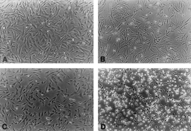
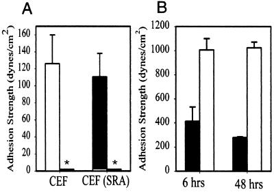
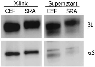
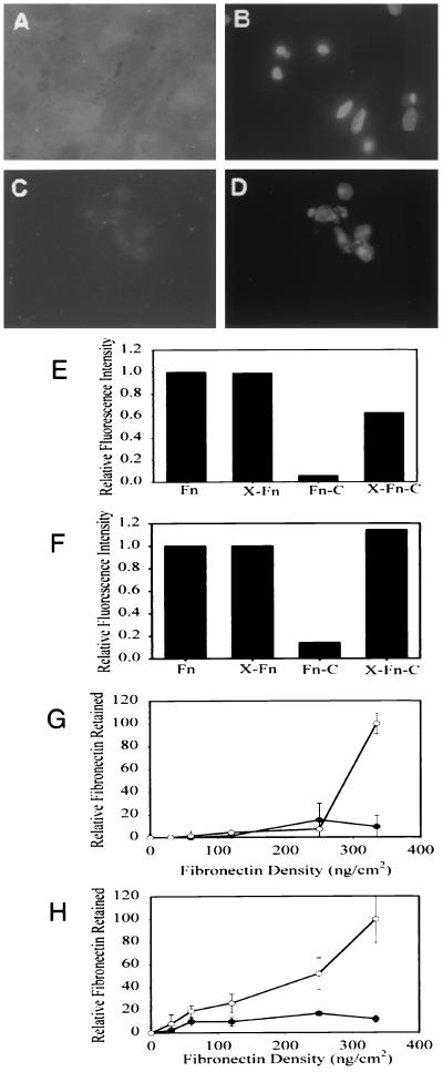
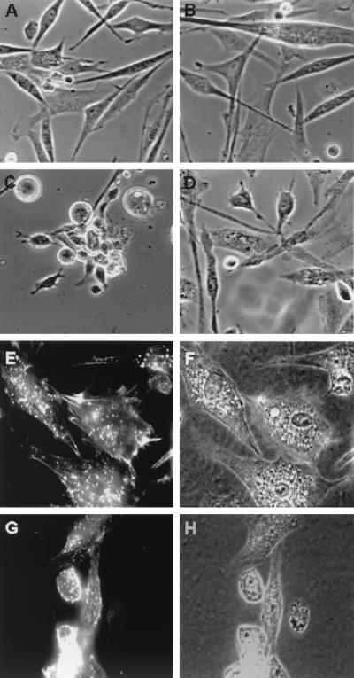
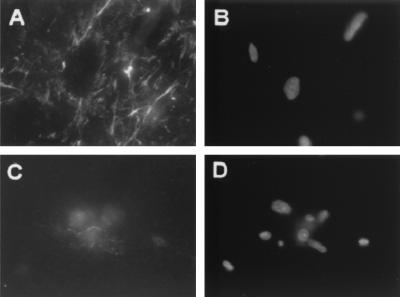
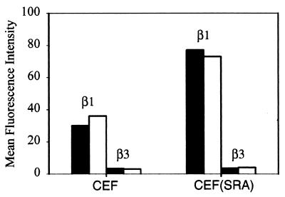
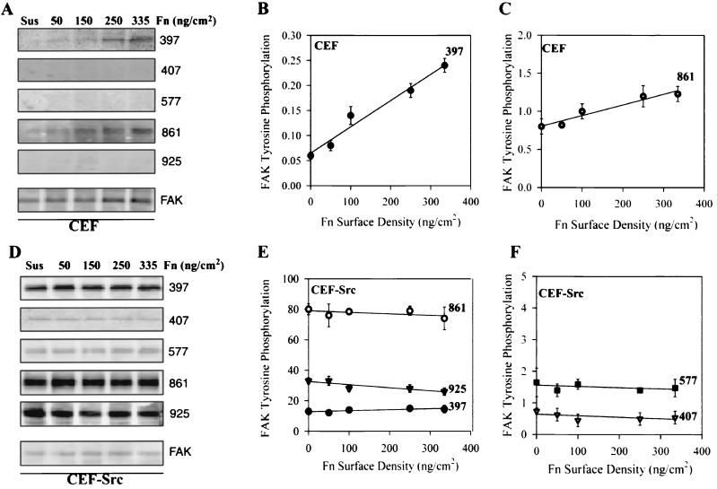
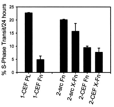
Adhesion of cells to extracellular matrix is mediated by integrin family receptors. The process of receptor-ligand binding is dependent on metabolic energy and is regulated by intracellular signals, termed inside-out signals. The strength of the initial alpha5beta1-mediated adhesion of v-src-transformed chicken embryo fibroblasts (v-srcCEF) was similar to that of normal CEF. A chemically cross-linked fibronectin substrate was able to restore cell spreading and the ability of v-srcCEF to assemble a fibronectin matrix. Over time, v-srcCEF showed decreased adhesion due to the reduction of alpha5beta1-fibronectin bonds consequent on the reduction of substrate-bound fibronectin due to the secretion of proteases by v-srcCEF. Excess synthesis of hyaluronic acid by v-srcCEF also reduced the alpha5beta1-fibronectin bonds and contributed to cell detachment at later times in culture. Thus, the adhesion defects were not due to a failure of alpha5beta1 function and adhesion of the v-srcCEF was alpha5beta1 dependent. Integrin-mediated adhesion also produces signals that affect cell proliferation and cell differentiation. An early consequence of these "outside-in" signals was the phosphorylation of FAK Y397 in direct proportion to the number of alpha5beta1-fibronectin bonds formed. In contrast, v-srcCEF had an increased level of phosphorylation on five different tyrosines in FAK, and none of these phosphorylation levels were sensitive to the number of alpha5beta1-fibronectin bonds. In the absence of serum, CEF proliferation was sensitive to changes in alpha5beta1-mediated adhesion levels. Transformation by v-src increased the serum-free proliferation rate and made it insensitive to alpha5beta1-mediated adhesion. Thus, the v-srcCEF were insensitive to the normal outside-in signals from alpha5beta1 integrin.
Figures
Similar articles
-
Integrin-dependent PLC-gamma1 phosphorylation mediates fibronectin-dependent adhesion.J Cell Sci. 2005 Feb 1;118(Pt 3):601-10. doi: 10.1242/jcs.01643. Epub 2005 Jan 18. J Cell Sci. 2005. PMID: 15657076
-
v-Src-induced degradation of focal adhesion kinase during morphological transformation of chicken embryo fibroblasts.Oncogene. 1995 Jun 1;10(11):2247-52. Oncogene. 1995. PMID: 7784071
-
SRC-mediated phosphorylation of focal adhesion kinase couples actin and adhesion dynamics to survival signaling.Mol Cell Biol. 2004 Sep;24(18):8113-33. doi: 10.1128/MCB.24.18.8113-8133.2004. Mol Cell Biol. 2004. PMID: 15340073 Free PMC article.
-
Integrin-mediated signaling in normal and malignant cells: a role of protein tyrosine kinases.Biochim Biophys Acta. 1996 Jun 7;1287(2-3):73-6. doi: 10.1016/0304-419x(96)00008-x. Biochim Biophys Acta. 1996. PMID: 8672530 Review. No abstract available.
-
Collaboration of fibronectin matrix with other extracellular signals in morphogenesis and differentiation.Curr Opin Cell Biol. 2016 Oct;42:1-6. doi: 10.1016/j.ceb.2016.03.014. Epub 2016 Apr 7. Curr Opin Cell Biol. 2016. PMID: 27062478 Free PMC article. Review.
Cited by
-
Src kinase mediates productive endocytic sorting of reovirus during cell entry.J Virol. 2011 Apr;85(7):3203-13. doi: 10.1128/JVI.02056-10. Epub 2011 Jan 19. J Virol. 2011. PMID: 21248042 Free PMC article.
-
Coordinate regulation of estrogen-mediated fibronectin matrix assembly and epidermal growth factor receptor transactivation by the G protein-coupled receptor, GPR30.Mol Endocrinol. 2009 Jul;23(7):1052-64. doi: 10.1210/me.2008-0262. Epub 2009 Apr 2. Mol Endocrinol. 2009. PMID: 19342448 Free PMC article.
-
A novel mode for integrin-mediated signaling: tethering is required for phosphorylation of FAK Y397.Mol Biol Cell. 2003 Oct;14(10):4306-15. doi: 10.1091/mbc.e03-01-0046. Epub 2003 Sep 5. Mol Biol Cell. 2003. PMID: 12960434 Free PMC article.
-
Effects of Atypical Protein Kinase C Inhibitor (DNDA) on Lung Cancer Proliferation and Migration by PKC-ι/FAK Ubiquitination Through the Cbl-b Pathway.Onco Targets Ther. 2020 Feb 24;13:1661-1676. doi: 10.2147/OTT.S224866. eCollection 2020. Onco Targets Ther. 2020. PMID: 32158232 Free PMC article.
-
FRNK blocks v-Src-stimulated invasion and experimental metastases without effects on cell motility or growth.EMBO J. 2002 Dec 2;21(23):6289-302. doi: 10.1093/emboj/cdf631. EMBO J. 2002. PMID: 12456636 Free PMC article.
References
-
- Ali I U, Maunter V, Lanza R, Hynes R O. Restoration of normal morphology, adhesion and cytoskeleton in transformed cells by addition of a transformation-sensitive surface protein. Cell. 1977;11:115–126. - PubMed
-
- Bell S M, Brackenbury R W, Leslie N D, Degen J L. Plasminogen activator gene expression is induced by the src oncogene product and tumor promoters. J Biol Chem. 1990;265:1333–1338. - PubMed
-
- Boschek C B, Jockusch B M, Friis R R, Back R, Grundmann E, Bauer H. Early changes in the distribution and organization of microfilament proteins during cell transformation. Cell. 1981;24:175–184. - PubMed
-
- Chen W-T, Chen J-M, Parsons S J, Parsons J T. Local degradation of fibronectin at sites of expression of the transforming gene product pp60src. Nature. 1985;316:156–158. - PubMed
MeSH terms
Substances
LinkOut - more resources
Full Text Sources
Molecular Biology Databases
Miscellaneous