. 2001 Nov;21(21):7307-19.
doi: 10.1128/MCB.21.21.7307-7319.2001.

Distinct RNP complexes of shuttling hnRNP proteins with pre-mRNA and mRNA: candidate intermediates in formation and export of mRNA

Affiliations

Distinct RNP complexes of shuttling hnRNP proteins with pre-mRNA and mRNA: candidate intermediates in formation and export of mRNA

S Mili et al. Mol Cell Biol. 2001 Nov.

Abstract

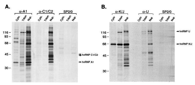
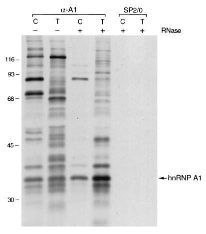
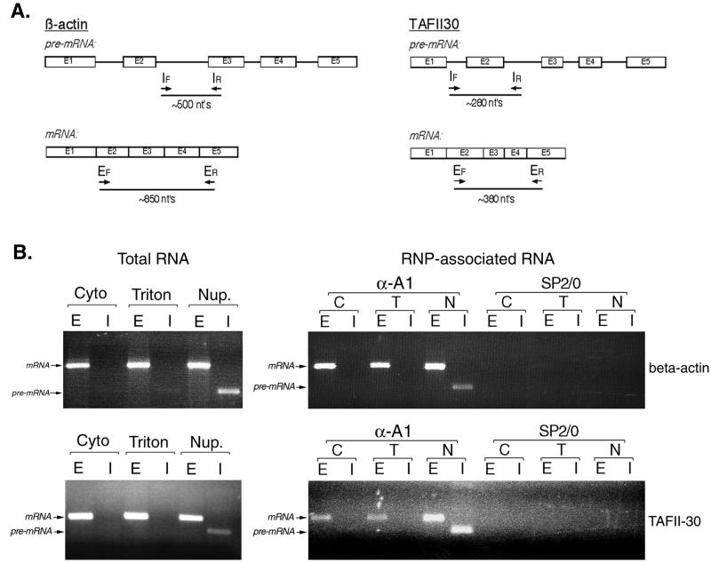
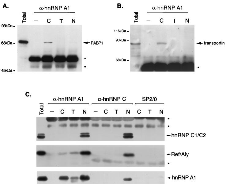
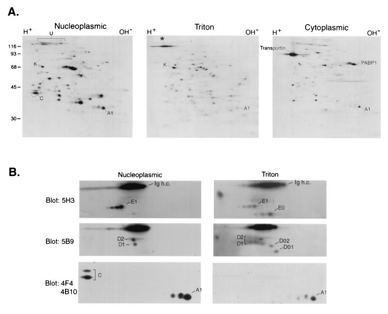
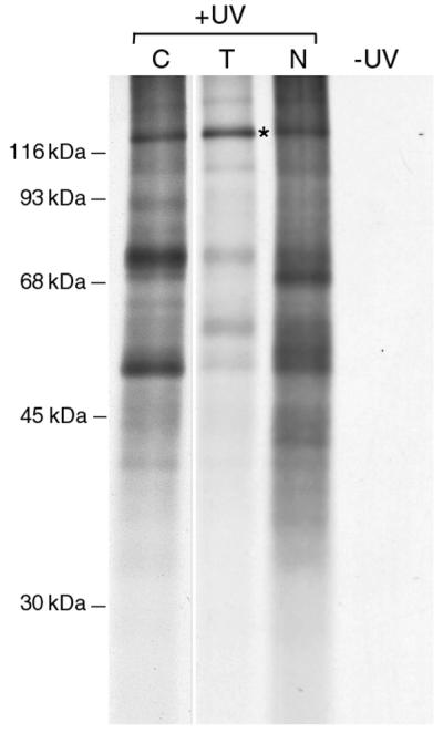
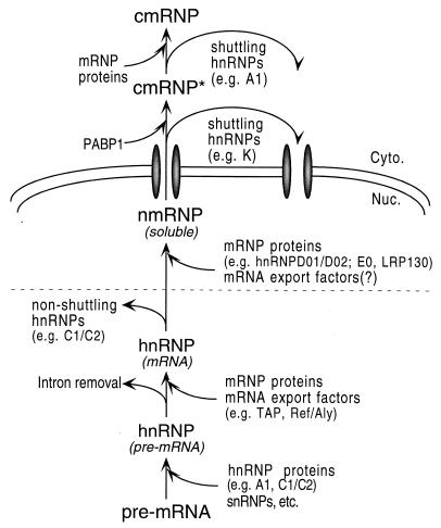
Nascent pre-mRNAs associate with hnRNP proteins in hnRNP complexes, the natural substrates for mRNA processing. Several lines of evidence indicate that hnRNP complexes undergo substantial remodeling during mRNA formation and export. Here we report the isolation of three distinct types of pre-mRNP and mRNP complexes from HeLa cells associated with hnRNP A1, a shuttling hnRNP protein. Based on their RNA and protein compositions, these complexes are likely to represent distinct stages in the nucleocytoplasmic shuttling pathway of hnRNP A1 with its bound RNAs. In the cytoplasm, A1 is associated with its nuclear import receptor (transportin), the cytoplasmic poly(A)-binding protein, and mRNA. In the nucleus, A1 is found in two distinct types of complexes that are differently associated with nuclear structures. One class contains pre-mRNA and mRNA and is identical to previously described hnRNP complexes. The other class behaves as freely diffusible nuclear mRNPs (nmRNPs) at late nuclear stages of maturation and possibly associated with nuclear mRNA export. These nmRNPs differ from hnRNPs in that while they contain shuttling hnRNP proteins, the mRNA export factor REF, and mRNA, they do not contain nonshuttling hnRNP proteins or pre-mRNA. Importantly, nmRNPs also contain proteins not found in hnRNP complexes. These include the alternatively spliced isoforms D01 and D02 of the hnRNP D proteins, the E0 isoform of the hnRNP E proteins, and LRP130, a previously reported protein with unknown function that appears to have a novel type of RNA-binding domain. The characteristics of these complexes indicate that they result from RNP remodeling associated with mRNA maturation and delineate specific changes in RNP protein composition during formation and transport of mRNA in vivo.


Figures

FIG. 1
FIG. 1
Immunopurification of complexes containing hnRNP proteins from different subcellular fractions. (A) HeLa cells were labeled with [35S]methionine and fractionated as described in the text. Immunopurifications were carried out from each fraction with monoclonal antibodies to hnRNP A1 (4B10) or hnRNP C1/C2 (4F4) or nonimmune parent myeloma immunoglobulins (SP2/0). The positions of hnRNP A1 and hnRNP C1/C2 in the gel are indicated on the right. The asterisk indicates the 130-kDa protein discussed in the text. Cyto., cytosol; Triton, Triton-extracted fraction; Nup., nucleoplasmic fraction. (B) Immunopurifications carried out under conditions identical to those shown in panel A, using monoclonal antibodies to hnRNP K/J (12G4) and hnRNP U (3G6). The positions of hnRNP K/J and hnRNP U in the gel are indicated on the right. Positions of molecular mass standards (in kilodaltons) are indicated on the left.
FIG. 2
FIG. 2
RNase sensitivity of hnRNP A1-associated complexes. Complexes associated with hnRNP A1 were isolated from the soluble cytosolic fraction (lanes C) or Triton-soluble nuclear fraction (lanes T), as shown in Fig. 1, without (lanes −) or with (lanes +) prior digestion of the fractions with RNase A. The position of hnRNP A1 in the gel is indicated on the right. Identical immunopurifications were carried out with nonimmune parent myeloma immunoglobulins (SP2/0). Positions of molecular mass standards (in kilodaltons) are indicated on the left.
FIG. 3
FIG. 3
Subcellular distribution and RNP association of specific pre-mRNAs and mRNAs. (A) Diagram depicting the positions in the actin and TAFII30 pre-mRNA and mRNA of the primers used for RT-PCR analysis. Subscripts F and R refer to the forward and reverse primers, respectively, used in the reactions. (B) Left panel, RT-PCR analysis of the distribution of pre-mRNA and mRNA for the transcripts shown in panel A, using total RNA from the various subcellular fractions as a template. Right panel, analysis of hnRNP A1-associated RNA in complexes isolated from the different subcellular fractions. Lanes E, RT-PCR products when all primers correspond to exon sequences. Lanes I, use of at least one intron-specific primer in the RT-PCR. Abbreviations are as in Fig. 1 and 2.
FIG. 4
FIG. 4
Immunoblot analysis of immunopurified RNPs. HeLa cells were fractionated and RNP complexes were immunopurified from each fraction with monoclonal antibodies to hnRNP A1 (4B10) or hnRNP C1/C2 (4F4) or nonimmune antibodies (SP2/0). The proteins in the complexes were resolved by SDS-PAGE, transferred to nitrocellulose, and probed with monoclonal antibodies to PABP1 (A) or to transportin (B) or with antibodies against hnRNP A1 (4B10), hnRNP C (4F4), and REF (C). Lanes C, T, and N, cytosol, Triton-extracted, and nucleoplasmic fractions, respectively. Asterisks denote the positions of the heavy or light chains from antibodies used for immunopurification. Lanes −, mock immunopurifications in which cellular material was omitted.
FIG. 5
FIG. 5
Two-dimensional gel electrophoresis of hnRNP A1-associated RNP complexes. (A) Proteins in complexes immunopurified with 4B10 from each subcellular fraction of [35S]methionine-labeled cells were resolved by two-dimensional gel electrophoresis as described in the text and visualized by autoradiography. The positions of proteins discussed in the text are indicated. (B) Immunoblot analysis. Nucleoplasmic and Triton-soluble nuclear RNPs resolved by two-dimensional gel electrophoresis as for panel A were immunoblotted with the indicated monoclonal antibodies. The positions of the hnRNP D, E, A1, and C1/C2 proteins and corresponding alternatively spliced isoforms are indicated. Ig h.c., position of the heavy chains from antibodies used for immunopurification.
FIG. 6
FIG. 6
Selection of proteins bound to poly(A)+ RNA in vivo in the different subcellular fractions. HeLa cells were exposed to UV light to induce covalent protein-RNA cross-links, and the cells were fractionated into cytoplasmic (lane C), Triton-extracted (lane T), and nucleoplasmic (lane N) fractions. The cross-linked complexes were selected from each fraction by oligo(dT) chromatography under protein-denaturing conditions. Bound proteins were released from the cross-linked complexes by digestion with RNase, resolved by SDS-PAGE, and visualized by autoradiography. The position of LRP130 is indicated with an asterisk. A shorter exposure of the lane corresponding to cytoplasmic cross-linked proteins is shown here for clarity.
FIG. 7
FIG. 7
Model for the sequential changes in protein composition of complexes associated with pre-mRNA and mRNA during maturation and nuclear export of mRNA. See text for details.

References

    1. Adam S A, Sterne-Marr R, Gerace L. Nuclear protein import in permeabilized mammalian cells requires soluble cytoplasmic factors. J Cell Biol. 1990;111:807–816. - PMC - PubMed
    1. Altschul S F, Madden T L, Schaffer A A, Zhang J, Zhang Z, Miller W, Lipman D J. Gapped BLAST and PSI-BLAST: a new generation of protein database search programs. Nucleic Acids Res. 1997;25:3389–3402. - PMC - PubMed
    1. Arao Y, Kuriyama R, Kayama F, Kato S. A nuclear matrix-associated factor, SAF-B, interacts with specific isoforms of AUF/hnRNP D. Arch Biochem Biophys. 2000;380:228–236. - PubMed
    1. Bachi A, Braun I C, Rodrigues J P, Panté N, Ribbeck K, von Kobbe C, Kutay U, Wilm M, Görlich D, Carmo-Fonseca M, Izaurralde E. The C-terminal domain of TAP interacts with the nuclear pore complex and promotes export of specific CTE-bearing RNA substrates. RNA. 2000;6:136–158. - PMC - PubMed
    1. Biemann K. Mass spectrometry of peptides and proteins. Annu Rev Biochem. 1992;61:977–1010. - PubMed

Publication types

MeSH terms

Substances

LinkOut - more resources