. 2001 Nov;21(21):7366-79.
doi: 10.1128/MCB.21.21.7366-7379.2001.

Absence of Dbp2p alters both nonsense-mediated mRNA decay and rRNA processing

Affiliations

Absence of Dbp2p alters both nonsense-mediated mRNA decay and rRNA processing

A T Bond et al. Mol Cell Biol. 2001 Nov.

Abstract

Dbp2p, a member of the large family of DEAD-box proteins and a yeast homolog of human p68, was shown to interact with Upf1p, an essential component of the nonsense-mediated mRNA decay pathway. Dbp2p:Upf1p interaction occurs within a large conserved region in the middle of Upf1p that is largely distinct from its Nmd2p and Sup35/45p interaction domains. Deletion of DBP2, or point mutations within its highly conserved DEAD-box motifs, increased the abundance of nonsense-containing transcripts, leading us to conclude that Dbp2p also functions in the nonsense-mediated mRNA decay pathway. Dbp2p, like Upf1p, acts before or at decapping, is predominantly cytoplasmic, and associates with polyribosomes. Interestingly, Dbp2p also plays an important role in rRNA processing. In dbp2Delta cells, polyribosome profiles are deficient in free 60S subunits and the mature 25S rRNA is greatly reduced. The ribosome biogenesis phenotype, but not the mRNA decay function, of dbp2Delta cells can be complemented by the human p68 gene. We propose a unifying model in which Dbp2p affects both nonsense-mediated mRNA decay and rRNA processing by altering rRNA structure, allowing specific processing events in one instance and facilitating dissociation of the translation termination complex in the other.


Figures
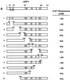
FIG. 1
FIG. 1
Mapping Upf1p-Dbp2p interaction domains. (A) Schematic representation of full-length Upf1p (amino acids 1 to 971) with known motifs denoted by shaded boxes and the Nmd2p interaction domain underlined. Interaction of full-length GAL4(AD)-UPF1, and fragments thereof, with lexA(DB)-DBP2 (amino acids 272 to 546) was assayed by measuring the extent of resistance to 3-AT. (B) Schematic representation of full-length Dbp2p (amino acids 1 to 546) with known RNA helicase domains and RGG motifs denoted by Roman numerals and shaded boxes. GAL4(AD)-UPF1 (full-length) interaction with lexA(DB)-DBP2 was assayed by measuring the extent of resistance to 3-AT. (C) GAL4(AD)-UPF1 or GAL4(AD) interaction with lexA(DB)-DBP2 (amino acids 272 to 546) or lexA(DB)-p68 (amino acids 253 to 614) assayed by determining the extent of resistance to 3-AT and by the production of β-galactosidase activity in individual clones analyzed on X-Gal plates. For the 3-AT assay, the highest concentration of 3-AT (on SC-his-leu-trp plates) that still allowed substantial cellular growth is noted; -his, cells could grow in the absence of histidine but were unable to grow in the presence of 5 mM 3-AT.
FIG. 1
FIG. 1
Mapping Upf1p-Dbp2p interaction domains. (A) Schematic representation of full-length Upf1p (amino acids 1 to 971) with known motifs denoted by shaded boxes and the Nmd2p interaction domain underlined. Interaction of full-length GAL4(AD)-UPF1, and fragments thereof, with lexA(DB)-DBP2 (amino acids 272 to 546) was assayed by measuring the extent of resistance to 3-AT. (B) Schematic representation of full-length Dbp2p (amino acids 1 to 546) with known RNA helicase domains and RGG motifs denoted by Roman numerals and shaded boxes. GAL4(AD)-UPF1 (full-length) interaction with lexA(DB)-DBP2 was assayed by measuring the extent of resistance to 3-AT. (C) GAL4(AD)-UPF1 or GAL4(AD) interaction with lexA(DB)-DBP2 (amino acids 272 to 546) or lexA(DB)-p68 (amino acids 253 to 614) assayed by determining the extent of resistance to 3-AT and by the production of β-galactosidase activity in individual clones analyzed on X-Gal plates. For the 3-AT assay, the highest concentration of 3-AT (on SC-his-leu-trp plates) that still allowed substantial cellular growth is noted; -his, cells could grow in the absence of histidine but were unable to grow in the presence of 5 mM 3-AT.
FIG. 2
FIG. 2
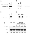
Nonsense-containing transcripts are stabilized in a dbp2Δ strain. Total RNA from yeast strains of the indicated genotypes was isolated and analyzed by Northern hybridization. The blots were hybridized with radioactive probes that detected the CYH2 pre-mRNA and mRNA (A and E); the can1–100 mRNA (B); the mini-pgk1 nonsense-containing mRNA (C); and full-length PGK1 mRNAs harboring either an early (panel D, lanes 1 and 3) or late (panel D, lanes 2 and 4) nonsense codon. The transcripts analyzed in panels C and D were expressed from plasmids. In panel E, galactose was added at t0 to cells growing in raffinose, and samples were taken every 2 h. The ratios of CYH2 pre-mRNA/mRNA are indicated below each lane. WT, wild-type.
FIG. 3
FIG. 3
Mutations in conserved domains of Dbp2p affect the abundance of nonsense-containing transcripts. (A) Schematic diagram of Dbp2p showing the amino acids that were changed. (B) Northern analysis of total RNA from wild-type, dbp2Δ, and dbp2Δ cells transformed with a CEN plasmid containing one of the four dbp2 alleles. Blots were hybridized with a CYH2 probe as described in the legend to Fig. 2. Ratios of CYH2 pre-mRNA/mRNA are indicated below each lane. WT, wild type.
FIG. 4
FIG. 4
Nonsense-containing mRNAs are predominantly full-length in dbp2Δ cells. Total RNA was isolated from yeast strains of the indicated genotypes, and the 5′ end of the CYH2 pre-mRNA was analyzed by primer extension. DNA sequencing reactions with the same primer (run on lanes G, A, T, and C) were used to determine the positions of the primer extension products. The major transcriptional start sites for the CYH2 pre-mRNA and decapped mRNA are indicated by an arrow. The atypical extension products detected in the xm1Δ strain are marked by asterisks. WT, wild type.
FIG. 5
FIG. 5
Translational fidelity is altered in dbp2Δ cells. Cultures of the identified genotypes were diluted serially and spotted on SC-arg plates containing 0, 50, 75, 100, or 200 μg of canavanine per ml. Cells were analyzed after 7 days of growth at 30°C. WT, wild type.
FIG. 6
FIG. 6
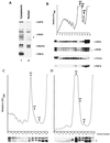
Dbp2p copurifies with nuclei and cofractionates with polyribosomes. (A) Western blot analysis of cytoplasm and purified nuclei isolated from strain BJ2168. Samples loaded represented equivalent numbers of cells. (B) An extract of SWP154 cells (51) was fractionated on a 15 to 50% sucrose gradient that was subsequently analyzed by Western blotting. (Top) Absorbance at 260 nm (OD260), with sedimentation proceeding from right to left. The 80S, 60S, and 40S ribosome peaks are indicated by arrows. (Bottom) Western blot analysis of gradient fractions 1 to 9 and the pellet fraction (P) included the entire sample, whereas fractions 10 to 13 included only one-fifth of the sample. The blots in both panels were serially stripped and reprobed with the indicated antibodies. The asterisk denotes Dbp2p. The upper band in the α-DEAD panels is a mixture of the comigrating proteins Dbp1p and Ded1p (4). (C and D) Cultures of SJ21R (PRT1) (C) and TP11B-4-1 (prt1-1) (D) were grown at 23°C and shifted to 37°C for 30 min. Extracts were prepared, fractionated, and analyzed as for panel B.
FIG. 7
FIG. 7
Amino acid incorporation in wild-type (WT) and mutant strains. (A) Incorporation of 35S-labeled amino acids was measured in HFY1200 (wild type), yATB100 (dbp2Δ), yATB101 (K163R), yATB102 (E268D), yATB103 (T300A), and yATB104 (R447K) as described in Materials and Methods. The values are given as the percentage incorporation, with the wild type taken as 100%, and are the averages of at least five samples. The error bars denote the standard deviations of the five separate samples. (B) Cells were subjected to galactose induction for different lengths of time, and incorporation of 35S-labeled amino acids was measured as described in Materials and Methods. Data are expressed as the percentage of incorporation at t0 and are the averages of triplicate samples. Squares depict HFY1200 cells (wild type), circles depict yATB100 cells (dbp2Δ), and triangles depict yATB200 cells (dbp2Δ+p68).
FIG. 8
FIG. 8
Polysome profiles are altered in dbp2Δ cells. Cytoplasmic extracts were prepared from wild-type (A and C) and dbp2Δ (B and D) cells and fractionated on either 15 to 47% (A and B) or 7 to 25% (C and D) sucrose gradients as described in Materials and Methods. The A260 trace of the gradients is shown, with the polysome and 80S, 60S, and 40S ribosome peaks indicated. Cytoplasmic extracts prepared from yATB200 (dbp2Δ+p68) cells, grown in raffinose (E) or induced for 4 h in galactose (F), were fractionated on 7 to 47% sucrose gradients.
FIG. 9
FIG. 9
Accumulation of rRNA precursor species in dbp2Δ cells. (A) Schematic of the yeast 35S pre-rRNA and its principal processing sites, derived from a figure in reference . The locations of the oligonucleotide probes used in this study are numbered ( to 6) and highlighted by black bars. (B) Schematic of the processing of the 35S pre-rRNA and its principal products. (C to G) Northern analysis of rRNAs. Total cellular RNA was isolated from HFY1200 (wild type [WT]), yATB100 (dbp2Δ), and yATB101 (K163R) cells and fractionated by Northern blotting, and the same blot was stripped and reprobed repeatedly. (C) Oligonucleotides 2 and 6, complementary to sites within the mature 18S and 25S rRNA sequences, respectively, were used to probe the blot. (D) The blot was probed with oligonucleotide 1, which is complementary to a portion of the 5′ ETS. (E) The blot was probed with oligonucleotide 3, which hybridizes between site A2 and A3 in ITS1. (F) The blot was probed with oligonucleotide 4, which hybridizes between site A3 and B1 in ITS1. (G) The blot was probed with oligonucleotide 5, which hybridizes upstream of C2 in ITS2. (H) Total cellular RNA was isolated from HFY1200 (wild type) and yATB100 (dbp2Δ) cells and from yATB200 (dbp2Δ+p68) cells at 0, 1, 2, 4, and 8 h after induction with galactose. Oligonucleotides 2 and 6, complementary to sites within the mature 18S and 25S rRNA sequences, respectively, were used to probe the blot.
FIG. 9
FIG. 9
Accumulation of rRNA precursor species in dbp2Δ cells. (A) Schematic of the yeast 35S pre-rRNA and its principal processing sites, derived from a figure in reference . The locations of the oligonucleotide probes used in this study are numbered ( to 6) and highlighted by black bars. (B) Schematic of the processing of the 35S pre-rRNA and its principal products. (C to G) Northern analysis of rRNAs. Total cellular RNA was isolated from HFY1200 (wild type [WT]), yATB100 (dbp2Δ), and yATB101 (K163R) cells and fractionated by Northern blotting, and the same blot was stripped and reprobed repeatedly. (C) Oligonucleotides 2 and 6, complementary to sites within the mature 18S and 25S rRNA sequences, respectively, were used to probe the blot. (D) The blot was probed with oligonucleotide 1, which is complementary to a portion of the 5′ ETS. (E) The blot was probed with oligonucleotide 3, which hybridizes between site A2 and A3 in ITS1. (F) The blot was probed with oligonucleotide 4, which hybridizes between site A3 and B1 in ITS1. (G) The blot was probed with oligonucleotide 5, which hybridizes upstream of C2 in ITS2. (H) Total cellular RNA was isolated from HFY1200 (wild type) and yATB100 (dbp2Δ) cells and from yATB200 (dbp2Δ+p68) cells at 0, 1, 2, 4, and 8 h after induction with galactose. Oligonucleotides 2 and 6, complementary to sites within the mature 18S and 25S rRNA sequences, respectively, were used to probe the blot.
FIG. 10
FIG. 10
The dbp2 null mutation leads to reduced synthesis of the mature 25S rRNA. Strains HFY1200 (wild type) and yATB100 (dbp2Δ) were grown in SC-met medium. Cells were pulse-labeled (p) for 1 min with [methyl-3H]methionine and then chased (c) for 2, 5, and 15 min with unlabeled methionine. Total RNA was isolated, and equal counts per minute were run on a 1.2% agarose-formaldehyde gel, transferred to a Zeta-probe membrane, and visualized by fluorography. The positions of the pre-rRNAs and mature rRNAs are indicated.

Similar articles

Cited by

References

    1. Applequist S E, Selg M, Raman C, Jack H M. Cloning and characterization of HUPF1, a human homolog of the Saccharomyces cerevisiae nonsense mRNA-reducing UPF1 protein. Nucleic Acids Res. 1997;25:814–821. - PMC - PubMed
    1. Atkin A L, Altamura N, Leeds P, Culbertson M R. The majority of yeast UPF1 co-localizes with polyribosomes in the cytoplasm. Mol Biol Cell. 1995;6:611–625. - PMC - PubMed
    1. Atkin A L, Schenkman L R, Eastman M, Dahlseid J N, Lelivelt M J, Culbertson M R. Relationship between yeast polyribosomes and Upf proteins required for nonsense mRNA decay. J Biol Chem. 1997;272:22163–22172. - PubMed
    1. Barta I, Iggo R. Autoregulation of expression of the yeast Dbp2p ‘DEAD-box’ protein is mediated by sequences in the conserved DBP2 intron. EMBO J. 1995;14:3800–3808. - PMC - PubMed
    1. Belk J P, He F, Jacobson A. Overexpression of truncated Nmd3p inhibits protein synthesis in yeast. RNA. 1999;5:1055–1070. - PMC - PubMed

Publication types

MeSH terms