. 2001 Nov;21(21):7380-90.
doi: 10.1128/MCB.21.21.7380-7390.2001.

claudin-18, a novel downstream target gene for the T/EBP/NKX2.1 homeodomain transcription factor, encodes lung- and stomach-specific isoforms through alternative splicing

Affiliations

claudin-18, a novel downstream target gene for the T/EBP/NKX2.1 homeodomain transcription factor, encodes lung- and stomach-specific isoforms through alternative splicing

T Niimi et al. Mol Cell Biol. 2001 Nov.

Abstract

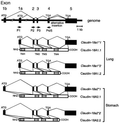
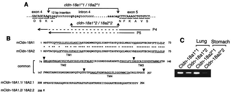
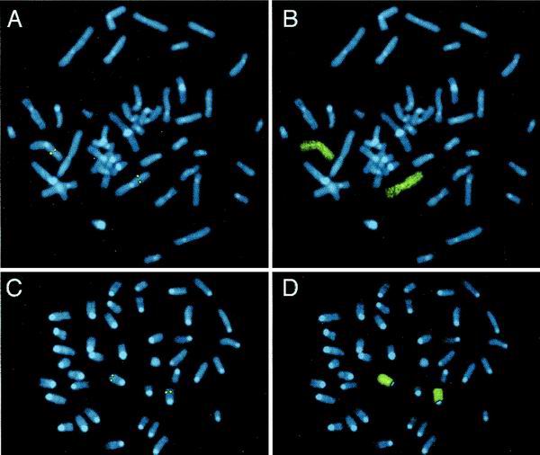
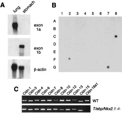
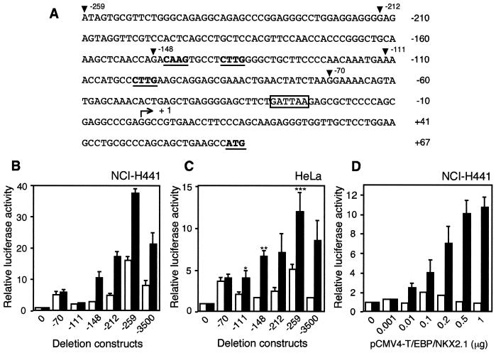
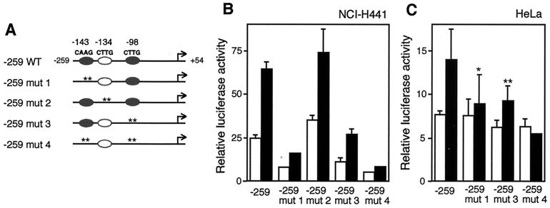
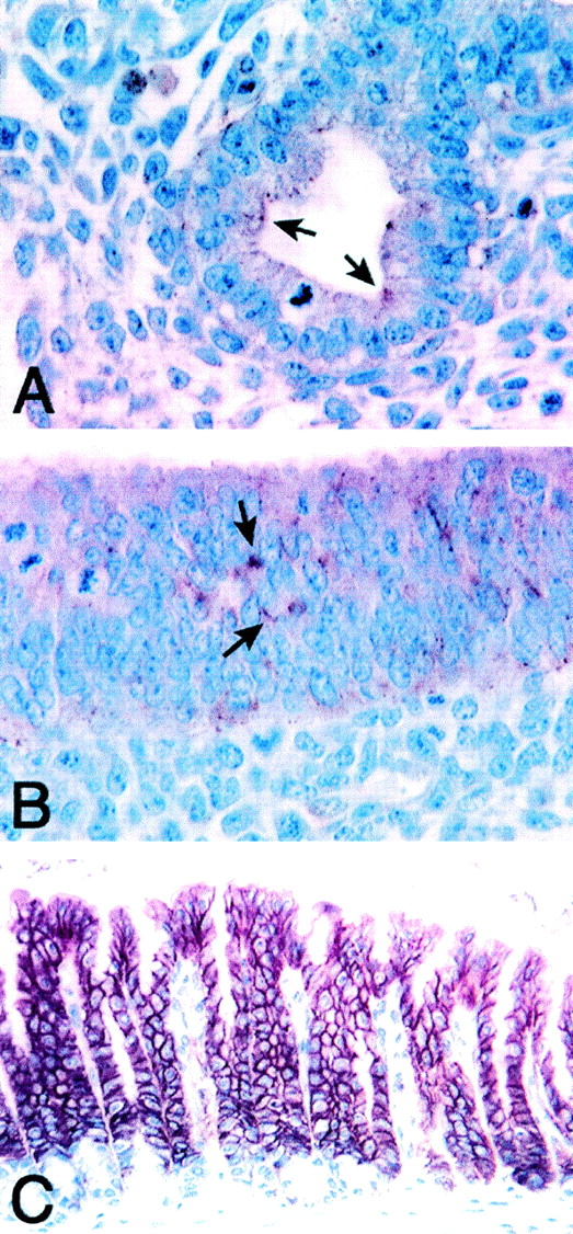
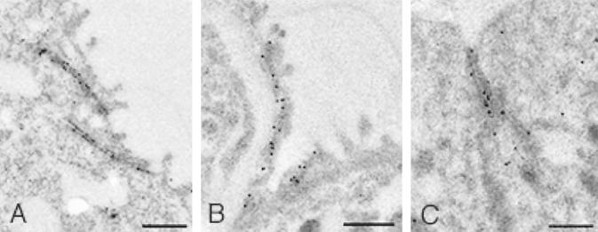
T/EBP/NKX2.1, a member of the NKX family of homeodomain-containing transcription factors, regulates the expression of a number of genes in lung and thyroid. Here we describe the isolation and characterization of a novel target gene, termed claudin-18, that is down-regulated in the lungs of T/ebp/Nkx2.1-null mouse embryos. The gene product exhibits an amino acid sequence similar to those of the claudin multigene family of proteins that constitute tight junction strands in epithelial cells. The gene was localized by fluorescence in situ hybridization to mouse chromosome 9 at region 9E3-F1 and to human chromosome 3 at region 3q21-23. The claudin-18 gene has two promoters, each with its own unique exon 1 that is spliced to common exons 2 through 5. Alternative usage of these promoters leads to production of lung and stomach-specific transcripts. The downstream lung-specific promoter contains two T/EBP/NKX2.1 binding sites responsible for trans activation of the gene by T/EBP/NKX2.1 in lung cells. Only claudin-18 was down-regulated in T/ebp/Nkx2.1-null embryo lungs among 11 claudin transcripts examined. Furthermore, the claudin-18 transcript has an alternative 12-bp insertion derived from the 5' end of intron 4, which produces a C-terminally truncated isoform in lung and stomach. Immunohistochemistry demonstrated complete membrane localization of claudin-18 with small focal dots in the lung and stomach epithelial cells. Immunogold electron microscopy analysis revealed that claudin-18 is concentrated at the cell-cell borders of epithelial cells. These unique features suggest a potentially important role for claudin-18 in the structure and function of tight junctions in lung and stomach.


Figures

FIG. 1
FIG. 1
Schematic representation of four types of mouse claudin-18 transcripts and polypeptides. Organization of exons and introns and predictions of how the four types of transcripts are produced are shown. Solid boxes represent exons. The positions of the translation initiation and termination codons defining open reading frames are indicated. The alternatively inserted 12-bp sequence after exon 4 is depicted by an open box. Arrows indicate the positions of primers used for RT-PCR analysis (P1 to P5). Shaded boxes represent four transmembrane domains (TM1 to TM4). The nomenclature for the genes and their respective polypeptides is shown on the right for lung and stomach forms.
FIG. 2
FIG. 2
(A) Alternative splicing of the mouse claudin-18 gene between exons 4 and 5. The intron 4,5′-splice donor gt, and 3′-splice acceptor ag are in boldface. Amino acid residues are shown below each underlined codon, and the termination codon is marked with an asterisk. The claudin (cldn)-18a1∗1/18a2∗1 gene encodes the amino acid sequence DSNFK at the exon-intron junction, whereas the claudin-18a1∗2/18a2∗2 gene encodes the amino acid sequence DSK. The P4 primer has a continuous sequence of exons 4 and 5 without alternative insertion, and P5 is an alternative insertion-specific primer. The primer sequences are indicated by solid lines. Primer pairs P3-P4 and P3-P5 (Fig. 1) produce fragments specific to claudin-18a1∗1/2∗1 and claudin-18a1∗2/2∗2, respectively. (B) Deduced amino acid sequences of four types of claudin-18 polypeptides. The locations of the putative transmembrane domains are underlined and indicated as TM1 to TM4. Amino acids that are identical in claudin-18A1 and -18A2 are indicated by asterisks. The exon-intron junctions are indicated by arrowheads. (C) RT-PCR confirmation of expression of the four types of transcripts. Products obtained with primer pairs P3-P4 and P3-P5 using mouse adult lung and stomach mRNAs are shown.
FIG. 3
FIG. 3
Chromosomal localization. Human (A) and mouse (C) chromosome spreads after hybridization with species-specific biotin-labeled claudin-18 gene probes are shown. Both chromosomes 3 in human and chromosomes 9 in mouse exhibit symmetrical fluorescent signals at regions 3q21-23 and 9E3-F1, respectively, as depicted by enhanced DAPI-induced chromosome banding. To confirm the identity of chromosomes with specific signals, metaphases from panels A and C were rehybridized with human chromosome painting probe for human chromosome 3 (B) and mouse chromosome 9 (D).
FIG. 4
FIG. 4
Expression of claudin-18 transcripts. (A) Northern blot analysis using exon-specific probes in adult mouse lung and stomach. Ten micrograms of total RNA from the indicated tissues was consecutively hybridized with each probe shown on the right. (B) Dot blot analysis of claudin-18 mRNA in human tissues. RNA sources are as follows: A1, whole brain; A2, amygdala; A3, caudate nucleus; A4, cerebellum; A5, cerebral cortex; A6, frontal lobe; A7, hippocampus; A8, medulla oblongata; B1, occipital lobe; B2, putamen; B3, substantia nigra; B4, temporal lobe; B5, thalamus; B6, subthalamic nucleus; B7, spinal cord; C1, heart; C2, aorta; C3, skeletal muscle; C4, colon; C5, bladder; C6, uterus; C7, prostate; C8, stomach; D1, testis; D2, ovary; D3, pancreas; D4, pituitary gland; D5, adrenal gland; D6, thyroid gland; D7, salivary gland; D8, mammary gland; E1, kidney; E2, liver; E3, small intestine; E4, spleen; E5, thymus; E6, peripheral leukocyte; E7, lymph node; E8, bone marrow; F1, appendix; F2, lung; F3, trachea; F4, placenta; G1, fetal brain; G2, fetal heart; G3, fetal kidney; G4, fetal liver; G5, fetal spleen; G6, fetal thymus; and G7, fetal lung. (C) Differential expression of claudin mRNAs in wild-type and T/ebp/Nkx2.1-null embryo lungs. Total RNAs prepared from E12.5 embryonic lungs of wild-type (WT) or T/ebp/Nkx2.1-null mutant (−/−) mice were analyzed by RT-PCR using various claudin primers as indicated. Primer pair P1-P2 (shown in Fig. 1) was used for claudin-18a1.
FIG. 5
FIG. 5
T/EBP/NKX2.1 activates transcription of the mouse claudin-18a1 gene. (A) Sequence of the mouse claudin-18a1 gene promoter. Arrowheads indicate the positions of the pGL3-259, -212, -148, -111 and -70 deletion constructs. The minimal T/EBP/NKX2.1 binding consensus sequences (5′-CAAG-3′) (19) and ATG initiation codon are shown in boldface and underlined. A TATA-like box is boxed. The bent arrow with + 1 indicates the major transcription start site. (B and C) Deletion analysis of the mouse claudin-18a1 gene promoter. The relative luciferase activities in NCI-H441 cells (B) or HeLa cells (C) transiently transfected with the indicated deletion constructs are shown. Activities obtained in the presence of cotransfected pCMV4-T/EBP/NKX2.1 (black bars) or pCMV4 (white bars) are relative to the activity of pGL3-Basic (construct 0). Values represent the means ± SD from three separate experiments. ∗, significantly different from the control pCMV4 vector at a P value of <0.05, ∗∗, significantly different from construct pGL3-111 with the expression plasmid at a P value of <0.01; ∗∗∗, significantly different from construct pGL3-212 with the expression plasmid at a P value of <0.05. (D) Dose-dependent increase of claudin-18a1 reporter activity with pCMV4-T/EBP/NKX2.1. Increasing concentrations of pCMV4-T/EBP/NKX2.1 (black bars) or pCMV4 (white bars) were cotransfected with the construct pGL3-259 in NCI-H441 cells. Luciferase activities are relative to the activity of construct pGL3-259 without any cotransfected vectors. Values represent the means ± SD from three separate experiments.
FIG. 6
FIG. 6
Mutation of T/EBP/NKX2.1 binding sites reduces trans activation by T/EBP/NKX2.1. (A) Schematic representation of wild-type (−259 WT) and mutant (−259 mut 1 to 4) constructs. Closed ovals represent potential T/EBP/NKX2.1 binding sites. Open ovals depict functionless T/EBP/NKX2.1 binding consensus sites. Site-specific mutations are indicated by asterisks (each asterisk corresponds to a nucleotide). The mutated sequences are shown in Fig. 7A. (B and C) The deletion −259 construct with or without various site-specific mutations was transiently transfected into NCI-H441 cells (B) or HeLa cells (C) in the presence of coexpressed pCMV4-T/EBP/NKX2.1 (black bars) or pCMV4 (white bars). Values represent the means ± SD from three separate experiments. ∗, significantly different from construct −259 with the expression vector at a P value of <0.01; ∗∗, significantly different from construct −259 with the expression vector at a P value of <0.05.
FIG. 7
FIG. 7
The T/EBP/NKX2.1 minimal consensus sequence in the mouse claudin-18a1 gene promoter specifically binds to NCI-H441 nuclear proteins. (A) Sequences of probes and competitors used in electrophoretic gel mobility shift analysis. Oligo C is taken from the rat thyroglobulin promoter (rTG) (−82 to −56 bp), which had been identified as a T/EBP/NKX2.1 binding site (7). Putative T/EBP/NKX2.1 consensus sequences (19) are shown in boldface and underlined. (B and C) Electrophoretic mobility shift assay of the T/EBP/NKX2.1 binding site. NCI-H441 nuclear extracts were incubated with 32P-labeled probe I (B) or probe II (C). The specifically retarded complex is shown by an arrowhead. Competition assays were performed with 100-fold (C) or 100- and 500-fold (B) excesses of unlabeled specific (lanes s), nonspecific (lanes ns), mutated (lanes mut 1 and mut 2), or oligo C (lanes c) oligonucleotides. For antibody supershift analysis, TTF1-specific monoclonal antibody was added to the reaction mixture (lanes Ab +). Asterisks indicate the position of the supershifted complex.
FIG. 8
FIG. 8
Immunolocalization of claudin-18 in mouse lung and stomach. E13.5 whole normal mouse embryo (A and B) and normal adult mouse stomach (C) were fixed in Bouin's fixatives and were subjected to immunohistochemistry. (A) Lung bronchial epithelial cells. (B) Glandular stomach epithelial cells. Positive staining is shown by arrows. (C) Glandular stomach epithelial layers. Magnification, ×300.
FIG. 9
FIG. 9
Ultrastructural localization of claudin-18A1.1 and claudin-18A2.1 in mouse lung and stomach, respectively. IEM of E15.5 embryo (A) and adult (B) normal mouse lungs and of adult stomach (C) is shown. Note that the immunogold labeling is localized at TJs. Bars, 0.5 μm.

Similar articles

Cited by

References

    1. Bohinski R J, Di Lauro R, Whitsett J A. The lung-specific surfactant protein B gene promoter is a target for thyroid transcription factor 1 and hepatocyte nuclear factor 3, indicating common factors for organ-specific gene expression along the foregut axis. Mol Cell Biol. 1994;14:5671–5681. - PMC - PubMed
    1. Bruno M D, Bohinski R J, Huelsman K M, Whitsett J A, Korfhagen T R. Lung cell-specific expression of the murine surfactant protein A (SP-A) gene is mediated by interactions between the SP-A promoter and thyroid transcription factor-1. J Biol Chem. 1995;270:6531–6536. - PubMed
    1. Cardoso W V. Lung morphogenesis revisited: old facts, current ideas. Dev Dyn. 2000;219:121–130. - PubMed
    1. Cereijido M, Shoshani L, Contreras R G. Molecular physiology and pathophysiology of tight junctions. I. Biogenesis of tight junctions and epithelial polarity. Am J Physiol Gastrointest Liver Physiol. 2000;279:G477–G482. - PubMed
    1. Cereijido M, Valdes J, Shoshani L, Contreras R G. Role of tight junctions in establishing and maintaining cell polarity. Annu Rev Physiol. 1998;60:161–177. - PubMed

MeSH terms