. 2001 Nov;21(21):7429-41.
doi: 10.1128/MCB.21.21.7429-7441.2001.

Downregulation of the Ras-mitogen-activated protein kinase pathway by the EphB2 receptor tyrosine kinase is required for ephrin-induced neurite retraction

Affiliations

Downregulation of the Ras-mitogen-activated protein kinase pathway by the EphB2 receptor tyrosine kinase is required for ephrin-induced neurite retraction

S Elowe et al. Mol Cell Biol. 2001 Nov.

Abstract

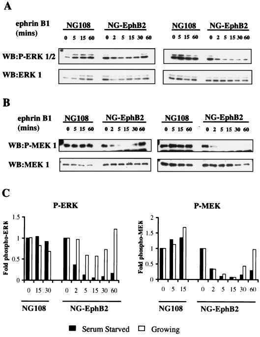
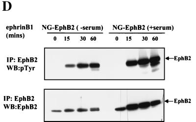
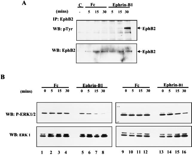
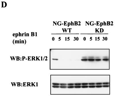
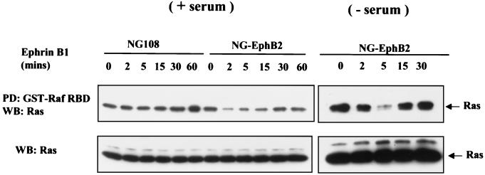
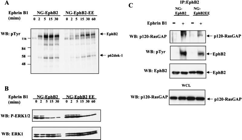
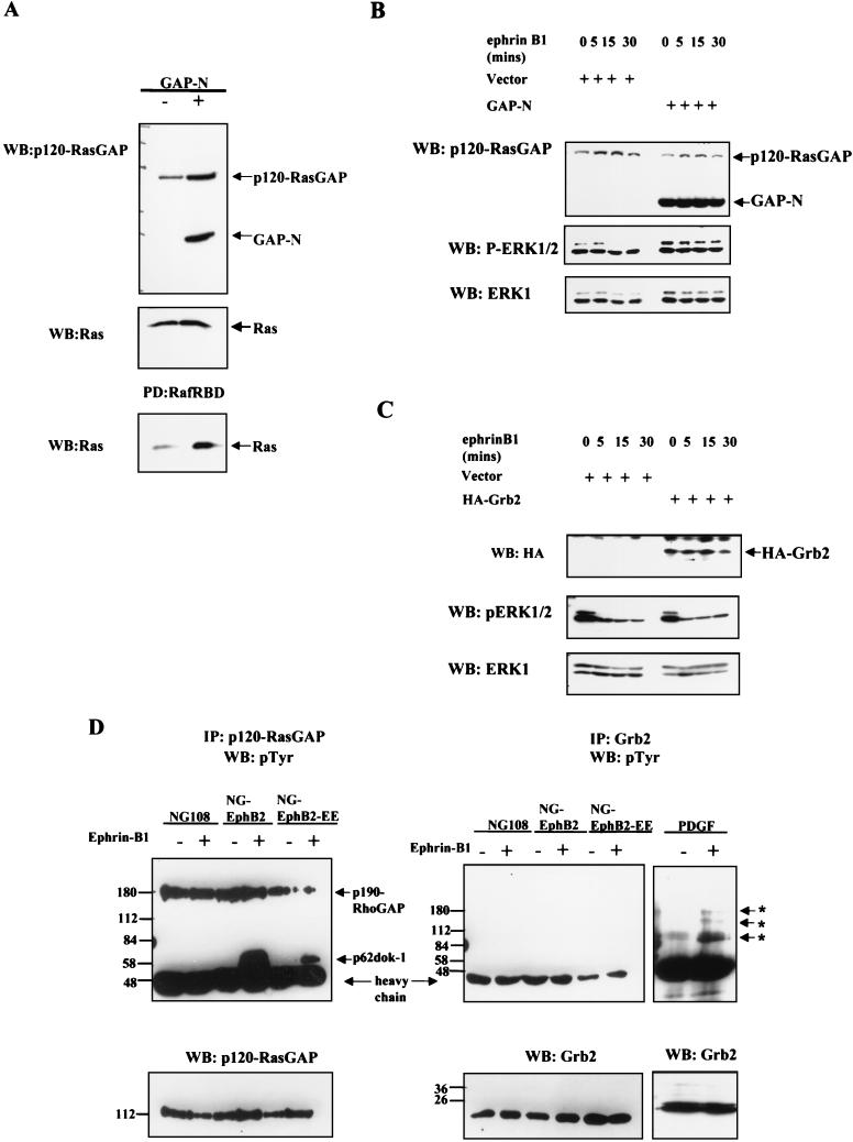
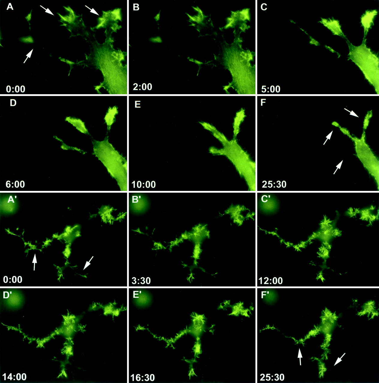
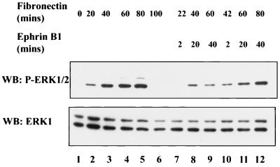
Activation of the EphB2 receptor tyrosine kinase by clustered ephrin-B1 induces growth cone collapse and neurite retraction in differentiated NG108 neuronal cells. We have investigated the cytoplasmic signaling events associated with EphB2-induced cytoskeletal reorganization in these neuronal cells. We find that unlike other receptor tyrosine kinases, EphB2 induces a pronounced downregulation of GTP-bound Ras and consequently of the extracellular signal-regulated kinase (ERK) mitogen-activated protein kinase (MAPK) pathway. A similar inhibition of the Ras-MAPK pathway was observed on stimulation of endogenous EphB2 in COS-1 cells. Inactivation of Ras, induced by ephrin B1 stimulation of NG108 neuronal cells, requires EphB2 tyrosine kinase activity and is blocked by a truncated form of p120-Ras GTPase-activating protein (p120-RasGAP), suggesting that EphB2 signals through the SH2 domain protein p120-RasGAP to inhibit the Ras-MAPK pathway. Suppression of Ras activity appears functionally important, since expression of a constitutively active variant of Ras impaired the ability of EphB2 to induce neurite retraction. In addition, EphB2 attenuated the elevation in ERK activation induced by attachment of NG108 cells to fibronectin, indicating that the EphB2 receptor can modulate integrin signaling to the Ras GTPase. These results suggest that a primary function of EphB2, a member of the most populous family of receptor tyrosine kinases, is to inactivate the Ras-MAPK pathway in a fashion that contributes to cytoskeletal reorganization and adhesion responses in neuronal growth cones.


Figures

FIG. 1
FIG. 1
Ephrin-B1 stimulation of EphB2 leads to the down regulation of the ERK1/2 MAPK signaling pathway. (A and B) Serum-starved (right panels) or growing (10% FBS) (left panels) parental NG108 or NG-EphB2 cells were stimulated with 2 μg of clustered Fc-ephrin-B1 per ml for the indicated time points and lysed directly in 2x SDS-PAGE sample buffer. The lysates were electrophoresed and blotted (WB) with antibodies against phosphorylated ERK1/2 (A, top panels), or phosphorylated MEK1 (B, top panels). The blots were stripped and reprobed for total ERK1 or MEK1 (bottom panels, A and B, respectively). (C) Graphical representation of phospho-ERK1/2 and phospho -MEK1 from panels A and B relative to total levels. (D) Time course of EphB2 tyrosine phosphorylation in NG108 cells. NG108 cells were serum starved (lanes −serum) or grown in the presence of serum (lanes +serum) and stimulated with 2 μg of clustered Fc-ephrin-B1 per ml as indicated. Immunoprecipitated (IP) EphB2 was then resolved by SDS-PAGE and probed with anti-pTyr antibodies (upper panel). Blots were subsequently stripped and reprobed with crude anti-EphB2 sera (lower panel).
FIG. 1
FIG. 1
Ephrin-B1 stimulation of EphB2 leads to the down regulation of the ERK1/2 MAPK signaling pathway. (A and B) Serum-starved (right panels) or growing (10% FBS) (left panels) parental NG108 or NG-EphB2 cells were stimulated with 2 μg of clustered Fc-ephrin-B1 per ml for the indicated time points and lysed directly in 2x SDS-PAGE sample buffer. The lysates were electrophoresed and blotted (WB) with antibodies against phosphorylated ERK1/2 (A, top panels), or phosphorylated MEK1 (B, top panels). The blots were stripped and reprobed for total ERK1 or MEK1 (bottom panels, A and B, respectively). (C) Graphical representation of phospho-ERK1/2 and phospho -MEK1 from panels A and B relative to total levels. (D) Time course of EphB2 tyrosine phosphorylation in NG108 cells. NG108 cells were serum starved (lanes −serum) or grown in the presence of serum (lanes +serum) and stimulated with 2 μg of clustered Fc-ephrin-B1 per ml as indicated. Immunoprecipitated (IP) EphB2 was then resolved by SDS-PAGE and probed with anti-pTyr antibodies (upper panel). Blots were subsequently stripped and reprobed with crude anti-EphB2 sera (lower panel).
FIG. 2
FIG. 2
Ephrin-B1 stimulation of COS-1 cells leads to tyrosine phosphorylation of endogenous EphB2 and down regulation of ERK1/2 phosphorylation. (A) COS-1 cells were serum starved for 14 h and were either left untreated (lane C) or stimulated with 8 μg of clustered human Fc (lanes Fc) or Fc-ephrin-B1 (lanes Ephrin-B1) per ml. The cells were subsequently washed twice with PBS and lysed in PLC buffer as indicated in Materials and Methods. Lysates equalized for protein concentration were subjected to immunoprecipitation (IP) with anti-EphB2 antibodies, and immunoprecipitates were subsequently separated by SDS-PAGE and blotted with anti-pTyr antibodies (upper panel). The membranes were then stripped and reprobed with anti-EphB2 antibodies (bottom panel). (B) Serum-starved (left panels) and growing (right panels) COS-1 cells were challenged for the indicated time with 8 μg/ml clustered Fc or Fc-ephrin-B1 and subsequently lysed and immunoblotted (WB) with anti-phospho-ERK1/2 antibodies as indicated in Fig. 1. The membranes were then stripped and reprobed with anti-ERK1 antibodies (B, bottom panels).
FIG. 3
FIG. 3
Kinase activity of EphB2 is required for down regulation of ERK1/2 signaling. (A) Schematic representation of the EphB2 mutant structures, indicating juxtamembrane tyrosines JX1 (Y604), JX2 (Y610) and the conserved SAM domain tyrosine (Y929). (B and C) EphB2-ΔC, a C-terminal truncation, ends at V951 in the SAM domain. NG108 or NG-EphB2 clones expressing WT or mutant receptors were stimulated with 2 μg of clustered Fc-ephrin-B1 per ml for the indicated time points. The cells were harvested directly in 2× SDS sample buffer, and lysates were electrophoresed and immunoblotted (WB) with antibodies against phosphorylated ERK1/2 (B, top panel) or phosphorylated MEK1 (C, top panel). Immunoblots were stripped and reprobed with anti-ERK1 (B, bottom panel) or anti-MEK1 (C, bottom panel). (D) NG108 cells expressing WT EphB2 and kinase-inactive EphB2 (KD) were stimulated with clustered Fc-ephrin-B1 and treated as indicated for panels B and C. Lysates were then probed with anti-phospho-ERK1/2 (upper panel), and anti-ERK1 (lower panel).
FIG. 3
FIG. 3
Kinase activity of EphB2 is required for down regulation of ERK1/2 signaling. (A) Schematic representation of the EphB2 mutant structures, indicating juxtamembrane tyrosines JX1 (Y604), JX2 (Y610) and the conserved SAM domain tyrosine (Y929). (B and C) EphB2-ΔC, a C-terminal truncation, ends at V951 in the SAM domain. NG108 or NG-EphB2 clones expressing WT or mutant receptors were stimulated with 2 μg of clustered Fc-ephrin-B1 per ml for the indicated time points. The cells were harvested directly in 2× SDS sample buffer, and lysates were electrophoresed and immunoblotted (WB) with antibodies against phosphorylated ERK1/2 (B, top panel) or phosphorylated MEK1 (C, top panel). Immunoblots were stripped and reprobed with anti-ERK1 (B, bottom panel) or anti-MEK1 (C, bottom panel). (D) NG108 cells expressing WT EphB2 and kinase-inactive EphB2 (KD) were stimulated with clustered Fc-ephrin-B1 and treated as indicated for panels B and C. Lysates were then probed with anti-phospho-ERK1/2 (upper panel), and anti-ERK1 (lower panel).
FIG. 4
FIG. 4
EphB2 activation by ephrin-B1 causes a transient decrease in the levels of activated p21Ras. (A) Parental NG108 or NG-EphB2 cells in the presence of serum (left panels) or serum starved (right panels) were stimulated for the indicated times with Fc-ephrin-B1 clusters (2 μg/ml), and lysates were mixed with GST-Raf RBD immobilized on glutathione beads in a pull-down (PD) assay. Samples were electrophoresed and immunoblotted (WB) for Ras (upper panel). To demonstrate equivalent protein levels at different time points, equal volumes of whole-cell lysate were electrophoresed and immunoblotted for Ras (lower panel).
FIG. 5
FIG. 5
Role for p120-RasGAP in signaling from EphB2 in NG108 cells. (A) NG-EphB2 and NG-EphB2-EE clones were stimulated with aggregated Fc-ephrinB1 ligand for the indicated times, and whole-cell lysates were separated by SDS-PAGE and immunoblotted (WB) for anti-pTyr. Tyrosine-phosphorylated EphB2 and p62dok-1 are indicated by arrows. (B) Cell lysates from (panel A) were immunoblotted for phosphorylated ERK1/2 (upper panel) and reprobed for ERK1 (lower panel). (C) Serum-starved NG-EphB2 and NG-EphB2-EE cells were incubated in the presence or absence of 2 μg of aggregated ephrin-B1 per ml for 20 min and lysed as indicated in Materials and Methods. EphB2 immunoprecipitates were blotted for p120-RasGAP (upper panel) and reprobed with anti-pTyr antibodies (second panel) and anti-EphB2 (third panel). For control, equivalent amounts of whole-cell lysate (WCL) were immunoblotted for p120-RasGAP to indicate equal input (bottom panel).
FIG. 6
FIG. 6
p120-RasGAP but not Grb2 participates in signaling from EphB2 in NG-108 cells. (A) Lyates from WT Rat2 cells or Rat2 cells stably expressing GAP-N (top panel) were mixed with GST-Raf RBD immobilized on glutathione beads. Samples were electrophoresed and immunoblotted (WB) for Ras (bottom panel). Equivalent aliquots of whole-cell lysate were probed for Ras to demonstrate equal input (middle panel). (B and C) NG-EphB2 cells were transfected with empty vector or GAP-N, as indicated (B), or in parallel empty vector and HA-Grb2 (C). At 48 h after transfection, the cells were challenged with 2 μg of clustered Fc-ephrin-B1 per ml and assayed for expression of p120-RasGAP and GAP-N (B, upper panel), HA-Grb2 (C, upper panel), and phospho-ERK1/2 (B and C, middle panel), then reprobed for total ERK1 (B and C, bottom panel). (D) Parental NG108, NG-EphB2 and NG-EphB2-EE cells were incubated in the absence (−) or presence (+) of clustered Fc-ephrin-B1 for 20 mins. For the Grb2 immunoprecipitation (IP) control, Rat2 cells were incubated in the absence (−) or presence (+) of PDGF for 20 min. Lysates were subsequently immunoprecipitated with either p120-RasGAP (left panel) or Grb2 (middle and right panels) antibodies, and immunoblots were probed with anti-pTyr antibodies. Asterisks indicate tyrosine-phosphorylated proteins in the Grb2 immunoprecipitation control. Lower panels indicate total protein levels.
FIG. 7
FIG. 7
EphB2 down regulation of Ras signaling is required for neurite retraction in NG108 cells. Stable cell lines expressing EphB2 were transfected with either a YFP-actin (yellow fluorescence protein conjugated actin) construct alone (A to F) or YFP-actin and HAp21RasV12 (A′ to F′) and stimulated with 2 μg of clustered ephrin-B1 per ml. Live imaging of cells was performed using an inverted Olympus IX-70 fluorescence microscope for 25 min (A to F) or 40 min (A′ to F′), with frames taken at 30-s intervals. Time points in the stimulation are indicated in minutes:seconds.
FIG. 8
FIG. 8
Integrin-induced stimulation of ERK1/2 phosphorylation is down regulated in response to EphB2 signaling. NG-EphB2 cells were serum starved overnight and subsequently detached and held in suspension for approximately 1 h in the absence of serum. The cells were subsequently plated onto fibronectin-coated six-well plates for the specified times. After incubation of the cells in the absence (lanes 1 to 6) or presence (lanes 7 to 12) of 2 μg of Fc-ephrin-B1 per ml as indicated, the cells were lysed directly in 2× SDS-PAGE sample buffer, separated by electrophoresis, and immunoblotted (WB) with anti-phospho-ERK1/2 antibodies (upper panel) or anti-ERK1 antibodies (lower panel).

Similar articles

Cited by

References

    1. Adams R H, Wilkinson G A, Weiss C, Diella F, Gale N W, Deutsch U, Risau W, Klein R. Roles of ephrinB ligands and EphB receptors in cardiovascular development: demarcation of arterial/venous domains, vascular morphogenesis, and sprouting angiogenesis. Genes Dev. 1999;13:295–306. - PMC - PubMed
    1. Aletta J M, Greene L A. Growth cone configuration and advance: a time-lapse study using video-enhanced differential interference contrast microscopy. J Neurosci. 1988;8:1425–1435. - PMC - PubMed
    1. Bar-Sagi D, Feramisco J R. Microinjection of the ras oncogene protein into PC12 cells induces morphological differentiation. Cell. 1985;42:841–848. - PubMed
    1. Becker E, Huynh-Do U, Holland S, Pawson T, Daniel T O, Skolnik E Y. Nck-interacting Ste20 kinase couples Eph receptors to c-Jun N-terminal kinase and integrin activation. Mol Cell Biol. 2000;20:1537–1545. - PMC - PubMed
    1. Bentley D, O'Connor T P. Cytoskeletal events in growth cone steering. Curr Opin Neurobiol. 1994;4:43–48. - PubMed

Publication types

MeSH terms