. 2001 Nov;21(21):7509-22.
doi: 10.1128/MCB.21.21.7509-7522.2001.

The HOX homeodomain proteins block CBP histone acetyltransferase activity

Affiliations

The HOX homeodomain proteins block CBP histone acetyltransferase activity

W F Shen et al. Mol Cell Biol. 2001 Nov.

Abstract

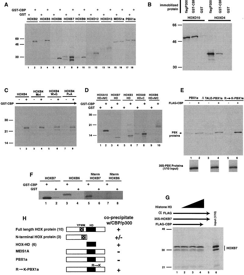
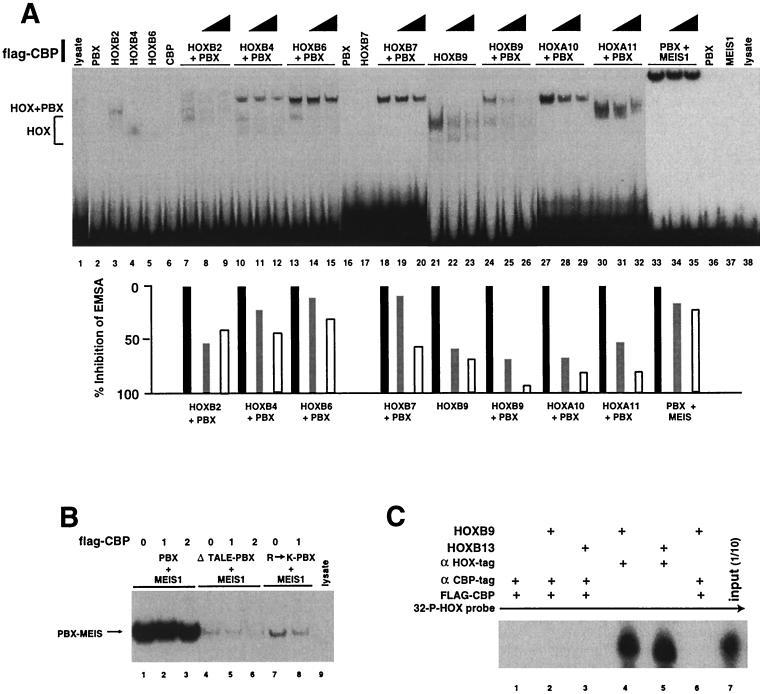
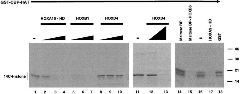
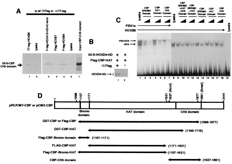
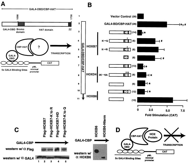
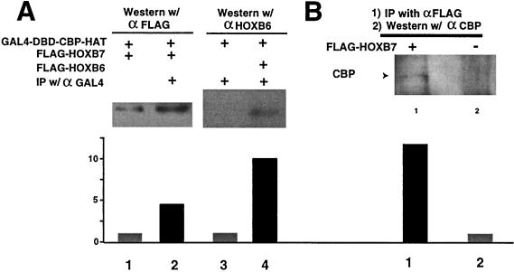
Despite the identification of PBC proteins as cofactors that provide DNA affinity and binding specificity for the HOX homeodomain proteins, HOX proteins do not demonstrate robust activity in transient-transcription assays and few authentic downstream targets have been identified for these putative transcription factors. During a search for additional cofactors, we established that each of the 14 HOX proteins tested, from 11 separate paralog groups, binds to CBP or p300. All six isolated homeodomain fragments tested bind to CBP, suggesting that the homeodomain is a common site of interaction. Surprisingly, CBP-p300 does not form DNA binding complexes with the HOX proteins but instead prevents their binding to DNA. The HOX proteins are not substrates for CBP histone acetyltransferase (HAT) but instead inhibit the activity of CBP in both in vitro and in vivo systems. These mutually inhibitory interactions are reflected by the inability of CBP to potentiate the low levels of gene activation induced by HOX proteins in a range of reporter assays. We propose two models for HOX protein function: (i) HOX proteins may function without CBP HAT to regulate transcription as cooperative DNA binding molecules with PBX, MEIS, or other cofactors, and (ii) the HOX proteins may inhibit CBP HAT activity and thus function as repressors of gene transcription.


Figures

FIG. 1
FIG. 1
HOX proteins coprecipitate with CBP and p300 and interact through the HD. Coprecipitation experiments were performed by incubating 35S-labeled HOX or TALE HD proteins, prepared by in vitro transcription-translation, with immobilized GST-CBP (bromo, HAT, and CH3 domains) (see Fig. 4D for structures), Flag-p300, or GST or with M2 control beads. Following extensive washing, the pellets were solubilized and run on SDS-PAGE gels followed by autoradiography. (A) Representative members of HOX paralog groups bind to CBP. In contrast, two TALE HD proteins, PBX and MEIS, exhibit reduced binding compared to background (lanes 15 to 18; also panel E, lanes 1 and 2). (B) HOX proteins also bind to the full-length p300 protein. (C) The highly conserved YPWM motif within the HOX proteins is not required for CBP binding. Changing the M, W, or P does not prevent HOXB4 interactions with CBP. (D) The HOX HD is sufficient for CBP binding. The constructs are described in Materials and Methods, but all contain the HD alone or with short N-terminal or C-terminal flanking regions. (E) The TALE motif does not prevent PBX binding to CBP, but changing an arginine to lysine within HD helix 3 increases CBP binding to PBX. Δ-TALE PBX represents a mutant PBX protein in which the three-amino-acid loop within the HD has been deleted. R→K-PBX1a represents a mutant PBX protein in which arginine-55 (underlined) within an RYKK motif (numbering according to the Antennapedia HD system) has been changed to lysine-55 to match the KXKK motif found in all of the HOX proteins. (F) HOXB7 N-terminal protein (Nterm HOXB7), but not the HOXB6 N-terminal region (Nterm HOXB6), coprecipitates with CBP. 35S-labeled HOXB7 N-terminal protein (lane 5 versus 6) coprecipitates with GST-CBP. In contrast, HOXB6 N-terminal protein does not bind GST-CBP (lane 7 versus 8). Full-length HOXB6 and HOXB7 proteins were included as positive controls for coprecipitation with GST-CBP (lane 1 to 4). (G) Histone H3 does not effectively compete with HOXB7 for binding to CBP. Approximately 10- to 100-fold excess of purified histone H3 was added to the coprecipitation mixture of Flag-CBP and 35S-labeled HOXB7. The order of addition did not affect the data shown. (H) Summary of the interactions of Hox proteins with CBP/p300. The number of different HOX proteins tested is shown in parentheses. w/, with; +, precipitation; −, no precipitation.
FIG. 2
FIG. 2
CBP prevents DNA binding by HOX proteins. (A and B) EMSA analysis was performed with 32P-labeled oligonucleotides containing cooperative binding sites for PBX and the respective HOX or MEIS proteins. (A) CBP blocks HOX protein DNA binding in EMSA. HOX, PBX, and MEIS proteins were preincubated with 1 or 4 μl of a Flag epitope-tagged CBP protein containing the bromo, HAT, and CH3 domains, synthesized by in vitro transcription-translation, or equivalent amounts of control lysate. The lysate controls did not shift any of the oligonucleotide probes (lanes 1 and 38 and data not shown). Some of the HOX proteins shift DNA alone (lanes 3, 4, and 21), while other HOX proteins do not (lanes 5 and 17). All of the HOX proteins form moderate or strong EMSA bands with PBX (lanes 7, 10, 13, 18, 24, 27, and 30). In each case, addition of approximately 0.5- to 2-fold molar ratio of Flag-CBP resulted in a dose-dependent decrease in the intensities of the EMSA bands, ascribed to binding by either HOX alone or the HOX-PBX complexes. However, Flag-CBP did not compete with DNA-bound PBX-MEIS1a complexes (lanes 33 to 35). The probes used contained the following consensus binding sites: TGATTGAT for lysate (lane 1), PBX1a (lane 2), CBP (lane 6), and HOXB2 through HOXB6 (lanes 3 to 5 and 7 to 15); TGATTTAC for HOXB7 through HOXA11 (lanes 16 to 32); and TGATTGACAG for PBX with MEIS (lanes 33 to 37) and lysate (lane 38). Densitometry scans of EMSA bands are shown below the lanes. Solid bars, no Flag-CBP; shaded and open bars, 1 and 4 μl of Flag-CBP, respectively. (B) The TALE motif does not alter CBP-PBX interactions, but changing R-55 to K-55 increases PBX-CBP binding. PBX and MEIS proteins form a heterodimeric EMSA complex on a consensus DNA target (compare lanes 1 and 9). Removing the three-amino-acid loop (TALE) from PBX1a weakened protein-DNA binding, as measured by EMSA (compare lanes 1 to 3 with 4 to 6), but did not enhance interactions with CBP, which would be demonstrated by reduced gel shift with increasing Flag-CBP (compare lanes 5 and 6 with 4). In contrast, changing arginine-55 to lysine-55 (underlined) to produce a KYKK motif, as described in the legend to Fig. 1, results in a PBX protein that shows reduced gel shift band intensity in the presence of Flag-CBP (compare lanes 7 and 8). This result indicates interaction of these proteins, as reflected by competitive inhibition of R→K PBX molecule binding to DNA by CBP. (C) Immunoprecipitation of CBP-HOX complexes does not coprecipitate DNA prebound to the HOX protein. 32P-labeled DNA in preformed HOXB9-DNA or HOXB13-DNA complexes could be precipitated with antiserum to the HOX epitope tag (lanes 4 and 5). Following addition of Flag-CBP, precipitation with antiserum to the Flag epitope did not yield DNA (lanes 2 and 3), confirming that CBP does not bind to HOX proteins in preformed HOX-DNA binding complexes in vitro. In lane 6, the proteins were preincubated prior to the addition of the DNA, demonstrating that the order of addition did not influence complex formation. Prolonged exposure of the autoradiograph yielded extremely faint bands in lanes 2 and 3, which represented less than 1% of the DNA bound by the HOX proteins. Lane 1, immunoprecipitation control; lane 7, 1/10 input oligonucleotide. +, present.
FIG. 3
FIG. 3
The HOX proteins competitively inhibit CBP HAT activity through the homeodomain but are not substrates. Purified preparations of immobilized GST-CBP HAT domain were incubated with histone H3 substrate in the presence of 14C-labeled acetyl coenzyme A. Assays were performed in the absence (−) of HOX proteins (lanes 1, 11, and 16); with purified bacterially expressed preparations of the full-length, T7-tagged HOXB1 (0.1- to 0.3-fold protein ratio to H3) (lanes 5 to 7); or, in two separate experiments, with HOXD4 protein (0.1- to 1.0-fold protein ratio to H3) (lanes 8 to 10, 12, and 13) or a maltose-binding-HOXB6 fusion protein (0.2-fold ratio to H3) (lane 15). The band of labeled histone, representing CBP HAT activity, was diminished in a dose-dependent manner by the addition of the full-length HOXB1, HOXD4, or HOXB6 protein. Note that new bands representing acetylation of the HOX proteins, which would migrate in the region from approximately 30 to 55 kDa, were not detected, indicating that these proteins are not substrates for CBP HAT. Similar experiments were performed with GST-CBP HAT or Flag-p300 and the HOX proteins in the absence of histone to confirm that HOX proteins were not acetylated (not shown). Affinity-purified bacterially expressed HOXA10 and HOXA9 HD fragments (0.1- to 0.3-fold or 0.2-fold protein ratio to H3) also acted as competitive inhibitors of CBP HAT activity (lanes 2 to 4 and 17, respectively). Again, no bands for 14C-labeled HD fragments, which would migrate at approximately 8 to 10 kDa, were observed. Control affinity-purified maltose binding protein (BP) or GST did not inhibit CBP HAT activity at concentrations similar to those used for the HOX fusion proteins (lanes 14 and 18).
FIG. 4
FIG. 4
Different CBP domains interact with HOX proteins. (A) The CBP CH3 domain interacts with HOX proteins. Coimmunoprecipitation was performed using 35S-labeled CBP CH3 domain together with immobilized T7 or Flag epitope-tagged HOX proteins (lanes 1, 3, 4, and 5) or control beads (lanes 2 and 6). The CBP CH3 polypeptide was specifically precipitated by full-length HOXB9 (compare lanes 1 and 2), as well as HOXB4 and HOXB7 (compare lane 4 or 5 with 6) or the HOXA10 HD plus a short C-terminal (C-term) region (compare lanes 3 and 6). (B) The HOXD4 homeodomain binds to the CBP HAT domain. 35S-labeled T7 epitope-tagged HOXD4-HD and unlabeled Flag-CBP HAT domain protein fragments, synthesized by in vitro translation, were preincubated and subjected to coprecipitation in the presence (+; lane 1) or absence (−; lane 2) of antiserum to the Flag epitope. (C) HOXB9 interacts with the CBP HAT, bromo, and CH3 domains. CBP domains (shown schematically in panel D) were expressed as Flag epitope-tagged proteins by using an in vitro transcription-translation system and used in competitive EMSA assays at approximately one to threefold molar ratio to the HOXB9 protein, as described in Materials and Methods. Each domain was capable of specifically blocking HOXB9-DNA interactions, as reflected by diminution of the EMSA band intensities. (D) CBP expression clones.
FIG. 5
FIG. 5
HOX proteins block in vivo CBP HAT activity. (A) Schematic representation of the assay system used to demonstrate the in vivo importance of CBP HAT activity for reporter gene transcription. A previously described system for demonstrating the importance of HAT activity for the transcriptional effects of CBP (24) was studied in the presence and absence of HOX proteins using transient-transfection assays in 293T cells. (B) HOX proteins block in vivo CBP HAT-mediated gene transcription. In this transient-transfection system, expression of a GAL4-CBP HAT fusion protein stimulates reporter gene expression from a plasmid containing multiple GAL4 binding sites (compare bars 1 and 2). Cotransfection of full-length Flag-HOXB7 (bar 3), HOXD4 (bar 7), or Flag-HOXB6 (bar 10) resulted in complete inhibition of the GAL4-CBP-induced activity (∗, P < 0.001). Flag-HOXB7 proteins containing mutations at Lys-55 within helix 3 of the HD (bars 4 and 5) showed partial inhibition of CBP HAT (#, P < 0.03 for K→Q HOXB7; P = 0.06 for K→R HOXB7), suggesting the importance of this amino acid for CBP interactions. In contrast, a HOXD4 protein containing Lys-Ala substitutions at positions 55, 57, and 58 of the HD (bar 8) still inhibited CBP HAT. Consistent with this finding, the observation that the N-terminal region of HOXD4 lacking the HD (bar 9) also blocks in vivo CBP HAT suggests that this protein does not require the HD for interaction with CBP. However, the N-terminal region of HOXB6 does not block CBP HAT activity (bar 11), suggesting that the HD is required for this HOX-CBP interaction. The isolated Flag-HOXB7 HD fragment (bar 6) exhibited reduced effectiveness in blocking CBP HAT-mediated activation in a statistically significant manner (#, P < 0.03), suggesting that the HOXB7 N-terminal flanking region is required for full in vivo functional interaction with CBP. Statistical differences were calculated compared to the vector control (bar 2). Means and standard deviations are reported. The approximate locations of point mutations in HOXB7 and HOXD4 are denoted by X above the HD. (C) Western blot analysis demonstrating the expression of approximately equal amounts of the various HOX proteins and equal expression of the GAL4-CBP protein in transient-transfection assays. Parallel transfection dishes from the experiments shown in panel B were subjected to Western blotting with antiserum to either the epitope Tag (HOXB7) or the GAL4 fusion moiety (GAL4-CBP HAT) or with antiserum against the N-terminal region of the HOXB6 protein (HOXB6-Nterm). w/, with. (D) Model for HOX protein inhibition of CBP HAT-mediated transcriptional activation. CBP HAT potentiates gene transcription by an unknown mechanism previously shown to depend on CBP HAT activity (panel A). In the model, HOX proteins are proposed to bind to and inhibit the CBP HAT domain activity.
FIG. 6
FIG. 6
HOX proteins bind CBP in vivo. (A) Coimmunoprecipitation of exogenous CBP with exogenous Flag-tagged HOXB7 and HOXB6. Transiently transfected GAL4 DBD-CBP HAT was precipitated from whole 293T cell lysates using antiserum to the GAL4 fusion partner. Following resolution of the precipitated proteins by SDS-PAGE, exogenous Flag-HOXB7 or HOXB6, brought down by coprecipitation, was visualized using specific antiserum to the Flag epitope fused to HOXB7 (lane 2) or antiserum to HOXB6 (lane 4). Minimal or no protein bands were visualized in control lysates lacking the antiserum for GAL4 (lane 1) or HOXB6 (lane 3) protein. (B) Coimmunoprecipitation of transiently transfected HOXB7 and endogenous CBP. Following precipitation of Flag-HOXB7 protein from whole 293T cell lysates with antiserum to the Flag epitope, endogenous CBP protein was detected by Western blotting of precipitated proteins with CBP-specific antiserum (lane 1), while cell lysates containing a control expression vector showed no band for CBP protein (lane 2). Densitometry scans of electrochemiluminescence band intensities are shown below the lanes. +, present; −, absent. y axis shows relative intensity.

Similar articles

Cited by

References

    1. Boyes J, Byfield P, Nakatani Y, Ogryzko V. Regulation of activity of the transcription factor GATA-1 by acetylation. Nature. 1998;396:594–598. - PubMed
    1. Brownell J E, Allis C D. Special HATs for special occasions: linking histone acetylation to chromatin assembly and gene activation. Curr Opin Genet Dev. 1996;6:176–184. - PubMed
    1. Burglin T. A comprehensive classification of homeobox genes. In: Duboule D, editor. Guidebook to the homeobox genes. Oxford, England: Oxford University Press; 1994. pp. 25–71.
    1. Chakravarti D, Ogryzko V, Kao H Y, Nash A, Chen H, Nakatani Y, Evans R M. A viral mechanism for inhibition of p300 and PCAF acetyltransferase activity. Cell. 1999;96:393–403. - PubMed
    1. Chan S K, Popperl H, Krumlauf R, Mann R S. An extradenticle-induced conformational change in a HOX protein overcomes an inhibitory function of the conserved hexapeptide motif. EMBO J. 1996;15:2476–2487. - PMC - PubMed

Publication types