. 2001 Oct 15;21(20):7928-36.
doi: 10.1523/JNEUROSCI.21-20-07928.2001.

Protein kinase c increases the apparent affinity of the release machinery to Ca2+ by enhancing the release machinery downstream of the Ca2+ sensor

Affiliations

Protein kinase c increases the apparent affinity of the release machinery to Ca2+ by enhancing the release machinery downstream of the Ca2+ sensor

X S Wu et al. J Neurosci. .

Abstract

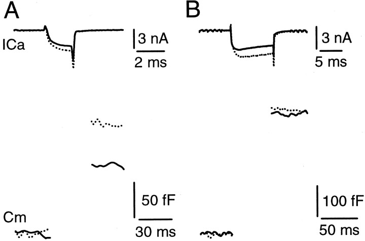
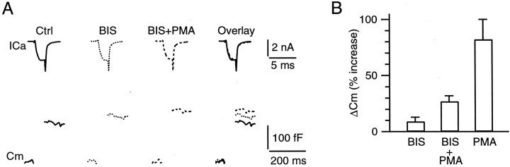
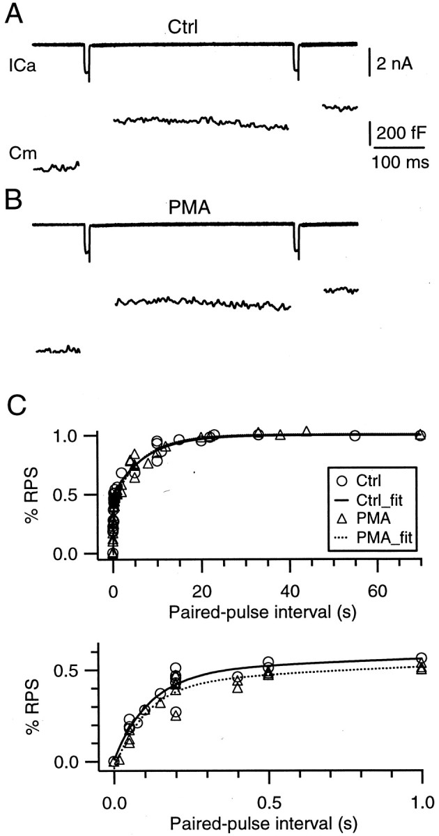
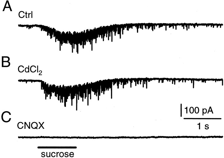
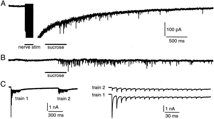
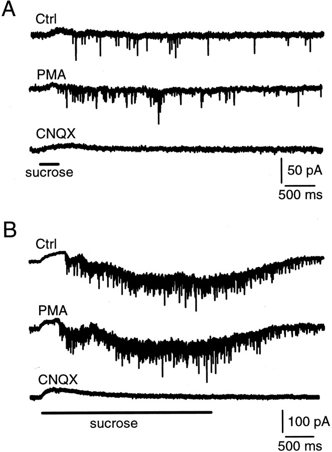
Modulation of the release probability of releasable vesicles in response to Ca(2+) influx (Prob(Ca)) is involved in mediating several forms of synaptic plasticity, including short-term depression, short-term augmentation, and potentiation induced by protein kinases. Given such an important role, however, the mechanism underlying modulation of the Prob(Ca) is unclear. We addressed this question by investigating how the activation of protein kinase C modulates the Prob(Ca) at a calyx-type nerve terminal in rat brainstem. Various lengths of step depolarization were applied to the nerve terminal to evoke different amounts of Ca(2+) currents and capacitance jumps, the latter of which reflect vesicle release. The relationship between the capacitance jump and the Ca(2+) current integral was sigmoidal and was fit well with a Hill function. The sigmoidal relationship was shifted significantly to the left during the application of the PKC activator 12-myristate 13-acetate (PMA), suggesting that PMA increases the apparent affinity of the release machinery to Ca(2+). This effect was blocked in large part by the application of the PKC inhibitor bisindolylmaleimide, suggesting that the effect is mediated mainly by the activation of PKC. We also found that PMA increased the rate of miniature EPSCs evoked by the application of hypertonic sucrose solution, which triggers release downstream of the Ca(2+) influx. Taken together, our results suggest that PKC enhances the apparent affinity of the release machinery to Ca(2+) by a mechanism downstream of the binding between Ca(2+) and its sensor. These results have provided the first example of the mechanisms underlying modulation of the Prob(Ca).


Figures

Fig. 1.
Fig. 1.
The capacitance jumps recorded in different extracellular Ca2+ concentrations. A, The Ca2+ current (top) and the capacitance jump (bottom) evoked by a 2 msec step from −80 to +10 mV in extracellular solutions containing 2 mm(solid trace) and 4 mm (dotted trace) Ca2+. Both the Ca2+ current and the capacitance jump were increased significantly in 4 mm Ca2+.B, The Ca2+ current (top) and the capacitance jump (bottom) evoked by a 10 msec step from −80 to +10 mV in extracellular solutions containing 2 mm (solid trace) and 4 mm (dotted trace) Ca2+. Only the Ca2+ current, but not the capacitance jump, was increased significantly in 4 mmCa2+. Data in A and Bwere obtained from the same calyx.
Fig. 2.
Fig. 2.
PMA increases the capacitance jump by increasing the apparent affinity of the release machinery to Ca2+, but not the releasable pool size.A, B, Sample traces of Ca2+ currents (top) and capacitance jumps (bottom) induced by a 2 msec (A) and a 10 msec (B) step depolarization from −80 to +10 mV before (left, solid trace) and during the application of 100 nm PMA for 10 min (middle, dotted trace). They are superimposed on the right for comparison. C, The relationship between the capacitance jump (ΔCm) and the charge of the Ca2+ current (ICa) obtained before (Ctrl, circles) and during the application of PMA (triangles) for at least 10 min (n = 12 synapses). Before the data were pooled from different synapses, the data were normalized to the value obtained during the 10 msec step depolarization to +10 mV in the control condition. Both the data in control and in the presence of PMA were fit with a Hill equation (see Eq. 1 in Results). The application of PMA did not change two parameters in the equation, theRPS and the n, but decreased the EC50 from 0.30 (solid curve,Ctrl_fit) to 0.16 (dotted curve,PMA_fit).
Fig. 3.
Fig. 3.
A PKC antagonist BIS blocks in large part the effect of PMA on the capacitance jump. A, Sample recordings of presynaptic Ca2+ currents (ICa, top) and capacitance changes (Cm, bottom) evoked by a 2 msec presynaptic step from −80 to +10 mV in the control (left), in the presence of BIS (1 μm) for 10 min (middle left), and in the presence of BIS (1 μm) plus PMA (100 nm) for 10 min (middle right). These traces are superimposed on theright for comparison. B, Percentage of increase in the capacitance jump (ΔCm) evoked by a 2 msec step depolarization to +10 mV during the application of 1 μm BIS (n = 6), 1 μmBIS plus 100 nm PMA (n = 6), and 100 nm PMA (n = 6). The percentages refer to the changes in capacitance jumps during the application of the drug or drugs normalized to the value obtained before any drug application at the same synapse. Data are expressed as means ± SE.
Fig. 4.
Fig. 4.
PMA does not affect the rate of vesicle mobilization. A, B, Sample recordings of Ca2+ currents (top) and capacitance jumps (bottom) induced by a pair of 10 msec step depolarizations (from −80 to +10 mV) with an interval of 400 msec before (A, Ctrl) and during (B) the application of 100 nm PMA. The labels and scales in A apply to B.C, The ratio between the second and the first ΔCm as a function of the paired pulse interval obtained in control and in the presence of PMA (100 nm). Data were obtained from experiments similar to those shown inA and B from 11 synapses. In control, the data were fit with a double-exponential function with time constants of 0.11 and 7.14 sec, respectively (solid curve). PMA does not affect these time constants and the relative contribution of these two components (dotted curve). The topand bottom panels show the same data in different scales.
Fig. 5.
Fig. 5.
Hypertonic sucrose application evokes mEPSCs.A, Shown are the mEPSCs induced by a 1 sec puff application of hypertonic sucrose solution (2 m sucrose plus the bath solution in the pipette). The puff application time is marked in C. B, The Ca2+ channel blocker CdCl2 (200 μm) did not block sucrose-induced mEPSCs.C, The non-NMDA glutamate receptor blocker CNQX (10 μm) blocked sucrose-induced mEPSCs. Calibration applies to all panels. Data in A–C were obtained from the same synapse.
Fig. 6.
Fig. 6.
Release evoked by hypertonic sucrose solution and nerve stimulation share the same vesicle pool. A, A train of nerve stimulations (20 V, 0.1 msec at 100 Hz for 200 msec) was applied, followed at 300 msec after the train by a puff application of sucrose solution (2 m in the pipette) for 500 msec. The EPSC evoked by the train was truncated to see the mEPSCs clearly. Calibration also applies to B. B, The EPSC is evoked by a puff the same as in A but without a conditioning train of stimulation. C, Shown are the EPSCs evoked by two identical electrical trains (20 V, 0.1 msec at 100 Hz for 200 msec) applied to the nerve with an interval of 1 sec (left). The EPSCs evoked by the second train (right, top) were smaller than those evoked by the first train (right, bottom). The stimulation artifacts were blanked.
Fig. 7.
Fig. 7.
PMA enhances the rate of mEPSCs evoked by hypertonic sucrose solution. A, The mEPSCs evoked by a puff application of hypertonic sucrose solution (2 m) for 300 msec in the control (top), in the presence of 100 nm PMA (middle), and in the presence of 10 μm CNQX (bottom). B, The mEPSC evoked by a puff application of hypertonic sucrose solution (2m) for 3 sec in the control (top), in the presence of 100 nm PMA (middle), and in the presence of 10 μm CNQX (bottom). The initial rise of the current is an artifact of the puff application because it was not blocked by CNQX (bottom trace). All data in this figure were obtained from the same synapse.

Similar articles

Cited by

References

    1. Albillos A, Dernick G, Horstmann H, Almers W, Alvarez de Toledo G, Lindau M. The exocytotic event in chromaffin cells revealed by patch amperometry. Nature. 1997;389:509–512. - PubMed
    1. Bachoo M, Heppner T, Fiekers J, Polosa C. A role for protein kinase C in long-term potentiation of nicotinic transmission in the superior cervical ganglion of the rat. Brain Res. 1992;585:299–302. - PubMed
    1. Betz A, Ashery U, Rickmann M, Augustin I, Neher E, Sudhof TC, Rettig J, Brose N. Munc13-1 is a presynaptic phorbol ester receptor that enhances neurotransmitter release. Neuron. 1998;21:123–136. - PubMed
    1. Blackmer T, Larsen EC, Takahashi M, Martin TF, Alford S, Hamm HE. G-protein βγ subunit-mediated presynaptic inhibition: regulation of exocytotic fusion downstream of Ca2+ entry. Science. 2001;292:293–297. - PubMed
    1. Borst JGG, Helmchen F, Sakmann B. Pre- and postsynaptic whole-cell recordings in the medial nucleus of the trapezoid body of the rat. J Physiol (Lond) 1995;489:825–840. - PMC - PubMed

Publication types

MeSH terms