. 2001 Oct 15;21(20):7937-43.
doi: 10.1523/JNEUROSCI.21-20-07937.2001.

Oscillating on borrowed time: diffusible signals from immortalized suprachiasmatic nucleus cells regulate circadian rhythmicity in cultured fibroblasts

Affiliations

Oscillating on borrowed time: diffusible signals from immortalized suprachiasmatic nucleus cells regulate circadian rhythmicity in cultured fibroblasts

G Allen et al. J Neurosci. .

Abstract

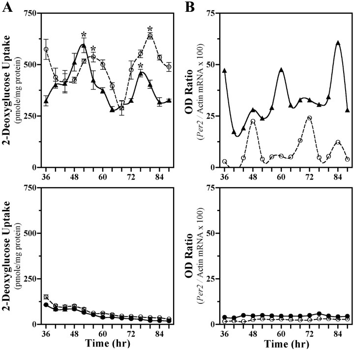
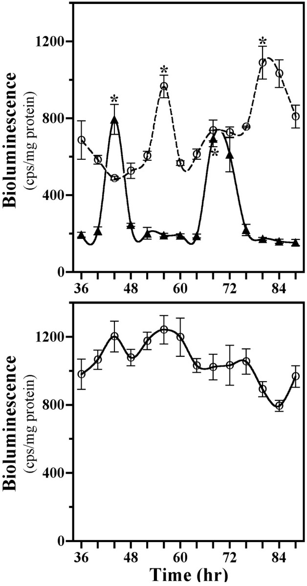
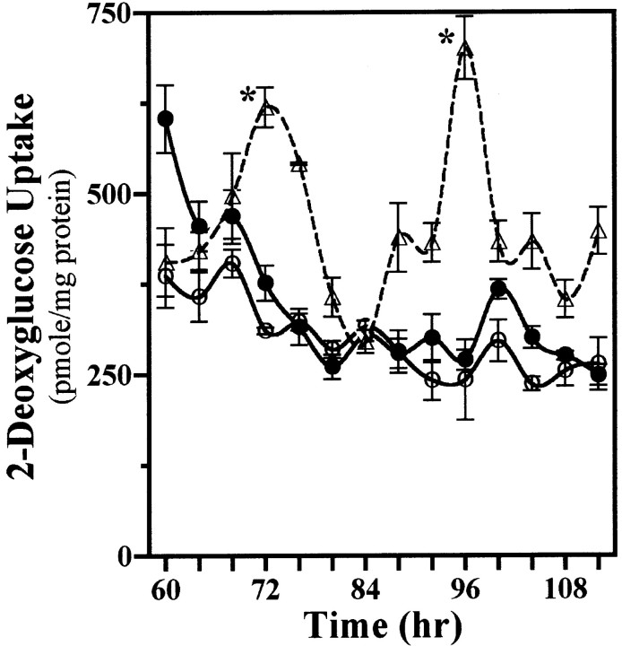
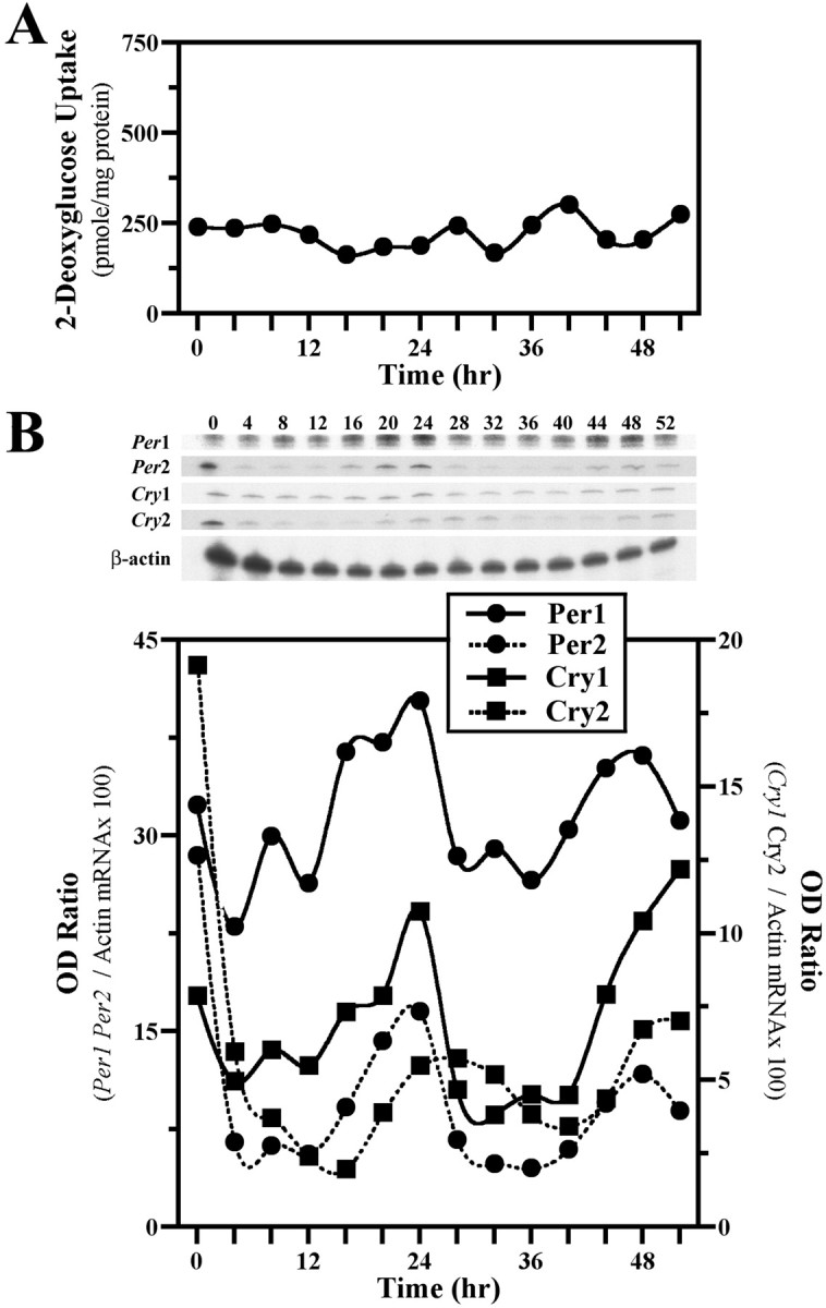
The capacity to generate circadian rhythms endogenously and to confer this rhythmicity to other cells was compared in immortalized cells derived from the suprachiasmatic nucleus (SCN) and a fibroblast line to differentiate SCN pacemaker properties from the oscillatory behavior of non-clock tissues. Only SCN2.2 cells were capable of endogenously generating circadian rhythms in 2-deoxyglucose uptake and Per gene expression. Similar to SCN function in vivo, SCN2.2 cells imposed rhythms of metabolic activity and Per gene expression on cocultured NIH/3T3 fibroblasts via a diffusible signal. The conferred rhythms in NIH/3T3 cells were phase delayed by 4-12 hr relative to SCN2.2 circadian patterns, thus resembling the phase relationship between SCN and peripheral tissue rhythms in vivo. Sustained metabolic rhythmicity in NIH/3T3 cells was dependent on continued exposure to SCN2.2-specific outputs. In response to a serum shock the NIH/3T3 fibroblasts exhibited recurrent oscillations in clock gene expression, but not in metabolic activity. These molecular rhythms in serum-shocked fibroblasts cycled in a phase relationship similar to that observed in the SCN in vivo; peak Per1 and Per2 mRNA expression preceded the rhythmic maxima in Cry1 and Cry2 mRNA levels by 4 hr. Despite these clock gene oscillations the serum-shocked NIH/3T3 cells failed to drive circadian rhythms of Per1 and Per2 expression in cocultures of untreated fibroblasts, suggesting that expression and circadian regulation of the Per and Cry genes are not sufficient to confer pacemaker function. Therefore, SCN-specific outputs are necessary to drive circadian rhythms of metabolic activity, and these output signals are not a direct product of clock gene oscillations.


Figures

Fig. 1.
Fig. 1.
SCN2.2 are distinguished by the capacity to drive metabolic and molecular rhythmicity in cocultured NIH/3T3 fibroblasts. Shown are temporal patterns of 2-deoxyglucose (2-DG) uptake (A) and Per2 expression (B) in cocultures (top) containing SCN2.2 cells on wells (n = 4; solid line, ▴) and NIH/3T3 fibroblasts on inserts (n = 4; dashed line, ○) and in cocultures (bottom) composed of NIH/3T3 cells on both wells (n = 6; solid line, ●) and inserts (n = 6; dashed line, ○). The symbols denote determinations of 2-DG uptake (mean ± SEM) and optical density (OD) ratios ofPer2/β-actin mRNA signal at 4 hr intervals. With the exception of Figure 4, determinations in this and subsequent figures are plotted as a function of time such that time 0denotes when cells located on companion wells and inserts were cocultured together first. Asterisks indicate sampling intervals during which peak values for 2-DG uptake were significantly greater (p < 0.05) than those observed during the preceding or succeeding minima.
Fig. 2.
Fig. 2.
Profiles of Per1/luctransgene expression in cocultures (top) containing SCN2.2 cells on wells (n = 3; solid line, ▴) and NIH/3T3 fibroblasts on inserts (n = 3; dashed line, ○) and in NIH/3T3 cells cultured alone (bottom) on wells (n = 3; solid line, ○). Thesymbols denote determinations ofPer1-driven luciferase bioluminescence (mean ± SEM) at 4 hr intervals. Asterisks indicate sampling intervals during which peak values for Per1-driven luciferase bioluminescence were significantly greater (p < 0.05) than those observed during the preceding or succeeding minima.
Fig. 3.
Fig. 3.
Conferred rhythmicity in NIH/3T3 cells persists only in the presence of SCN2.2 cells. Shown is a “kick-start” analysis of 2-DG uptake in SCN2.2 cells on inserts (n = 3; dashed line, ▵) and NIH/3T3 fibroblasts on wells (n = 3; solid line, ●) that were cocultured together for only 24 hr and maintained separately thereafter. NIH/3T3 fibroblasts cultured alone on wells (n = 3; solid line, ○) are shown for comparison. The symbols denote mean ± SEM determinations of 2-DG uptake at 4 hr intervals.Asterisks indicate sampling intervals during which peak values were significantly greater (p < 0.05) than those observed during the preceding or succeeding minima.
Fig. 4.
Fig. 4.
Serum shock induces molecular, but not metabolic, rhythmicity in NIH/3T3 fibroblasts. Shown are temporal patterns of 2-DG uptake (A) and Clock gene expression (B) in cultures of NIH/3T3 cells after a 2 hr exposure to a 50% serum shock. Determinations of 2-DG uptake and OD ratios of mPer1, mPer2, mCry1, or mCry2/β-actin mRNA signal at 4 hr intervals are plotted such that time 0 coincides with the conclusion of the serum shock treatment.
Fig. 5.
Fig. 5.
Serum-shocked NIH/3T3 fibroblasts express molecular oscillations but cannot drive rhythmicity in cocultured cells. Shown are temporal patterns of mPer1(top) and mPer2(bottom) mRNA expression in NIH/3T3–NIH/3T3 cocultures. NIH/3T3 cells on wells (SS-NIH/3T3; solid line, ●) were exposed to a 2 hr serum shock (50%) and then cocultured with untreated NIH/3T3 fibroblasts on inserts (UT-NIH/3T3;dashed line, ○). The symbols denote OD ratios of mPer1 or mPer2/β-actin mRNA signal at 4 hr intervals.

References

    1. Aguilar-Roblero R, Morin LP, Moore RY. Morphological correlates of circadian rhythm restoration induced by transplantation of the suprachiasmatic nucleus in hamsters. Exp Neurol. 1994;130:250–260. - PubMed
    1. Akashi M, Nishida E. Involvement of the MAP kinase cascade in resetting of the mammalian circadian clock. Genes Dev. 2000;14:645–649. - PMC - PubMed
    1. Bae K, Jin X, Maywood ES, Hastings MH, Reppert SM, Weaver DR. Differential functions of mPer1, mPer2, and mPer3 in the SCN circadian clock. Neuron. 2001;30:525–536. - PubMed
    1. Balsalobre A, Damiola F, Schibler U. A serum shock induces circadian gene expression in mammalian tissue culture cells. Cell. 1998;93:929–937. - PubMed
    1. Balsalobre A, Brown SA, Marcacci L, Tronche F, Kellendonk C, Reichardt HM, Schutz G, Schibler U. Resetting of circadian time in peripheral tissues by glucocorticoid signaling. Science. 2000;289:2344–2347. - PubMed

Publication types

MeSH terms

LinkOut - more resources