. 2001 Oct 15;21(20):7954-68.
doi: 10.1523/JNEUROSCI.21-20-07954.2001.

Myr 8, a novel unconventional myosin expressed during brain development associates with the protein phosphatase catalytic subunits 1alpha and 1gamma1

Affiliations

Myr 8, a novel unconventional myosin expressed during brain development associates with the protein phosphatase catalytic subunits 1alpha and 1gamma1

K G Patel et al. J Neurosci. .

Abstract

Directed neuronal, astroglial, and oligodendroglial cell migrations comprise a prominent feature of mammalian brain development. Because molecular motor proteins have been implicated in a wide spectrum of processes associated with cell motility, we initiated studies to define the pool of myosins in migrating cerebellar granule neurons and type-1 neocortical astrocytes. Our analyses identified two isoforms of a novel unconventional myosin, which we have cloned, sequenced, and designated myr 8a and 8b (eighth unconventional myosin from rat). Phylogenetic analysis indicates that myr 8 myosins comprise a new class of myosins, which we have designated class XVI. The head domain contains a large N-terminal extension composed of multiple ankyrin repeats, which are implicated in mediating an association with the protein phosphatase 1 (PP1) catalytic subunits 1alpha and 1gamma. The motor domain is followed by a single putative light-chain binding domain. The tail domain of myr 8a is comparatively short with a net positive charge, whereas the tail domain of myr 8b is extended, bears an overall neutral charge, and reveals several stretches of poly-proline residues. Neither the myr 8a nor the myr 8b sequence reveals alpha-helical coiled-coil motifs, suggesting that these myosins exist as monomers. Both immunoblot and Northern blot analyses indicate that myr 8b is the predominant isoform expressed in brain, principally at developmental time periods. The structural features and restricted expression patterns suggest that members of this novel class of unconventional myosins comprise a mechanism to target selectively the protein phosphatase 1 catalytic subunits 1alpha and/or 1gamma in developing brain.


Figures

Fig. 1.
Fig. 1.
N-terminal extension of myr 8a shows eight ankyrin repeat sequences. Alignment of the myr 8a clone 4 sequence with the consensus sequence for ankyrin repeats reveals an array of eight ankyrin repeats in the N-terminal extension. Myr 8a amino acids that are identical to the ankyrin consensus sequence are indicated bygray boxes. Amino acid numbering for ankyrin repeats is as follows: 1, G59-S91; 2, S92-E124;3, D125-V157; 4, N158-S189;5, L190-D220; 6, D221-D253;7, G254-C286; 8, N287-K316. The consensus sequence for ankyrin repeats is taken from Michaely and Bennett (1992).
Fig. 2.
Fig. 2.
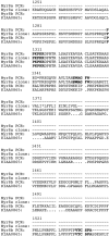
Comparative alignment of the amino acid sequences deduced from PCR products amplified using sense primers common to both myr 8a (clone 4) and cDNA KIAA0865 and antisense primers unique to either 3′-untranslated sequence of myr 8a or KIAA0865. PCR amplifications were performed using cDNA prepared from granule neurons isolated from postnatal day 10 rat cerebellum. Sequence 1 was generated using a sense primer common to both myr 8a and KIAA0865 with an antisense primer unique to 3′-untranslated sequence of myr 8a. This product is identical to the 3′ terminus of myr 8a (clone 4) including the stop codon. Sequence 2 was generated using the sense primer common to both myr 8a and KIAA0865 with the antisense primer unique to the KIAA0865 sequence. This product does not encode a stop codon, therefore extending the sequence of myr 8a (clone 4) in the C-terminal direction. This extended sequence, which identifies the initial segment of the C-terminal tail domain of myr 8b, is 71% identical to the sequence described for the human cDNA KIAA0865. The sense and antisense primers correspond to the nucleotides of the amino acids indicated inbold text.
Fig. 3.
Fig. 3.
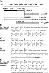
Relationships among myr 8a, myr 8b, and KIAA0865. Schematic diagram of inferred relationships between rat cDNA clone 4 and clone 18 and human cDNA clone KIAA0865 and the corresponding proteins myr 8a, myr 8b, and KIAA0865, respectively. Clone 4 contains a single open reading frame and codes for myr 8a. Clone 18 overlaps with the terminal 3′ sequence of clone 4 but lacks the stop codon identified in the clone 4 sequence and thus extends in the C-terminal direction. Together, clone 4 and clone 18 comprise myr 8b. The homologous human cDNA clone KIAA0865 overlaps most of clone 4 and all of clone 18. The overlapping region of identity for the rat myr 8 clone 4 and clone 18 and the human KIAA0865 sequences is indicated by the hatched pattern. At the 3′ terminus, the clone 4 (white) sequence diverges from clone 18 and KIAA0865 sequences (gray). The 5′ terminus of clone 4 (black) contains the ATP binding site and the ankyrin repeats (A). The human cDNA clone KIAA0865 maps to chromosome 13. Comparison of genomic and cDNA sequences reveals an intron sequence (lowercase letters) located between amino acids ANE and ALAR (B). The site where myr 8a and myr 8b sequences diverge corresponds to a consensus exon–intron boundary (indicated by arrow), starting at nucleotide 4002 and reading 5′-AG/GT-3′, that is identical to that demonstrated for KIAA0865. The myr 8b sequence, analogous to KIAA0865, is obtained by use of the splice junction (indicated by arrow) (C), whereas myr 8a is derived by crossing through the splice junction (indicated by arrow) (D). The intervening intron in KIAA0865 (starting at nt 2717999) does not include a stop codon in the same position as revealed for myr 8a but extends the coding sequence for an additional 16 amino acids. This human sequence, which contributes unique sequence to the KIAA0865 isoform, reveals a consensus polyadenylation sequence (AATAAA, nt 2718106–2718111) located within a ∼30 nucleotide region that is well conserved with that sequence identified for rat myr 8a (E). Actin, Actin binding site;Ank, ankyrin repeats; ATP, ATP binding site; IQ, IQ motif domain; M, Kozak start sites; asterisk indicates stop codon. The scale bar represents nucleotide number. The predicted nucleotide length and molecular masses are noted for myr 8a and myr 8b. The deduced amino acid sequence is shown in single-letter code below the nucleotide sequence. The consensus polyade- nylation signal (AATAAA) is underlined.Lowercase nucleotide sequence in Brepresents the intron sequence, whereas in D andE, the lowercase nucleotide sequence represents a 3′-untranslated sequence.
Fig. 4.
Fig. 4.
Unrooted phylogenetic tree of the myosin superfamily. Head domains of 40 myosin proteins available from public data bases were aligned to amino acids 213–511 of the chicken fast skeletal myosin (Gg FSK), a class II myosin, using the default CLUSTAL method settings (Lasergene/DNASTAR). The PHYLIP phylogeny package (http://evolution. genetics.washington.edu/phylip.html) (Felsenstein, 1993) was used to generate a genetic distance tree; 1000 bootstrap data sets were generated and analyzed with the programs PROTDIST and NEIGHBOR. The programs CONSENSE, FITCH, and DRAWTREE were used to produce the unrooted distance tree. The frequency of node placement for 1000 bootstrap trials is indicated as >90, >70, and >50% by solid circles, gray circles, and open circles, respectively. Sequence divergence is proportional to the length of the branches. The length of the bar equals 5% sequence divergence. Myosin classes are indicated by Roman numerals. The species represented areAcanthamoeba castellanii (Aca),Acetabularia cliftonii (Acl),Arabidopsis thaliana (At), Brugia malayi (Bm), Bos taurus(Bt), Caenorhabditis elegans(Ce), Dictyostelium discoidium(Dd), Drosophila melanogaster(Dm), Gallus gallus (Gg),Helianthus annus (Ha), Homo sapiens (Hs), Mus musculus(Mm), Plasmodium falciparum(Pf), Rattus norvegicus(Rn), Saccharomyces cerevisiae(Sc), Sus scrofa (Ss), andToxoplasma gondii (Tox). The GenBank accession numbers for the sequences by class are as follows—class I: Dd MIA (P22467), Dm MIA (S45573), Gg BBMI (U04049), MmIA (L00923), Rn myr 1a (X68199), Rn myr 2 (X74800), Rn myr 3 (X74815), Rn myr 4 (X71997); class II: Bm II (M74000), Dd II (p08799), Dm NM2 (P05661), Gg Fsk (P13538), Sc myo1(IIA) (S46773); class III: Dm Nina C (p10676); class IV: Aca HMW (j05678); class V: Gg p190 (z11718), Mm dilute (x57377), Rn myr 6 (u60416), Sc myo2 (VA) (p19524), Sc myo4 (VB) (p32492); class VI: Dm 95F (Q01989), Mm Snell's waltzer (u49739), Ss VI (a54818); class VII: Hs VIIa (u55208), Mm shaker-1 (U81453); class VIII: At ATM A (x67104), At ATM B (z34292), Ha my1 (U94781); class IX: Hs MIXb (u42391), Rn myr 5 (x77609), Rn myr 7 (AJ001713); class X: Bt X (U55042); class XI: At mya1 (z29389), At mya2 (z34294); class XII: Ce XII (z66563); class XIII: Acl M1 (U94397), Acl M2 (U94398); class XIV: Tox A (af006626), Tox B (af006627), Pf XIV (y09693); class XV: Mm myo15 (AF053130); class XVI: Rn myr 8 (AF209114).
Fig. 5.
Fig. 5.
Tissue-specific expression of myr 8 mRNA. Northern blots of poly(A) RNA (2.5 μg/lane) from type 1 astrocytes (A), total RNA (15 μg/lane) from cerebella at indicated developmental time points (B), poly(A) RNA (2.5 μg/lane) from neocortices at indicated developmental ages (C, D), and total RNA (15 μg/lane) from the indicated tissues obtained at postnatal day 10 (E,F) were hybridized with 32P-labeled probe 1 (A, B), probe 2 (C, E), probe 4 (F), and probe 5 (D). A prominent signal of ∼7.2 kb was detected in astrocytes and at all developmental ages for both neocortex and cerebellum, although the level of the ∼7.2 kb message peaked during the first and second postnatal weeks in both neocortex and cerebellum. The ∼7.2 kb myr 8 transcript (arrow) was detected principally in the CNS but was also observed to a minimal extent in adrenal and heart and skeletal muscle. Equal RNA loads were verified by evaluation of 18S ribosomal RNA after ethidium bromide staining. RNA size standards are indicated in kilobases. Ad, Adult; E, embryonic day; NB, newborn; P, postnatal day.
Fig. 6.
Fig. 6.
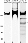
Antibodies directed against either N-terminal amino acids 2–52 or C-terminal amino acids 1763–1785 recognize principally a ∼210 kDa antigen. A cytoskeletal/membrane fraction was isolated from postnatal day 10 brain (A,C) and from neocortical type 1 astrocytes (B). Polypeptides were resolved by SDS-PAGE and processed for immunoblot analyses using antibodies directed toward the N-terminal amino acids 2–52 (A, C) or affinity-purified antibodies directed toward the C-terminal amino acids 1763–1785 located within the tail domain of myr 8b (B). Both antibodies detect a single broad immunoreactive band migrating with an apparent molecular mass of ∼210 kDa. Overexposure of immunoblots performed using the antibody directed against the N-terminal amino acids 2–52 reveals two minor band of ∼148 kDa (**) and ∼170 kDa (*).
Fig. 7.
Fig. 7.
Tissue distribution of myr 8 protein. Post-nuclear supernatants were prepared from the indicated tissues obtained at postnatal day 10, and proteins were resolved by SDS-PAGE and processed for immunoblot analysis using antibodies directed toward the N-terminal amino acids 2–52. A broad immunoreactive band migrating with an apparent molecular mass of ∼210 kDa was the principal antigen detected in both neural and non-neural tissues. a, Anterior; p, posterior. Molecular mass × 103 is indicatedvertically.
Fig. 8.
Fig. 8.
Myr 8b associates with the protein phosphatase catalytic subunits 1α and 1γ1. A crude cytoskeletal/membrane fraction prepared from postnatal day 10 cerebellum was solubilized in 1% Triton X-100, and unsolubilized components were sedimented by centrifugation. Aliquots of the detergent lysate were processed for immunoprecipitation using affinity-purified antibodies directed toward the C-terminal tail domain of myr 8b. Immunoprecipitated polypeptides were resolved by SDS-PAGE and processed for immunoblot analyses using polyclonal antiserum directed against multiple (1α, 1β, 1γ, 2A, 2B, and X) and individual (PP1α), (PP1β), (PP1γ), and (PP2A) protein phosphatase catalytic subunits and actin. Molecular mass × 103 is indicatedvertically.
Fig. 9.
Fig. 9.
Myr 8b cosediments with F-actin in an ATP-sensitive manner. Postnatal day 8 brain was homogenized in the presence of 0.5 mm ATP, and a soluble fraction was obtained by high-speed centrifugation. Supernatant samples (100 μg protein) were supplemented with glucose (50 mm) and 50 μg platelet nonmuscle actin (Control); glucose, platelet nonmuscle actin, and 1 mm ATP (ATP); and glucose, platelet nonmuscle actin, and 0.5 U hexokinase (Hexokinase). After incubation (15 min at 25°C), centrifugation yielded a supernatant (S) and a pellet (P), which were collected, resolved by SDS-PAGE, and processed for immunoblot analysis using antibodies directed toward the N-terminal amino acids 2–52. Molecular mass × 103 is indicatedvertically.
Fig. 10.
Fig. 10.
Myr 8b in brain exists in both soluble and sedimentable subcellular pools. Postnatal day 8 brain was homogenized in the absence (A) and presence (B) of 0.5 mm ATP and fractionated by velocity sedimentation. Equal protein loads were resolved by SDS-PAGE and processed for immunoblot analysis using antibodies directed toward the N-terminal amino acids 2–52. S1, P1: 1000 × g × 10 min supernatant (S) and pellet (P);S2, P2: 10,000 ×g × 15 min supernatant (S) and pellet (P); S3,P3: 100,000 × g × 60 min supernatant and pellet.
Fig. 11.
Fig. 11.
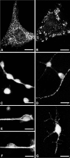
Immunofluorescent staining of myr 8b in primary cultures of astrocytes and neurons using affinity-purified antibodies directed toward the C-terminal tail domain of myr 8b. In neocortical and cerebellar type 1 astrocytes, myr 8b immunoreactivity was detected as intensely fluorescent puncta throughout the somal region and along the length of extended processes (A). Occasionally, immunoreactivity was organized in linear arrays or in large clusters located in a perinuclear position (B). In primary cultures of cerebellar granule neurons (C–E) and hippocampal neurons (G), myr 8b immunoreactivity was detected at a significant level in the cell body, but intense fluorescent puncta were also observed scattered along the entire length of both dendritic and axonal processes. In cell dissociates of neonatal cerebellum, granule neurons having the morphological features characteristic of migrating neurons displayed myr 8b immunoreactivity in a punctate manner throughout the cell body as well as along the length of the leading process (F). Scale bars: A, 10 μm; B, 10 μm; C, 18 μm;D, 15 μm; E, 10 μm; F, 10 μm; G, 18 μm.
Fig. 12.
Fig. 12.
Immunolocalization of myr 8b in developing and adult cerebellum using affinity-purified antibodies directed toward the C-terminal tail domain of myr 8b. Frozen, 6 μm-thick sagittal sections were used. At postnatal day 10, myr 8b immunoreactivity was detected principally around granule neurons located in the inner portion of the external granule layer. A lesser degree of immunoreactivity was detected within the soma and dendrites of Purkinje cells (A). In the adult cerebellum, myr 8b immunoreactivity continued to be detected in Purkinje cell bodies and associated dendritic processes, in the elongated processes of radial glial cells, and in astroglial cells and cell bodies of granule neurons located in the internal granule cell layer (B). EGL, External granule cell layer, IGL, internal granule cell layer;ML, molecular layer; PCL, Purkinje cell layer. Scale bars: A, 14 μm; B, 45 μm.

Similar articles

Cited by

References

    1. Adams RJ, Pollard TD. Binding of myosin I to membrane lipids. Nature. 1989;340:565–568. - PubMed
    1. Allen PB, Ouimet CC, Greengard P. Spinophilin, a novel protein phosphatase 1 binding protein localized to dendritic spines. Proc Natl Acad Sci USA. 1997;94:9956–9961. - PMC - PubMed
    1. Altman J, Bayer SA. Development of the cerebellar system: in relation to its evolution, structure and function. CRC; Boca Raton, FL: 1997.
    1. Altschul SF, Madden TL, Schaffer AA, Zhang J, Zhang Z, Miller W, Lipman DJ. Gapped BLAST and PSI-BLAST: a new generation of protein database search programs. Nucleic Acid Res. 1997;25:3389–3402. - PMC - PubMed
    1. Andersen SSL, Bi GQ. Axon formation: a molecular model for generation of neuronal polarity. BioEssays. 2000;22:172–179. - PubMed

Publication types

MeSH terms