. 2001 Oct 15;21(20):7985-92.
doi: 10.1523/JNEUROSCI.21-20-07985.2001.

An NMDA receptor signaling complex with protein phosphatase 2A

Affiliations

An NMDA receptor signaling complex with protein phosphatase 2A

S F Chan et al. J Neurosci. .

Abstract

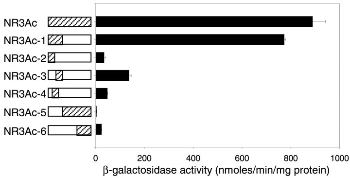
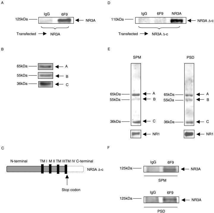
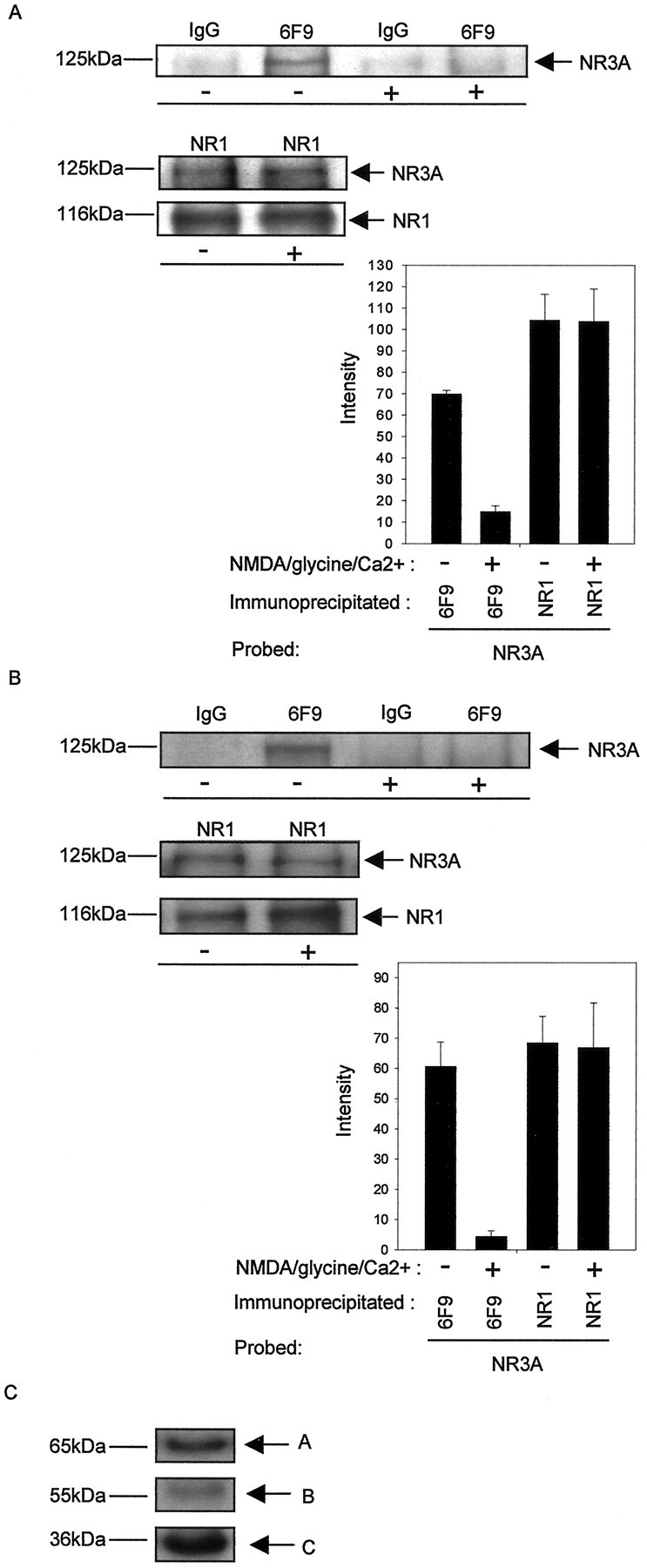
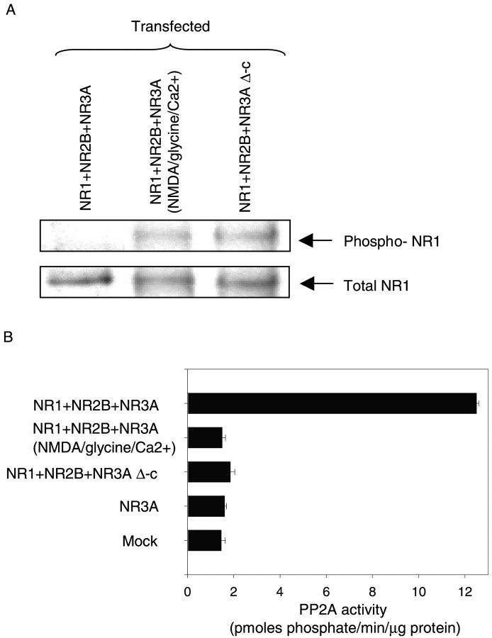
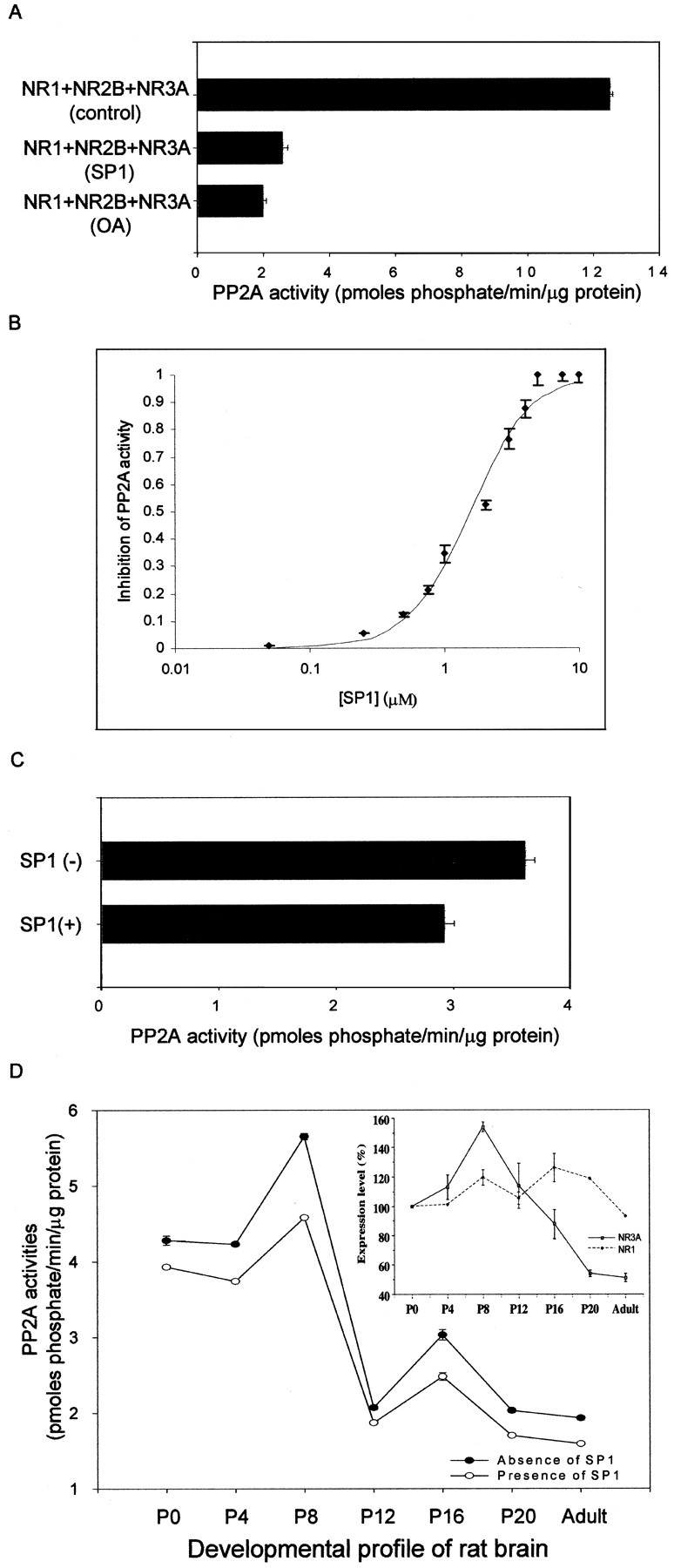
Regulation of protein phosphatase 2A (PP2A) activity and NMDA receptor (NMDAR) phosphorylation state contribute to the modulation of synaptic plasticity, yet these two mechanisms have not been functionally linked. The NMDAR subunit NR3A is equipped with a unique carboxyl domain that is different from other NMDAR subunits. We hypothesized that the NR3A C-terminal intracellular domain might serve as synaptic anchor for the phosphatase in the developing CNS. A cDNA library was screened by the yeast two-hybrid method using the NR3A carboxyl domain as the bait. The catalytic subunit of the serine-threonine PP2A was found to be associated with the NR3A carboxyl domain. Immunoprecipitation studies indicated that the NR3A subunit formed a stable complex with PP2A in the rat brain in vivo. Association of PP2A with NMDARs led to an increase in the phosphatase activity of PP2A and the dephosphorylation of serine 897 of the NMDAR subunit NR1. Stimulation of NMDARs led to the dissociation of PP2A from the complex and the reduction of PP2A activity. A peptide corresponding to the PP2A-NR3A binding domain functioned as a negative regulator of PP2A activity. These data suggest that NMDARs are allosteric modulators of PP2A, which in turn controls their phosphorylation state. The data delineate a mechanistic model of the dynamic regulation of a PP2A-NMDAR signaling complex, mediated by the interaction of NR3A and PP2A, and suggest a novel NMDAR-mediated signaling mechanism in addition to the traditional ionotropic functions of NMDARs.


Figures

Fig. 1.
Fig. 1.
Interaction of NR3A with PP2A. The entire carboxyl intracellular domain of NR3A (NR3Ac: Gly1-Ser165) and various deletion constructs (NR3Ac-1–NR3Ac-6) were used in a yeast two-hybrid assay.Hatched areas correspond to the constructs used in the assay; open boxes indicate the deletions that were made from the carboxyl intracellular domain. Corresponding β-galactosidase activities of each construct on interaction with PP2A are summarized in the histogram. β-galactosidase activities are given in nanomoles per minute per milligram of protein ± SEM.
Fig. 2.
Fig. 2.
PP2A–NR3A interaction mediated through the carboxyl intracellular domain of NR3A. A, Lysates from HEK 293 cells transiently transfected with NR3A were immunoprecipitated with control IgG antibody (anti-rat IgG) and anti-PP2A monoclonal antibody 6F9. Western blots were probed with monoclonal antibody anti-NR3A. Data are representative of six experiments showing similar results. B, Lysates from HEK 293 cells transiently transfected with NR3A were separated by SDS-PAGE, and the expression of PP2A was detected by probing with antibody PR65 (recognizing the A-subunit), antibody PR55α (recognizing the B-subunit), and antibody anti-PP2Ac (recognizing the C-subunit). C, Schematic drawing of NR3A Δ-c. The part of the carboxyl intracellular domain of NR3A that was deleted is indicated by the dotted line.TM I-IV designates transmembrane regions I-IV. M II designates membrane loop M II. D, Lysates from cells transfected with NR3A Δ-c were immunoprecipitated with control IgG antibody (anti-rat IgG) or monoclonal antibody 6F9. Western blots were probed with monoclonal antibody anti-NR3A to detect the expression of NR3A Δ-c. E, Co-immunoprecipitation of PP2A with NR3A in rat brain membrane fractions. Lysates from rat brain membrane fractions were separated by SDS-PAGE, and the expression of PP2A and NR1 was detected by probing with antibody PR65 (recognizing the A-subunit), antibody PR55α (recognizing the B-subunit), antibody anti-PP2Ac (recognizing the C-subunit), and anti-NR1. F, Immunoprecipitations from synaptic plasma membrane (SPM) and postsynaptic density (PSD) fractions from rat brain were performed using control IgG antibody and monoclonal antibody 6F9, which was used to immunoprecipitate PP2A (core enzyme and/or holoenzyme). Western blots were probed with monoclonal antibody anti-NR3A.
Fig. 3.
Fig. 3.
NMDAR activity-dependent association of PP2A with NR3A in transfected HEK 293 cells and neurons. A, HEK 293 cells expressing heteromeric NMDARs consisting of NR1 + NR2B + NR3A were incubated in the absence or presence of NMDA (200 μm nmda, 10 μm glycine, and 2.5 mmCa2+ in nominally Mg2+-free Hank's solution). Immunoprecipitations were performed using control IgG antibody, mono clonal antibody 6F9, and NR1. Top, Western blots probed with monoclonal antibody anti-NR3A and anti-NR1.Bottom, left, Intensity (mean gray level ± SD) of NR3A labeling in the presence (co-IP with 6F9, 14.9 ± 2.8; co-IP with NR1, 103.7 ± 15.3) and absence (co-IP with 6F9, 69.2 ± 2.3; co-IP with NR1, 104.3 ± 12.1) of NMDAR stimulation (co-IP with 6F9, p < 0.00005; co-IP with NR1,p < 0.5; n = 3 separate experiments). B, Cells from cerebrocortical neurons were incubated in the absence or presence of NMDA. The cell lysates were subsequently immunoprecipitated with control IgG antibody or monoclonal antibody 6F9 or NR1. Top, Western blots probed with monoclonal antibody anti-NR3A and anti-NR1. Bottom, left, Intensity (mean gray level ± SD) of NR3A labeling in the presence (co-IP with 6F9, 4.4 ± 1.9; co-IP with NR1, 66.9 ± 14.8) and absence (co-IP with 6F9, 60.6 ± 8.0; co-IP with NR1, 68.5 ± 8.8) of NMDAR stimulation (co-IP with 6F9,p < 0.005; co-IP with NR1, p< 0.5; n = 3 separate experiments).C, Lysates from cerebrocortical neurons were separated by SDS-PAGE, and the expression of PP2A was detected by probing with antibody PR65 (recognizing the PP2A-A subunit), antibody PR55α (recognizing the PP2A-B subunit), and antibody anti-PP2Ac (recognizing the PP2A-C subunit).
Fig. 4.
Fig. 4.
Dephosphorylation of the NMDAR subunit NR1 and activation of endogenous PP2A activity in transfected HEK 293 cells.A, HEK 293 cells were transiently transfected with a triple combination of NMDAR subunits consisting of NR1 + NR2B + NR3A and NR1 + NR2B + NR3A Δ-c. After incubation in the absence or presence of NMDA (200 μm nmda, 10 μm glycine and 2.5 mm Ca2+in nominally Mg2+-free Hank's solution), cells were harvested, and lysates of transfected cells were immunoprecipitated with monoclonal antibody anti-NR1. Blots were probed with monoclonal antibody anti-phospho-NR1 to detect the phosphorylated form of NR1. NR1 protein was detected by probing with monoclonal antibody anti-NR1. Three individual experiments using different batches of protein samples showed similar results. B, Lysates from transfected HEK 293 cells of NR1 + NR2B + NR3A, NR1 + NR2B + NR3A (treated with NMDA as above), and NR1 + NR2B + NR3A Δ-c were used to assay the endogenous PP2A activity using a phosphopeptide as a substrate. Endogenous PP2A activities are given in picomoles phosphate per minute per microgram of protein ± SD. Data are representative of three experiments showing similar results.
Fig. 5.
Fig. 5.
Blockade of endogenous PP2A activity by a synthetic peptide (SP1). A, Solubilized extract fractions from HEK 293 cells transfected with NR1 + NR2B + NR3A were used to assay the endogenous PP2A activity in the absence (12.5 ± 0.1 pmol · min−1 · μg−1) or presence (2.6 ± 0.2 pmol · min−1 · μg−1) of SP1 (5 μm; p < 0.1 × 10⁻⁸; n = 3 separate experiments). Incubation with the specific PP2A inhibitor 1 nm OA was used as control to block endogenous PP2A activity in transfected HEK 293 cells. B, Dose–response curve for the inhibition of PP2A activity by SP1. Solubilized extract fractions from HEK 293 cells transfected with NR1 + NR2B + NR3A were used to evaluate the IC50 of SP1 on PP2A activity. The maximum inhibitory effect of SP1 was normalized to 1. Each data point represents the average of three experiments ± SD.C, Solubilized extract fractions from cerebrocortical neurons were used to determine the endogenous PP2A activity in the absence (3.6 ± 0.09 pmol · min−1 · μg−1) or presence (2.9 ± 0.09 pmol · min−1 · μg−1) of SP1 (5 μm; p < 0.001;n = 3 separate experiments). D, Solubilized extract fractions from rat brain at different stages of development (P0, P4, P8, P12, P16, P20, and adult) were used to assay the endogenous PP2A activity in the absence or presence of 5 μm SP1, and each data point represents the average of three experiments ± SD (p < 0.005 at all points). The developmental protein expression profiles of NR3A and NR1 were determined, and the expression levels were normalized relative to P0 and expressed as percentage ± SD. Three individual experiments using different batches of protein samples showed similar results.

References

    1. Bliss TV, Collingridge GL. A synaptic model of memory: long-term potentiation in the hippocampus. Nature. 1993;361:31–39. - PubMed
    1. Ciabarra AM, Sullivan JM, Gahn LG, Pecht G, Heinemann S, Sevarino KA. Cloning and characterization of χ-1: a developmentally regulated member of a novel class of the ionotropic glutamate receptor family. J Neurosci. 1995;15:6498–6508. - PMC - PubMed
    1. Das S, Sasaki YF, Rothe T, Premkumar LS, Takasu M, Crandall JE, Dikkes P, Conner DA, Rayudu PV, Cheung W, Chen HS, Lipton SA, Nakanishi N. Increased NMDA current and spine density in mice lacking the NMDA receptor subunit NR3A. Nature. 1998;393:377–381. - PubMed
    1. Faux MC, Scott JD. More on target with protein phosphorylation: conferring specificity by location. Trends Biochem Sci. 1996;21:312–315. - PubMed
    1. Fraser D, Scott JD. Modulation of ion channels: a “current” view of AKAPs. Neuron. 1999;23:423–426. - PubMed

Publication types

MeSH terms