. 2001 Oct 15;21(20):8034-42.
doi: 10.1523/JNEUROSCI.21-20-08034.2001.

The neuronal form of adaptor protein-3 is required for synaptic vesicle formation from endosomes

Affiliations

The neuronal form of adaptor protein-3 is required for synaptic vesicle formation from endosomes

J Blumstein et al. J Neurosci. .

Abstract

Heterotetrameric adaptor complexes vesiculate donor membranes. One of the adaptor protein complexes, AP-3, is present in two forms; one form is expressed in all tissues of the body, whereas the other is restricted to brain. Mice lacking both the ubiquitous and neuronal forms of AP-3 exhibit neurological disorders that are not observed in mice that are mutant only in the ubiquitous form. To begin to understand the role of neuronal AP-3 in neurological disease, we investigated its function in in vitro assays as well as its localization in neural tissue. In the presence of GTPgammaS both ubiquitous and neuronal forms of AP-3 can bind to purified synaptic vesicles. However, only the neuronal form of AP-3 can produce synaptic vesicles from endosomes in vitro. We also identified that the expression of neuronal AP-3 is limited to varicosities of neuronal-like processes and is expressed in most axons of the brain. Although the AP-2/clathrin pathway is the major route of vesicle production and the relatively minor neuronal AP-3 pathway is not necessary for viability, the absence of the latter could lead to the neurological abnormalities seen in mice lacking the expression of AP-3 in brain. In this study we have identified the first brain-specific function for a neuronal adaptor complex.


Figures
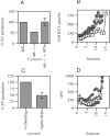
Fig. 1.
Fig. 1.
The in vitro budding of synaptic vesicles requires AP-3. PC12 N49A cells were labeled with [125I]-KT3 at 15°C. Endosomal membranes were incubated with mocha cytosol and an ATP-regenerating system. Budding reactions were performed at 37°C for 30 min.A, Mocha mice brain cytosol shows a 50% reduction in the production of synaptic vesicles from the donor endosome compartment compared with wild-type brain cytosol.Mocha cytosol supplemented with brain-purified AP-3 rescued the defect in budding, returning vesicle production to wild-type levels. The data shown represent an average ± SEM (n = 3). B, A representative example of the budding assay in which the fractions from the gradient, shown along the x-axis, have been collected from the bottom and counted. The no-cytosol control (■), mocha cytosol (♦), wild-type brain cytosol (●), and mocha brain cytosol plus brain-purified AP-3 (▴) were tested in this assay. The peak is at fractions 10 and 11 and represents the newly budded pool of synaptic vesicles; the label on the right is free antibody. C, The in vitro budding assays were performed with brain cytosol that was depleted for the ς3 subunit. The results show a 50% reduction in synaptic vesicle biogenesis compared with wild-type budding production (n = 3). D, A representative assay in which cytosol is depleted of ς3. The fractions collected from the gradient are shown along the x-axis. Here, a no-cytosol control (■), wild-type brain cytosol (♦), 4° rat brain cytosol (●), and brain cytosol that was immunodepleted by using the ς3 antibody (▴) were tested. When AP-3 was removed, the height of the peak was reduced, indicating reduced vesicle production.
Fig. 2.
Fig. 2.
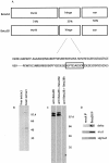
Production of β3B-specific antibody.A, The AP-3 subunits β3A and β3B are highly homologous. Within the hinge domain, the least homologous region between the ubiquitously and neuronally expressed β3 subunits, we chose a stretch of amino acids within β3B as our antigen. The GST fusion protein was used as the immunogen. B, Liver and brain extracts were run on SDS-PAGE gels and analyzed via immunoblot by antisera. This antiserum recognized a band of the approximate molecular weight of the β3B subunit, present only in brain extract. The antibody also nonspecifically recognized a lower-molecular-weight band present in both liver and brain extracts. C, Purified brain AP-3 was run on SDS-PAGE gels and probed with this antiserum. It recognized a protein of the correct molecular weight. Antisera also were preincubated with either purified β3A hinge (β3Ah) or with purified β3B hinge (β3Bh) and then used for Western blots. Anti-β3B recognized the neuronal subunit as well as antibody preincubated with the β3A hinge. Antibody preadsorbed with β3B hinge no longer could bind the neuronal subunits on blots, showing its specificity. The β3B subunit often appears as a doublet in purified AP-3, perhaps because of limited proteolysis during purification. D, The β3B antibody was also used to immunoprecipitate the other subunits of the AP-3 complex. Mock immunoprecipitations did not bring down any of the AP-3 subunits.
Fig. 3.
Fig. 3.
In vitro budding of synaptic vesicles depends on the neuronal form of AP-3. A,In vitro budding assays were performed as described. Budding assays were performed by using either mock-depleted cytosol (wild-type budding) or cytosol that was immunodepleted for β3B or for ς3. The inset shows immunoblots of either mock (+) or () immunodepleted cytosols. The top blot was probed for β3B in either mock or immunodepleted cytosol, and the bottom blot was probed for the ς3 subunits in either mock or immunodepleted cytosol. Cytosol that was immunodepleted showed essentially complete depletion. The depleted cytosols both showed a similar 50% reduction in the biogenesis of SLMVs compared with wild-type levels of synaptic vesicle production. B, In vitro budding assays also were performed by using cytosol from mice heterozygous for μ3B and for mice that lacked all expression of the μ3B subunit. Although the heterozygote cytosol showed robust SLMV biogenesis, the knock-out mouse cytosol showed a 50% reduction in budding compared with cytosol from its heterozygous littermate.
Fig. 4.
Fig. 4.
Neuronal AP-3 is not the major form of AP-3 in brain. A, Brain cytosol from heterozygotes of μ3B (+/−) and knock-outs for the neuronal μ3B subunit were run on SDS-PAGE gels. To determine whether there was significantly less AP-3 remaining in brains that lack the neuronal form, we probed for the δ subunit, present in all forms of AP-3. The levels of δ appear to be unchanged in the knock-out compared with the heterozygote, suggesting that the majority of brain AP-3 is in the ubiquitous form. We also probed with a pan-μ3 antibody that recognizes both μ3A and μ3B. The levels seen in the heterozygote of both ubiquitous and neuronal forms appeared to be no more than those in the knock-out, which contained only the ubiquitous form. Protein levels were standardized to levels of a variant of clathrin light chain A. B, Levels of the ς3 subunit, the other ubiquitously expressed subunit in all AP-3 complexes, also were compared in mock-depleted cytosol versus cytosol that was immunodepleted for β3B. Equal amounts of protein were run in each lane. Although β3B was removed in the depleted cytosol, levels of ς3 were unchanged from the amount in mock-depleted cytosol.
Fig. 5.
Fig. 5.
AP-3 is necessary to coat synaptic vesicles.A, Purified synaptic vesicles that are run over sucrose gradients sediment at ∼22% sucrose. The same vesicles that are incubated with wild-type brain cytosol, ATP-regenerating system, and GTPγS recruit coat and sediment at 30–32% sucrose. Cytosol that has been depleted for ς3-containing AP-3 complexes could not coat synaptic vesicles fully. Cytosol that had been depleted for β3B-containing AP-3 complexes, however, could provide coat to vesicles, which sedimented at 30–32% sucrose. B, A representative example of a coating assay analyzed on sucrose gradients showing the magnitude of the change in sedimentation properties. The fractions collected from the bottom of the gradient are shown along thex-axis. Conditions tested in the assay were synaptic vesicles without cytosol (■), mock-depleted rat brain cytosol (♦), and anti-β3B immunodepleted brain cytosol (●). Synaptic vesicles incubated without a source of coat, as in brain cytosol, did not undergo a density shift. Vesicles incubated with either mock-depleted rat brain cytosol or β3B-depleted rat brain cytosol did undergo a density shift. C, Synaptic vesicles could be coated fully after incubation in GTPγS with either brain cytosol that lacked μ3B or cytosol that did contain μ3B. D, Without any AP-3 in brain, as in the mocha mice (mh−/−), vesicles could not be coated. In vitro coating assays kept at 4°C, instead of incubation at 37°C, also could not recruit coat.
Fig. 6.
Fig. 6.
Neuronal AP-3 is localized to varicosities of neuronal-like processes. A, Differentiated PC12 cells were stained with the β3B antibody. Although there was no specific staining in the cell body (inset), we observed staining in the varicosities along the processes, yet it was absent at the tips.B, Differentiated cells were double stained for synaptotagmin; the staining was in contrast to that seen with the β3B antibody. Synaptotagmin staining was most intense at the tips of the processes. C, Differentiated PC12 cells also were stained for the δ subunit of AP-3, a subunit present in all AP-3 complexes. δ Staining is seen in varicosities, as with β3B, but in addition there is punctate staining in the cell body. D, A representative tip of a differentiated PC12 cell stained with synaptotagmin antibody. E, The same tip, which is enriched for synaptotagmin, lacks expression of β3B. F, A representative varicosity of a differentiated PC12 cell process enriched in β3B expression. G, A representative varicosity of a differentiated PC12 cell enriched in δ expression.
Fig. 7.
Fig. 7.
Neuronal AP-3 is expressed throughout axons in the brain. A, Adult rat brain sections were stained for β3B immunoreactivity. Neuronal AP-3 is seen in axons in most regions of the brain. B, A close-up view of β3B staining in the hippocampus shows intense staining in the lacunosum moleculare (LMol) as well as in the stratum oriens (Or), stratum radiatum (Rad), and the molecular layer of the dentate gyrus (Mol).C, Adjacent adult rat brain sections were stained for synaptophysin immunoreactivity. Synaptophysin also is expressed in most axonal pathways of the brain. D, A close-up view of synaptophysin staining in the hippocampus shows a different pattern of expression than that seen for neuronal AP-3. Synaptophysin has a more even level of expression throughout the hippocampus, although it appears to label the mossy fiber pathway more intensely than neuronal AP-3. E, Adult rat brain sections were stained with the β3B antiserum that had been preadsorbed with the GST fusion protein against which the antibody was made. No immunoreactivity was observed.F, A close-up view of the hippocampus also shows that no staining was observed in the negative control.
Fig. 8.
Fig. 8.
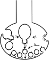
Neuronal AP-3-mediated pathway of synaptic vesicle biogenesis from endosomes. Synaptic vesicles that cluster in the active zone (the triangles at the plasma membrane) undergo a cycle of exocytosis and recycling. Synaptic vesicle proteins normally recycle via the AP-2/clathrin pathway of endocytosis (arrow A) but escape recovery at the plasma membrane and may recycle via the AP-3 pathway. Such synaptic vesicle proteins may be retrieved into specialized axonal endosomes that use neuronal AP-3 to bud synaptic vesicles (arrow B). The endosomal pathway of synaptic vesicle production also may function to recycle components of large dense core vesicles (LDCVs). LDCV proteins recycle via an endosomal intermediate, and some proteins may get sorted into synaptic vesicles. Neuronal AP-3 could recognize and bud such cargo into SLMVs from this endosomal intermediate (arrow C). Axonal endosomes that contain synaptic vesicle, as well as some LDCV membrane proteins, use neuronal AP-3 to produce synaptic vesicles, which are competent to fuse with the plasma membrane and release their contents (arrow D).

References

    1. Ahle S, Ungewickell E. Auxilin, a newly identified clathrin-associated protein in coated vesicles from bovine brain. J Cell Biol. 1990;111:19–29. - PMC - PubMed
    1. Altschuler Y, Barbas SM, Terlecky LJ, Tang K, Hardy S, Mostov KE, Schmid SL. Redundant and distinct functions for dynamin-1 and dynamin-2 isoforms. J Cell Biol. 1998;143:1871–1881. - PMC - PubMed
    1. Ball CL, Hunt SP, Robinson MS. Expression and localization of α-adaptin isoforms. J Cell Sci. 1995;108[Pt 8]:2865–2875. - PubMed
    1. Blagoveshchenskaya AD, Norcott JP, Cutler DF. Lysosomal targeting of P-selectin is mediated by a novel sequence within its cytoplasmic tail. J Biol Chem. 1998;273:2729–2737. - PubMed
    1. Clift-O'Grady L, Desnos C, Lichtenstein Y, Faundez V, Horng JT, Kelly RB. Reconstitution of synaptic vesicle biogenesis from PC12 cell membranes. Methods. 1998;16:150–159. - PubMed

Publication types

MeSH terms