. 2001 Oct 15;21(20):8043-52.
doi: 10.1523/JNEUROSCI.21-20-08043.2001.

GABA(B2) is essential for g-protein coupling of the GABA(B) receptor heterodimer

Affiliations

GABA(B2) is essential for g-protein coupling of the GABA(B) receptor heterodimer

M J Robbins et al. J Neurosci. .

Abstract

GABA(B) receptors are unique among G-protein-coupled receptors (GPCRs) in their requirement for heterodimerization between two homologous subunits, GABA(B1) and GABA(B2), for functional expression. Whereas GABA(B1) is capable of binding receptor agonists and antagonists, the role of each GABA(B) subunit in receptor signaling is unknown. Here we identified amino acid residues within the second intracellular domain of GABA(B2) that are critical for the coupling of GABA(B) receptor heterodimers to their downstream effector systems. Our results provide strong evidence for a functional role of the GABA(B2) subunit in G-protein coupling of the GABA(B) receptor heterodimer. In addition, they provide evidence for a novel "sequential" GPCR signaling mechanism in which ligand binding to one heterodimer subunit can induce signal transduction through the second partner of a heteromeric complex.


Figures

Fig. 1.
Fig. 1.
Intracellular loop sequence alignments and summary of GABAB1 and GABAB2 constructs.a, Alignment of the second and third intracellular loops of GABAB1 (GABAB1) and GABAB2(GABAB2) subunits with members of the mGluR family. The alignment was produced in Alscript using HMMALIGN and the G-protein-coupled receptors database (GPCRDB) (Barton, 1993; Horn et al., 1998). The shading indicates different levels of amino acid conservation, in which residues are colored if eight or more residues show conservation of amino acid properties: blue shading, small; yellow shading, hydrophobic; red characters, polar. Transmembrane helices (TMs, dark blue) are displayed for mGluR1 (as assigned in SwissProt, entry MGR1 HUMAN).Boxed letters indicate residues that are negatively charged. Conserved residues discussed in Results are shaded ingray. The alignment has been extracted from a larger alignment of family 3 GPCRs taken from GPCRDB (Horn et al., 1998).b, Enlarged alignment of the second intracellular loop of both GABAB1 and GABAB2 subunits. Bold red letters indicate residues mutated in this study.c, GABAB receptor subunit truncations and chimeras. Epitope tags are marked as red (c-myc) orblue (HA) boxes. Transmembrane domains 1–7 (TMD 1–7) are represented by black striped boxes.. The coiled-coil domains are depicted bygreen striped boxes.
Fig. 2.
Fig. 2.
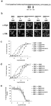
Residues within the second intracellular loop of GABAB2 are critical for agonist-induced GABAB receptor signaling. a, Schematic representation of GABAB2 il2. Arrowsindicate residues mutated. b, HEK293 cells were transiently transfected with wtGABAB1 (GB1) and wtGABAB2 (GB2), GB2K586E, GB2K590E, GB2K586E/M587E, or GB2tripleE. Cell surface expression of GABAB subunits was examined by immunofluorescence using an anti-myc antibody for GABAB1 (i–v) and an anti-HA antibody for GABAB2 (vi–x).c, Representative FLIPR analysis of intracellular Ca2+ changes after GABA stimulation of cells transiently transfected with Gqi5, wtGABAB1 (GB1), and wtGABAB2(GB2) GB2K586E, GB2K590E, GB2K586E/M587E, or GB2tripleE. Expression of single and double GABAB2 mutants resulted in decreased functional responses compared with wtGABAB2, with the GABAB2tripleE mutant showing no response at all. d, Representative FLIPR analysis of intracellular Ca2+ changes after GABA stimulation of cells transiently transfected with Gqi5, wtGABAB1 (GB1), and wtGABAB2(GB2), GB2K586A, GB2K590A, GB2K586A/M587A, or GB2tripleA. Single and double GABAB2 mutants resulted in decreased functional responses compared with wtGABAB2, with the GABAB2tripleA mutant completely unresponsive to GABA. e, Mutation of residues within the second intracellular loop of GABAB2 do not affect agonist or antagonist ligand binding. Membrane homogenates prepared from cells transiently transfected with wtGABAB1(GB1) and wtGABAB2(GB2), GB2K586E, GB2K590E, GB2K586E/M587E, or GB2tripleE specifically bound [3H]CGP54626. This could be completely displaced by GABA (10 mm). Data are expressed as means ± SEM (n = 3).
Fig. 3.
Fig. 3.
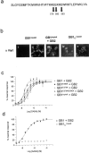
Acidic residues within the second intracellular loop of GABAB1 are not critical for agonist-induced GABAB receptor signaling. a, Schematic representation of GABAB1 il2.Arrows indicate residues mutated. b, HEK293 cells were transiently transfected with wtGABAB1(GB1), GB1E579K, GB1E583K, GB1E579K/E580K, GB1tripleK, or GB1C-tripleK alone or with wtGABAB2. Cell surface expression of GABAB1subunits was examined by immunofluorescence using an anti-myc antibody in the absence (i, iii,iv) or presence (ii, v) of a permeabilizing detergent. GABAB1 is only found at the cell surface when expressed with GABAB2, as shown with the GABAB1tripleK construct (i–iii). The C-terminally truncated GABAB1is expressed at the cell surface in the absence of GABAB2, as shown with the GABAB1C-tripleK mutant (iv, v). c, Representative FLIPR analysis of cells transiently transfected with Gqi5, wtGABAB2 (GB2), and wtGABAB1 (GB1), GB1E579K, GB1E583K, GB1E579K/E580K, or GB1tripleK. No differences in functional GABA responses were observed between mutant and wild-type GABAB1subunits. d, Representative FLIPR analysis of cells transiently transfected with Gqi5 and GB1C-tripleK. Expression of GB1C-tripleK alone did not give a functional response.
Fig. 4.
Fig. 4.
Basic residues within the second intracellular loop of GABAB1 are not critical for agonist-induced GABAB receptor signaling. a, Schematic representation of GABAB1 il2.Arrows indicate residues mutated. b, HEK293 cells were transiently transfected with wtGABAB2(GB2) and wtGABAB1 (GB1), GB1K577/578A, GB1K581/582A, GB1KQA, or GB1KQE. GABAB1 is only found at the cell surface when expressed with GABAB2, as shown by immunofluorescence using an anti-myc antibody in the absence (i–v) of a permeabilizing detergent. c, Representative FLIPR analysis of cells transiently transfected with Gqi5, wtGABAB2 (GB2), and wtGABAB1 (GB1),GB1K577/578A, GB1K581/582A, GB1KQA, or GB1KQE. Both mutant and wild-type GABAB1 subunits are functional when coexpressed with wtGABAB2.
Fig. 5.
Fig. 5.
Loss of functional coupling after exchange of GABAB1 and GABAB2 second intracellular loop charged residues. a, HEK293 cells were transiently transfected with GABAB1tripleK(GB1tripleK) and GABAB2tripleE(GB2tripleE). Cell surface expression of GABAB subunits was examined by immunofluorescence using an anti-myc antibody for GB1tripleK (i) and an anti-HA antibody for GB2tripleE (ii).b, Representative FLIPR analysis showing no functional response to GABA in cells transiently transfected with Gqi5, GB1tripleK, and GB2tripleE.
Fig. 6.
Fig. 6.
Coupling of wtGABAB2 and GABAB2 mutants to N-type Ca2+ channels in sympathetic neurons. a, Cell surface expression of wild-type and mutant GABAB2 subunits examined by immunofluorescence using an anti-HA antibody. b, Currents were recorded by stepping for 100 msec every 20 sec from −90 to 0 mV and leak-corrected by subtracting currents remaining after substituting 5 mm Co2+ for Ba2+. Records show superimposed leak-subtracted currents in the absence (black line) and presence (red line) of 50 μm baclofen for neurons injected 16–24 hr before recordings with 100–150 ng/μl wtGABAB2 (GB2), GB2K590E, or GB2tripleE. c, Bar charts show the mean inhibition of IBa amplitude by 50 μm baclofen, 10 msec after voltage stepping, in neurons injected with GB2, GB2K590E, or GB2tripleE. Error bars show SEM; nindicates number of cells. Note that GABAB2K590E still mediates inhibition of Ca2+ channel current, whereas GABAB2tripleE does not couple to Ca2+ channels. d, Plots show concentration dependence of Ca2+ current inhibition by baclofen in SCG neurons injected with GB2 (n = 5) or GB2K590E (n = 3). Curves were fitted to pooled data points (mean ± SEM) using Origin 5 software to the Hill equation y =ymax [infi] ·xnH/ (xnH+ KnH), where y is observed percentage of inhibition, ymax is the extrapolated maximal percentage of inhibition, x is baclofen concentration (micromolar), K is IC50 (micromolar), and nH is the Hill coefficient. For GABAB2K590E, IC50 of 1.04 ± 0.19 μm; nH of 1.23 ± 0.24; percentage of maximal inhibition, 42.03 ± 1.92%. For GABAB2, IC50 of 0.38 ± 0.06 μm;nH of 1.21 ± 0.19; percentage of maximal inhibition, 52.55 ± 2.02%. Note that GABAB2K590Einhibits Ca2+ current less effectively than GABAB2 and that plots could not be made for GABAB2tripleE because there was no detectable functional response.
Fig. 7.
Fig. 7.
GABAB2 expressed alone is unable to functionally couple to adenylate cyclase or bind GTPγS in response to GABA, but substitution of the GABAB2 N terminus with that of GABAB1 abolishes GABAB receptor function.a, Membranes prepared from cells expressing either GABAB2 (GB2) alone (squares) or both GABAB1 (GB1) and GABAB2together (circles, triangles) were incubated with either GABA (circles,squares) or baclofen (triangles) and subsequently with [35S]GTPγS. Data are presented as the percentage of increase in [35S]GTPγS binding over basal in response to agonist and are expressed as means ± SEM (n = 3). b, Cells expressing either GABAB2 alone or GABAB1 and GABAB2 together (symbols and legend as above) were incubated with forskolin and then either GABA or baclofen. Data are presented as the percentage of forskolin-stimulated cAMP levels remaining after incubation with agonist and are expressed as means ± SEM (n = 3). c, HEK293 cells were transiently transfected with GABAB1N/2(GB1N/2) and wtGABAB1(GB1), and cell surface expression of GABABsubunits was examined by immunofluorescence using either an anti-myc antibody for GABAB1N/2 in the absence (i) or presence (ii) of detergent or an anti-GABAB1 antibody for wtGABAB1(iii). d, Representative FLIPR analysis showing no functional response to GABA in cells transiently transfected with Gqi5, GABAB1N/2(GB1N/2), and wtGABAB1(GB1).
Fig. 8.
Fig. 8.
Proposed sideways signaling mechanism for the GABAB receptor heterodimer. GABAB1(orange) and GABAB2(green) subunits form heterodimers via C-terminal coiled-coil interactions, as well as N-terminal and possibly transmembrane domain interactions. The agonist GABA binds to the ligand binding domain within the GABAB1 N terminus, resulting in a conformational change in the GABAB2 subunit. This activated state of the receptor heterodimer allows recruitment of G-proteins, at least in part via an interaction with the positively charged residues in il2 of GABAB2 and subsequent activation of downstream signal transduction cascades.

References

    1. AbdAlla S, Lother H, Quitterer U. AT1-receptor heterodimers show enhanced G-protein activation and altered receptor sequestration. Nature. 2000;407:94–98. - PubMed
    1. Barton GJ. ALSCRIPT: a tool to format multiple sequence alignments. Protein Eng. 1993;6:37–40. - PubMed
    1. Bowen WP, Jerman JC. Nonlinear regression using spreadsheets. Trends Pharmacol Sci. 1995;16:413–417. - PubMed
    1. Bowery NG. GABAB receptor pharmacology. Annu Rev Pharmacol Toxicol. 1993;33:109–147. - PubMed
    1. Calver AR, Robbins MJ, Cosio C, Rice SQJ, Babbs AJ, Hirst WD, Boyfield I, Wood MD, Russell RB, Price GW, Couve A, Moss SJ, Pangalos MN. The C-terminal domains of the GABAB receptor subunits mediate intracellular trafficking, but are not required for receptor signaling. J Neurosci. 2001;21:1203–1210. - PMC - PubMed

Publication types

MeSH terms

LinkOut - more resources