. 2001 Oct 15;21(20):8053-61.
doi: 10.1523/JNEUROSCI.21-20-08053.2001.

Induction of alpha-synuclein aggregation by intracellular nitrative insult

Affiliations

Induction of alpha-synuclein aggregation by intracellular nitrative insult

E Paxinou et al. J Neurosci. .

Abstract

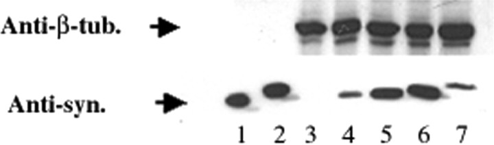
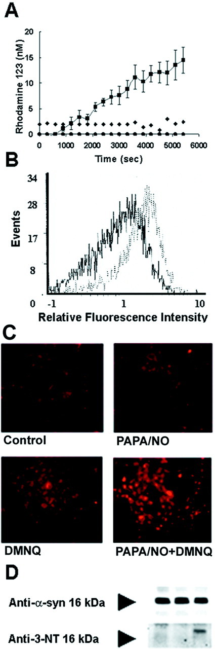
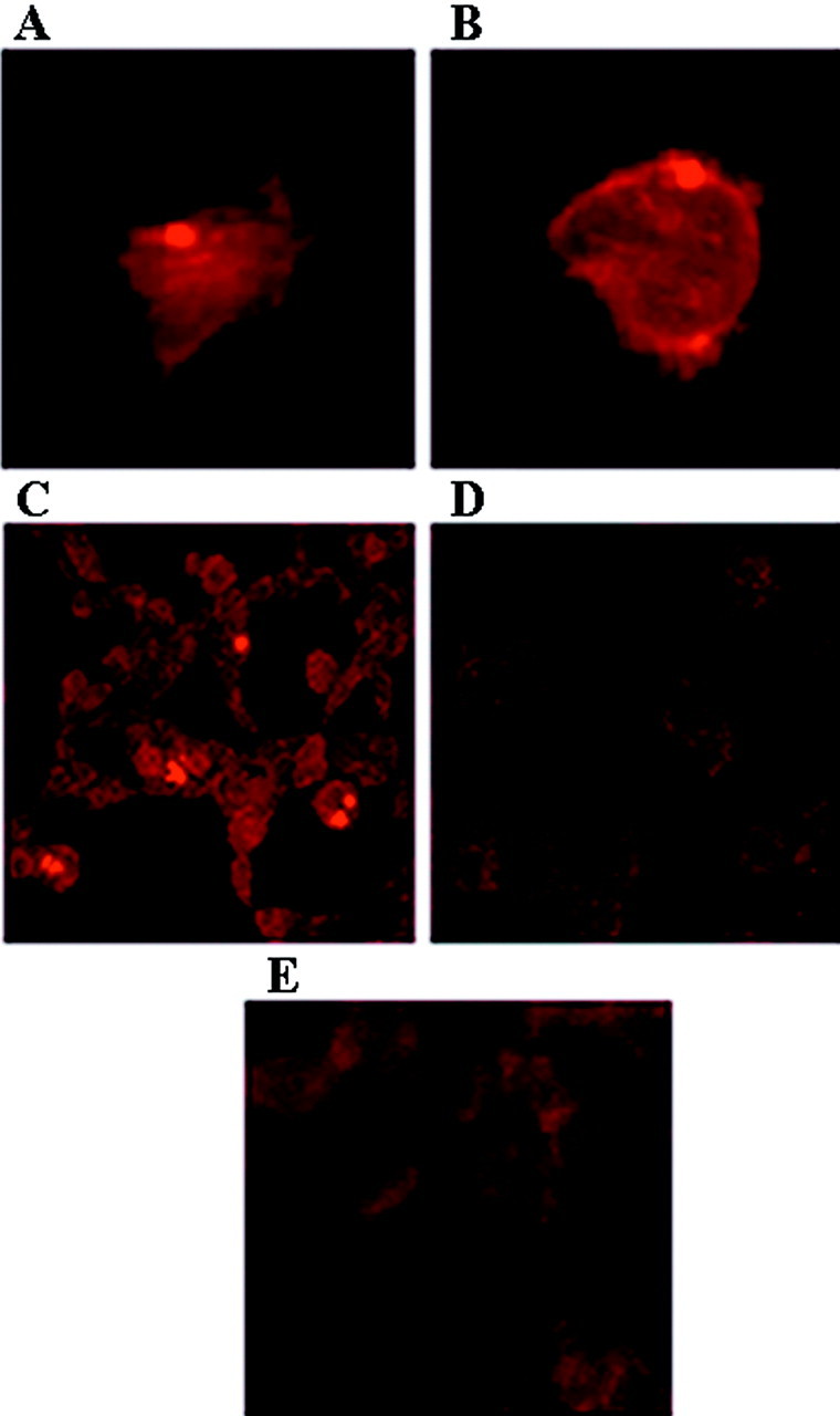
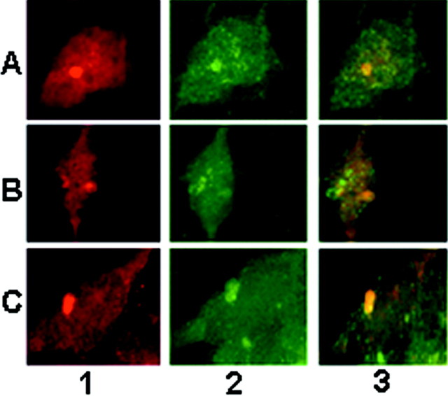
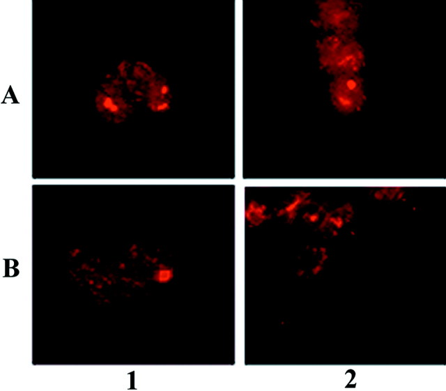
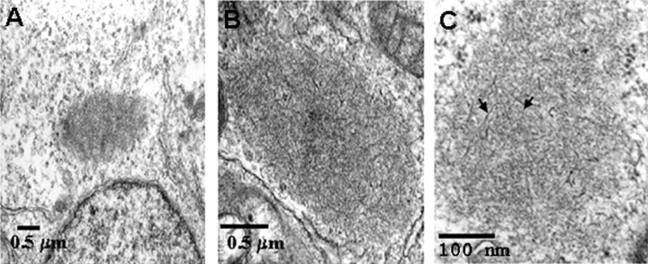
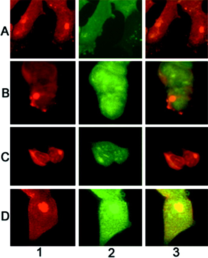
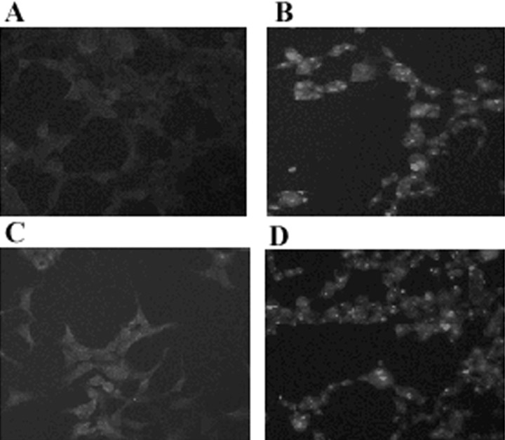
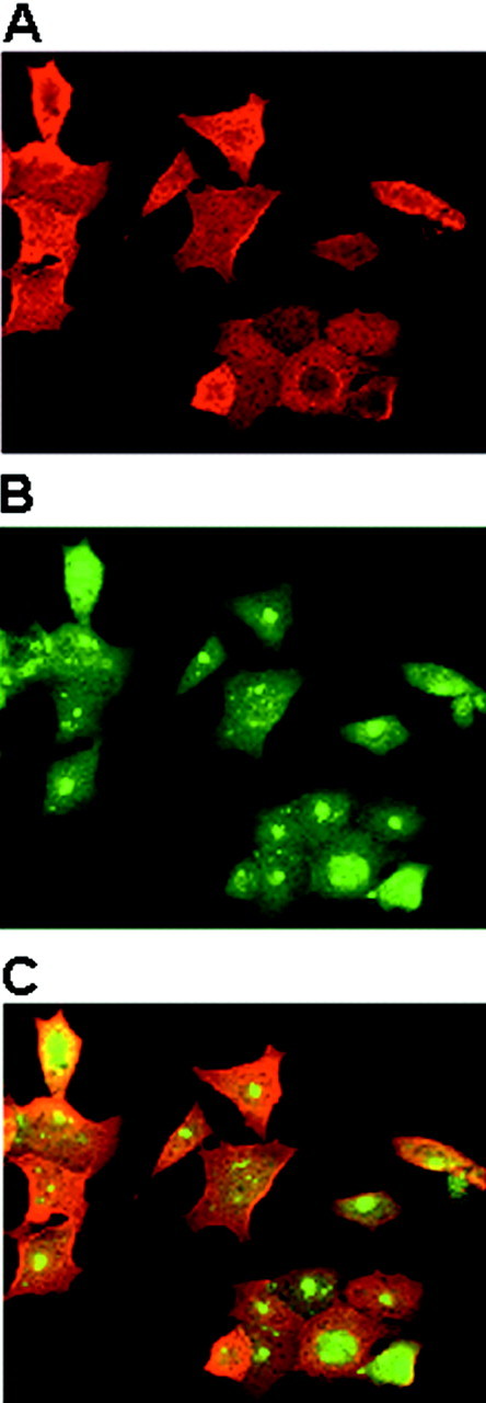
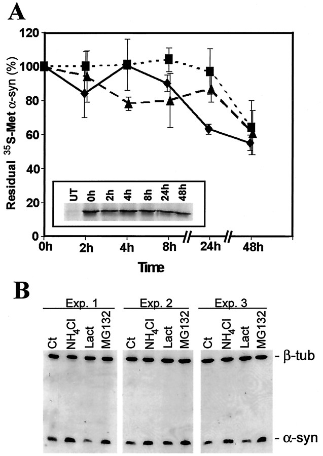
Brain lesions containing filamentous and aggregated alpha-synuclein are hallmarks of neurodegenerative synucleinopathies. Oxidative stress has been implicated in the formation of these lesions. Using HEK 293 cells stably transfected with wild-type and mutant alpha-synuclein, we demonstrated that intracellular generation of nitrating agents results in the formation of alpha-synuclein aggregates. Cells were exposed simultaneously to nitric oxide- and superoxide-generating compounds, and the intracellular formation of peroxynitrite was demonstrated by monitoring the oxidation of dihydrorhodamine 123 and the nitration of alpha-synuclein. Light microscopy using antibodies against alpha-synuclein and electron microscopy revealed the presence of perinuclear aggregates under conditions in which peroxynitrite was generated but not when cells were exposed to nitric oxide- or superoxide-generating compounds separately. alpha-Synuclein aggregates were observed in 20-30% of cells expressing wild-type or A53T mutant alpha-synuclein and in 5% of cells expressing A30P mutant alpha-synuclein. No evidence of synuclein aggregation was observed in untransfected cells or cells expressing beta-synuclein. In contrast, selective inhibition of the proteasome resulted in the formation of aggregates detected with antibodies to ubiquitin in the majority of the untransfected cells and cells expressing alpha-synuclein. However, alpha-synuclein did not colocalize with these aggregates, indicating that inhibition of the proteasome does not promote alpha-synuclein aggregation. In addition, proteasome inhibition did not alter the steady-state levels of alpha-synuclein, but addition of the lysosomotropic agent ammonium chloride significantly increased the amount of alpha-synuclein, indicating that lysosomes are involved in degradation of alpha-synuclein. Our data indicate that nitrative and oxidative insult may initiate pathogenesis of alpha-synuclein aggregates.


Figures

Fig. 1.
Fig. 1.
Expression of synucleins in stably transfected HEK 293 cell lines. Expression of human wild-type α-syn (lane 4), A53T mutant α-syn (lane 5), A30P mutant α-syn (lane 6), and human β-synuclein (lane 7) in stably transfected HEK 293 cells is shown, as demonstrated by Western blot analysis using the anti-synuclein antibody Syn102, which reacts equally to both syn proteins (Giasson et al., 2000b). Expression of α- and β-syn is not detected in untransfected HEK 293 cells (lane 3).Lanes 1 and 2 were loaded with 10 ng of recombinant human α- and β-syn, respectively. Lanes 3–7 were loaded with 10 μg of total cell lysates.
Fig. 2.
Fig. 2.
Formation of rhodamine 123 from the oxidation of DHR 123 in HEK 293/α-syn. A, HEK 293/α-syn cells were incubated for 2 hr at 37°C with 5 μm DHR 123. After extensive washing, rhodamine 123 fluorescence was monitored over time in untreated cells (triangles) and cells treated with 1 mm PAPA/NO alone (diamonds), DMNQ alone (circles), or 1 mm PAPA/NO plus 10 μm DMNQ (squares). B, Composite fluorescence histograms obtained by flow cytometric evaluation of HEK 293/α-syn cells after exposure to PAPA/NO plus DMNQ (broken trace) and control cells (solid trace). The mean fluorescence intensity value for each histogram, indicating the percentage of cells positive for rhodamine 123 fluorescence, was 6% for control and 29% for treated cells.C, Representative epifluorescence images (magnification, 17×) of cells after exposure to nitric oxide- and superoxide-generating compounds. D, Immunoprecipitation of α-syn using the anti-α-syn monoclonal antibody SYN-1 followed by Western blotting with SYN-1 or a polyclonal anti-3-nitrotyrosine antibody (Anti-3-NT). Cells were exposed to 1 mm PAPA/NO (first lane), 5 mm paraquat (second lane), or both 1 mm PAPA/NO and 5 mm paraquat (third lane).
Fig. 3.
Fig. 3.
Formation of α-syn intracellular aggregates on exposure to nitric oxide- and superoxide-generating compounds. HEK 293/α-syn cells were fixed and stained with monoclonal anti-α-syn antibodies Syn208 and Syn202. A–C, α-Syn inclusions were readily visible in cells exposed to 1 mm PAPA/NO plus 5 μm dopamine (A), 1 mmPAPA/NO plus 100 nm rotenone (B), and 1 mm PAPA/NO plus 5 mm paraquat (C). Only diffuse background staining was noted in untransfected (D) and β-syn-transfected (E) cells exposed to 1 mm PAPA/NO plus 5 mm paraquat. Magnification: A, B, 125×; C–E, 65×.
Fig. 4.
Fig. 4.
Nitration of α-syn intracellular aggregates on exposure to nitric oxide- and superoxide-generating compounds.A, HEK 293/α-syn exposed to 1 mm PAPA/NO plus 100 nm rotenone. B, HEK 293/A30P mutant α-syn exposed to 1 mm PAPA/NO plus 5 μmdopamine. C, HEK 293/A53T mutant α-syn exposed to 1 mm PAPA/NO plus 100 nm rotenone. Column 1, Cells were fixed and stained with monoclonal anti-α-syn antibody Syn 208. Column 2, Stained with a polyclonal anti-3-nitrotyrosine antibody. Column 3, Superimposed image. Magnification, 125×.
Fig. 5.
Fig. 5.
Intracellular aggregates of α-syn on exposure to nitric oxide- and superoxide-generating compounds are nitrated at Tyr39 as well as Tyr125 and Tyr136. A, HEK 293/α-syn exposed to 1 mm PAPA/NO plus 100 nm rotenone.B, HEK 293/α-syn exposed to 1 mm PAPA/NO plus 5 μm dopamine. Column 1, Cells were fixed and stained with monoclonal anti-nitrated α-syn antibody nSyn14, which recognizes nitrated tyrosine residue Tyr39 in the N terminus of α-syn. Column 2, Stained with monoclonal anti-nitrated α-syn antibody nSyn12, which recognizes nitrated tyrosine residues Tyr125 and Tyr136 in the C terminus of α-syn (Giasson et al., 2000a). Magnification, 98×.
Fig. 6.
Fig. 6.
Ultrastructure of inclusions induced by nitrative damage in HEK 293/α-syn cells. Electron microscopic examination of cytoplasmic inclusions in HEK 293/α-syn cells exposed to nitric oxide- and superoxide-generating compounds was performed. InC, The sample was treated with formic acid to enhance the appearance of fibrils (arrows) in the inclusion.
Fig. 7.
Fig. 7.
Paucity of ubiquitin immunoreactivity in α-syn aggregates. Cells were stained with the mouse anti-α-syn monoclonal antibodies Syn202 and Syn208 (column 1) or a rabbit anti-ubiquitin polyclonal antibody (column 2), and inclusions were visualized by immunofluorescence. Column 3, Overlay composite of images. Cells expressing wild-type α-syn (A, B, D) or A53T mutant α-syn (C) were exposed to PAPA/NO and paraquat (A), PAPA/NO and rotenone (B, C), or paraquat alone (D). Magnification, 125×.
Fig. 8.
Fig. 8.
Formation of protein aggregates after selective inhibition of the proteasome. Untransfected HEK 293 cells (A, B) or HEK 293/α-syn cells (C, D) were treated with 10 μm lactacystin-β-lactone for 1 hr and stained with anti-α-syn monoclonal antibody Syn202 (A, C) or a rabbit anti-ubiquitin antibody (B, D). Magnification, 40×.
Fig. 9.
Fig. 9.
Formation of protein aggregates after inhibition of the proteasome. HEK 293/α-syn cells were treated with 10 μm lactacystin-β-lactone for 1 hr and stained with the mouse anti-α-syn monoclonal antibody Syn202 (A) or with a rabbit anti-ubiquitin antibody (B). InC, the fluorescence fields in A andB were merged. Note that inhibition of the proteasome with lactacystin-β-lactone results in ubiquitin aggregates but not α-syn aggregates. Magnification, 65×.
Fig. 10.
Fig. 10.
Degradation of α-syn in HEK 293 cells.A, Pulse–chase analysis of wild-type (diamonds), A30P (triangles), and A53T (squares) α-syn turnover in HEK 293 stable transfectants. The residual [35S]Met is after chasing over 48 hr (n = 3). A, inset, Representative chase profile of [35S]Met-labeled wild-type α-syn.UT, Immunoprecipitation from untransfected HEK 293 cells. B, Western blot analysis using the anti-α-syn antibody LB509 showing three independent experiments. HEK 293/α-syn cells were untreated (Ct) or challenged with 25 mm NH4Cl, 10 μmlactacystin-β-lactone (Lact), or 2 μmMG132 for 24 hr. Equal amounts of protein (5 μg) were loaded in each separate lane of the gels as confirmed by the levels of β-tubulin (β-tub).

Similar articles

Cited by

References

    1. Alvarez B, Ferrer-Sueta G, Freeman BA, Radi R. Kinetics of peroxynitrite reaction with amino acids and human serum albumin. J Biol Chem. 1999;274:842–848. - PubMed
    1. Ancolio K, Alves da Costa C, Ueda K, Checler F. α-Synuclein and the Parkinson's disease-related mutant Ala53Thr-α-synuclein do not undergo proteasomal degradation in HEK293 and neuronal cells. Neurosci Lett. 2000;285:79–82. - PubMed
    1. Ara J, Przedborski S, Naini AB, Jackson-Lewis V, Trifiletti RR, Horwitz J, Ischiropoulos H. Inactivation of tyrosine hydroxylase following exposure to peroxynitrite and MPTP. Proc Natl Acad Sci USA. 1998;95:7659–7663. - PMC - PubMed
    1. Ayata C, Ayata G, Hara H, Matthews RT, Beal MF, Ferrante RJ, Endres M, Kim A, Christie RH, Waeber C, Huang PL, Hyman BT, Moskowitz MA. Mechanisms of reduced striatal NMDA excitotoxicity in type 1 nitric oxide synthase knock-out mice. J Neurosci. 1997;17:6908–6917. - PMC - PubMed
    1. Bennett MC, Bishop JF, Leng Y, Chock PB, Chase TM, Mouradian MM. Degradation of α-synuclein by proteasome. J Biol Chem. 1999;274:33855–33858. - PubMed

Publication types

MeSH terms

LinkOut - more resources