. 2001 Oct 15;21(20):8062-71.
doi: 10.1523/JNEUROSCI.21-20-08062.2001.

AMPA receptor channels with long-lasting desensitization in bipolar interneurons contribute to synaptic depression in a novel feedback circuit in layer 2/3 of rat neocortex

Affiliations

AMPA receptor channels with long-lasting desensitization in bipolar interneurons contribute to synaptic depression in a novel feedback circuit in layer 2/3 of rat neocortex

A Rozov et al. J Neurosci. .

Abstract

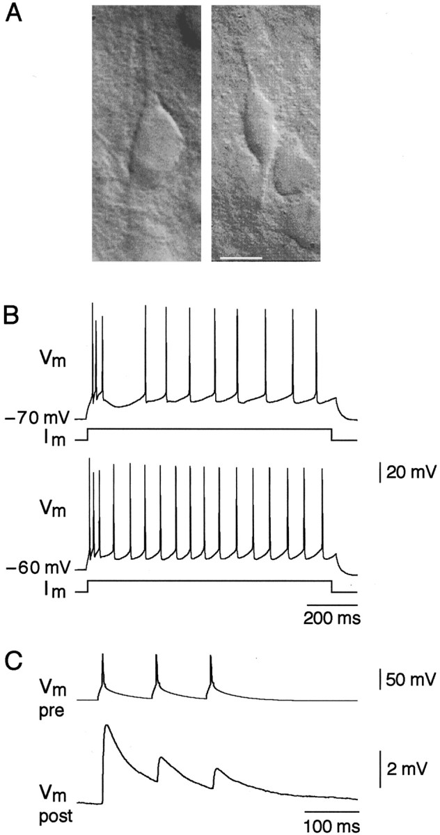
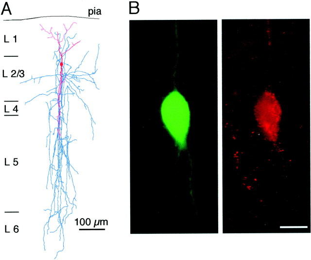
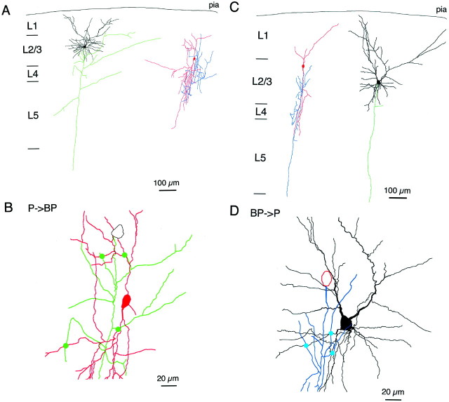
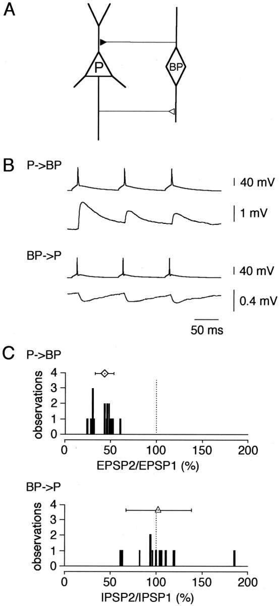
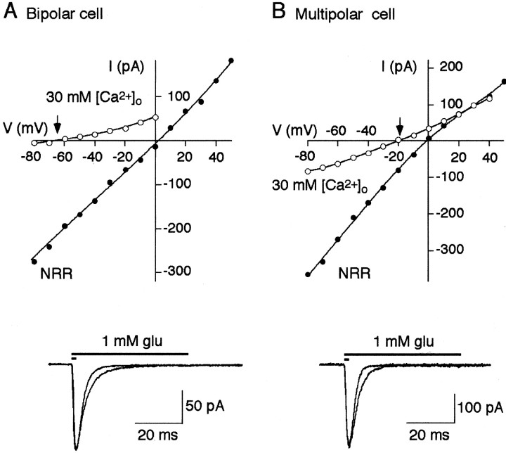
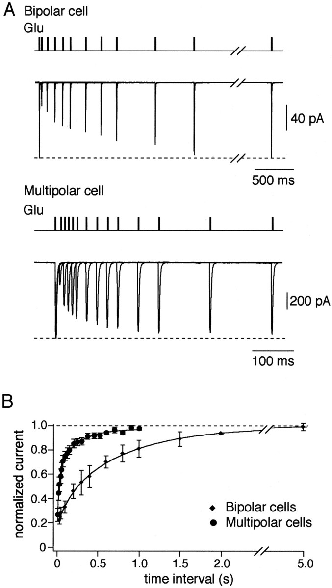
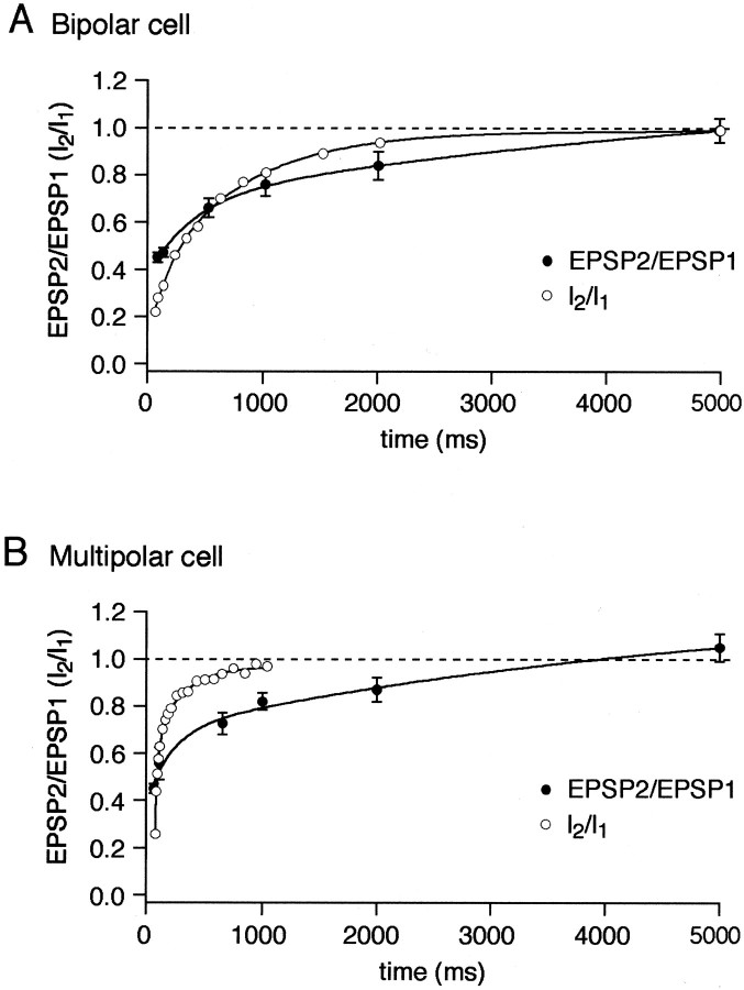
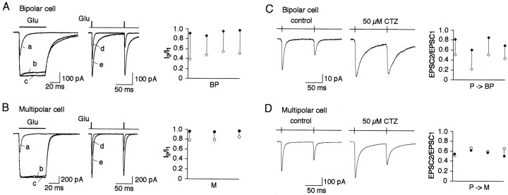
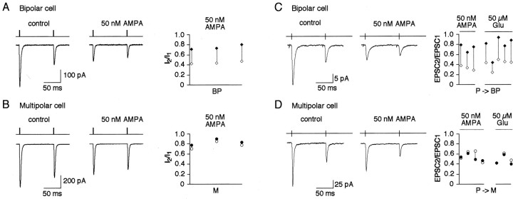
A novel, local inhibitory circuit in layer 2/3 of rat somatosensory cortex is described that connects pyramidal cells reciprocally with GABAergic vasoactive intestinal polypeptide-immunoreactive bipolar interneurons. In paired whole-cell recordings, the glutamatergic unitary responses (EPSPs or EPSCs) in bipolar cells evoked by repetitive (10 Hz) stimulation of a pyramidal cell show strong frequency-dependent depression. Unitary IPSPs evoked in pyramidal cells by repetitive stimulation of bipolar cells, on average, maintained their amplitude. This suggests that the excitatory synapses on bipolar cells act as a low-pass filter in the reciprocal pyramid-to-bipolar circuit. The EPSCs in bipolar cells are mediated predominantly by AMPA receptor (AMPAR) channels. AMPARs desensitize rapidly and recover slowly from desensitization evoked by a brief pulse of glutamate. In slices, reduction of AMPAR desensitization by cyclothiazide (50-100 microm) or conditioning steady-state desensitization induced by application of extracellular AMPA (50 nm) or glutamate (50 microm) strongly reduced synaptic depression. It is concluded that in the local circuits between pyramidal and bipolar cells the desensitization of AMPARs in bipolar cells contributes to low-pass feedback inhibition of layer 2/3 pyramidal neurons by bipolar cells.


Figures

Fig. 1.
Fig. 1.
Morphological and functional signature of bipolar interneurons. A, Representative IR-DIC image of a pyramidal cell (left) and a bipolar cell (right) in layer 2/3 region of rat neocortex. Scale bar, 10 μm. B, Action potential patterns of a bipolar cell after depolarizing current injection of the same value (100 pA) at resting potential (−70 mV; top trace) and at −60 mV (bottom trace). C, Short-term depression of EPSPs (bottom trace) in response to three action potentials (10 Hz; top trace) evoked in synaptically connected pyramidal cell.
Fig. 2.
Fig. 2.
Anatomical and immunocytochemical identification of bipolar interneurons. A, Dendritic (red) and axonal (blue) arbor morphology of a biocytin-labeled bipolar interneuron. B, Digital micrograph of a biocytin-labeled bipolar interneuron visualized by FITC-labeled avidin (left). VIP immunoreactivity of the same cell shown by CY-3 immunofluorescence (right). Scale bar, 10 μm.
Fig. 3.
Fig. 3.
Synaptic connections between bipolar and pyramidal neurons. A, Dendritic and axonal arbor morphology of a biocytin-labeled pyramidal neuron (left) making excitatory synaptic connection to bipolar interneuron (right). Two cells are shown separately for clarity.B, Four excitatory synaptic connections (green circles) between the axon of the pyramidal neuron (green) and dendrites of the bipolar interneuron (red). The same cells as in Ashown on expanded scale. C, Dendritic and axonal arbor morphology of a biocytin-labeled bipolar interneuron (left) making inhibitory synaptic connection to pyramidal neuron (right). D, Three inhibitory synaptic connections (blue circles) between the axon of the bipolar interneuron (blue) and dendrites of the pyramidal neuron (black). The same cells as inC shown on expanded scale.
Fig. 4.
Fig. 4.
Reciprocal innervation in layer 2/3 pyramid-bipolar-pyramid. A, Schematic diagram of reciprocal connections between pyramidal (P) and bipolar (BP) cells. B, Dual simultaneous recordings from reciprocally connected pyramidal and bipolar cells: EPSPs evoked by pyramidal cell terminals in postsynaptic bipolar cell (P → BP; top traces) and IPSPs evoked by bipolar cell terminals in postsynaptic pyramidal cell (BP → P;bottom traces). Presynaptic cells were stimulated at 10 Hz. C, Distribution of amplitude ratios of EPSP2/EPSP1 (top histogram) and IPSP2/IPSP1 (bottom histogram). Stimulation frequency, 10 Hz. Symbols above histograms give the mean (± SD) amplitude ratios [bipolar cells, 43 ± 10%, n = 18 (diamond); pyramidal cells, 102 ± 36%, n = 13 (triangle)].
Fig. 5.
Fig. 5.
Ca2+ permeability, deactivation, and desensitization time course of AMPAR channels.A, Top panel, Current–voltage relations for the glutamate-evoked currents recorded from nucleated patches pulled from bipolar cells in normal rat Ringer's solution (NRR;closed circles) and high Ca2+ (30 mm [Ca2+]o;open circles) solutions. Arrow indicates Ca2+/Cs+ reversal potential.Bottom panel, Currents recorded from nucleated patch in response to 2 and 50 msec glutamate pulses. Membrane potential, −60 mV. Extracellular solution contained 10 μmd-AP-5. B, Same as in A for multipolar cells.
Fig. 6.
Fig. 6.
Recovery from desensitization of AMPAR channels.A, Overlaid glutamate-evoked currents recorded using double-pulse protocol at variable interpulse intervals in nucleated patches pulled from bipolar (top traces) and multipolar (bottom traces) cells. Duration of glutamate (1 mm) pulses was 2 msec. Membrane potential, −60 mV.B, Time course of recovery from desensitization for the two types of interneurons measured as specified in A. Each point represents average of five to eight experiments.Solid lines represent double exponential fits for the data points.
Fig. 7.
Fig. 7.
Recovery from synaptic depression.A, Time course of recovery from depression of EPSPs in bipolar cells recorded using paired-pulse stimulation of pyramidal cells at different interpulse intervals. B, The same as in A for multipolar cells. Open symbolsindicate recovery from desensitization of the glutamate-evoked currents (I2/I1) for respective cells taken from Figure 6. Solid linesrepresent double exponential fits for the data points.
Fig. 8.
Fig. 8.
Effects of cyclothiazide on AMPAR desensitization and synaptic depression. A, Left, Overlaid glutamate-evoked currents recorded in the same nucleated patch pulled from a bipolar cell in control (a), with 50 μm (b), and 100 μm(c) CTZ. Duration of glutamate (1 mm) pulses was 50 msec. Middle, Overlaid glutamate-evoked currents recorded using double pulse protocol at 100 msec interpulse interval in the same nucleated patch pulled from a bipolar cell in control (d) and with 50 μm CTZ (e). Duration of glutamate pulses was 2 msec. Membrane potential, −60 mV. Right, Pairwise comparison of the current amplitude ratios (I2/I1) in control (open symbols) and in the presence of 50 μm CTZ (closed symbols) recorded from four nucleated patches. Connected symbols represent values obtained from the same patch. B, Same as inA for patches pulled from multipolar cells.C, Representative recordings of EPSCs evoked in the same bipolar cell after 10 Hz stimulation of a presynaptic pyramidal cell in control (left) and after application of 50 μm CTZ into extracellular solution (middle). Pairwise comparison of the amplitude ratios (EPSC2/EPSC1) in control (open symbols) and in the presence of CTZ (closed symbols) recorded from four cell pairs is shown on the right. Connected symbols represent values obtained from the same cell pairs.D, Same as in C for EPSCs in target multipolar cells.
Fig. 9.
Fig. 9.
Effects of AMPA and glutamate on synaptic depression. A, Glutamate-evoked currents recorded using double-pulse protocol at 100 msec interpulse interval in nucleated patch pulled from a bipolar cell in control (left) and in the presence of 50 nm AMPA (middle). Duration of glutamate pulses was 2 msec. Membrane potential, −60 mV. Pairwise comparison of the current amplitude ratios (I2/I1) in control (open symbols) and in the presence of 50 nm AMPA (closed symbols) is shown on theright. Connected symbols represent values obtained from the same patch. B, Same as inA for patches pulled from multipolar cells.C, Representative recordings of EPSCs evoked in the same bipolar cell after 10 Hz stimulation of a presynaptic pyramidal cell in control (left) and in the presence of 50 nmAMPA in extracellular solution (middle). Pairwise comparison of the amplitude ratios (EPSC2/EPSC1) in control (open symbols) and in the presence of 50 nmAMPA or 50 μm glutamate (closed symbols) is shown on the right. Connected symbolsrepresent values obtained from the same cell pairs. D, Same as in C for EPSCs in target multipolar cells.

References

    1. Angulo MC, Lambolez B, Audinat E, Hestrin S, Rossier J. Subunit composition, kinetic, and permeation properties of AMPA receptors in single neocortical nonpyramidal cells. J Neurosci. 1997;17:6685–6696. - PMC - PubMed
    1. Bellingham MC, Walmsley B. A novel presynaptic inhibitory mechanism underlies paired pulse depression at a fast central synapse. Neuron. 1999;23:159–170. - PubMed
    1. Brusa R, Zimmerman F, Koh D-S, Feldmeyer D, Gass P, Seeburg P, Sprengel R. Early-onset epilepsy and postnatal lethality associated with an editing-deficient GluR-B allele in mice. Science. 1995;270:1677–1680. - PubMed
    1. Cauli B, Audinat E, Lambolez B, Angulo MC, Ropert N, Tsuzuki K, Hestrin S, Rossier J. Molecular and physiological diversity of cortical nonpyramidal cells. J Neurosci. 1997;17:3894–3906. - PMC - PubMed
    1. Cauli B, Porter JT, Tsuzuki K, Lambolez B, Rossier J, Quenet B, Audinat E. Classification of fusiform neocortical interneurons based on unsupervised clustering. Proc Natl Acad Sci USA. 2000;97:6144–6149. - PMC - PubMed

MeSH terms

LinkOut - more resources