. 2001 Oct 15;21(20):8108-18.
doi: 10.1523/JNEUROSCI.21-20-08108.2001.

Neuroprotection through delivery of glial cell line-derived neurotrophic factor by neural stem cells in a mouse model of Parkinson's disease

Affiliations

Neuroprotection through delivery of glial cell line-derived neurotrophic factor by neural stem cells in a mouse model of Parkinson's disease

P Akerud et al. J Neurosci. .

Abstract

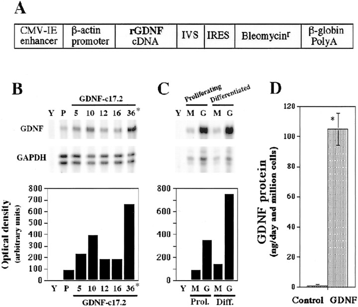
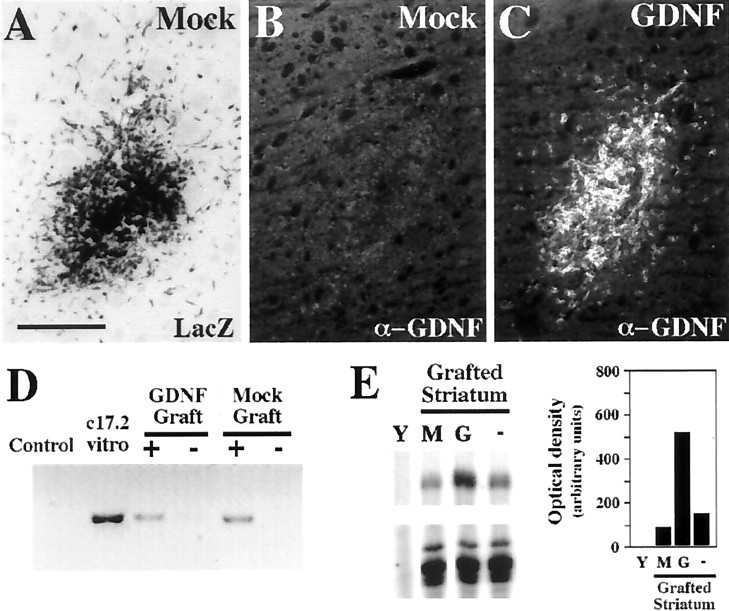
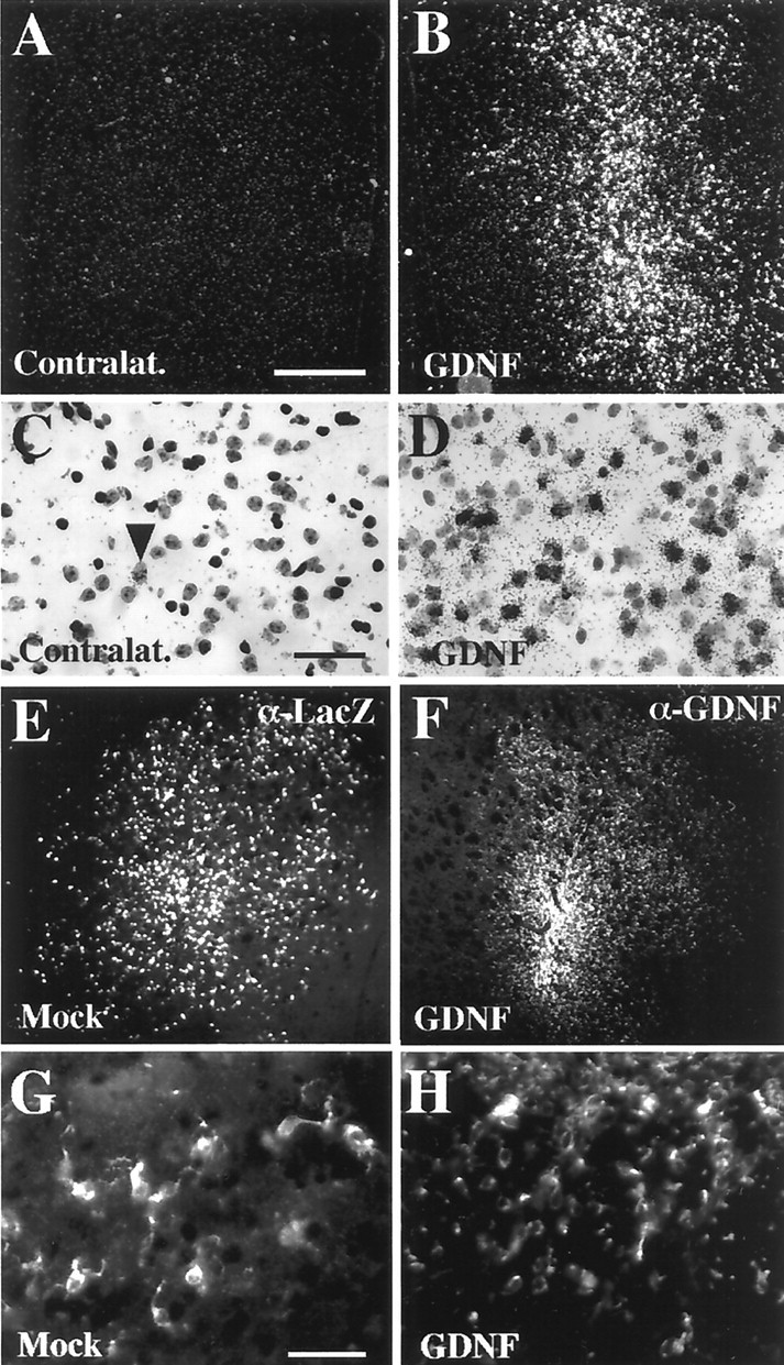
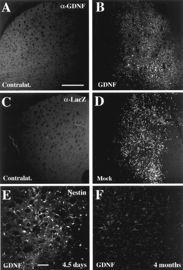
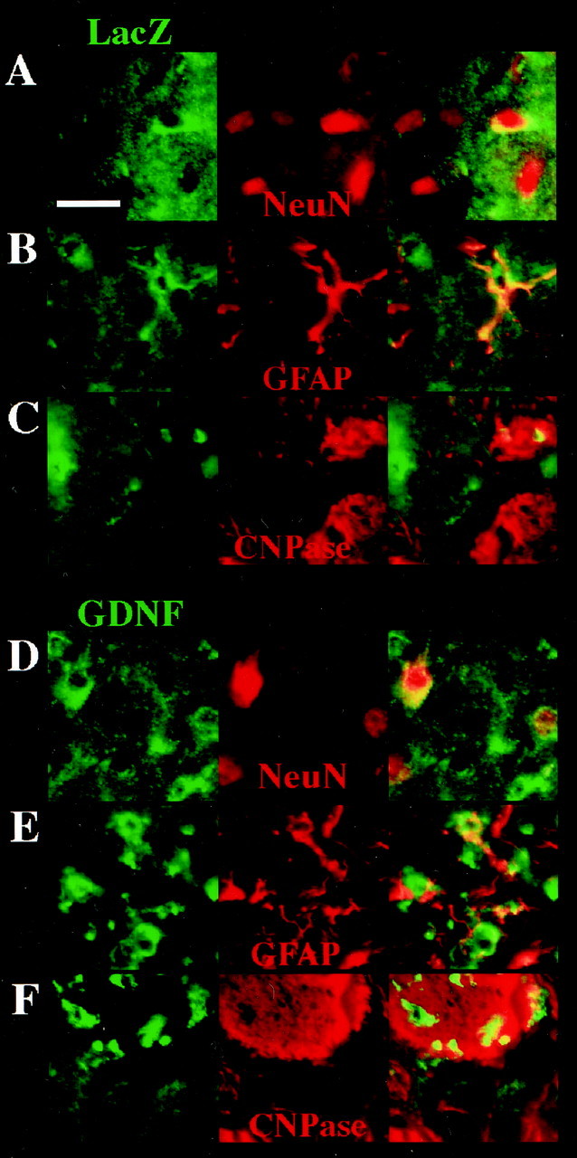
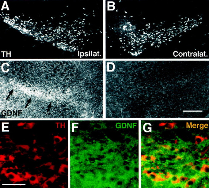
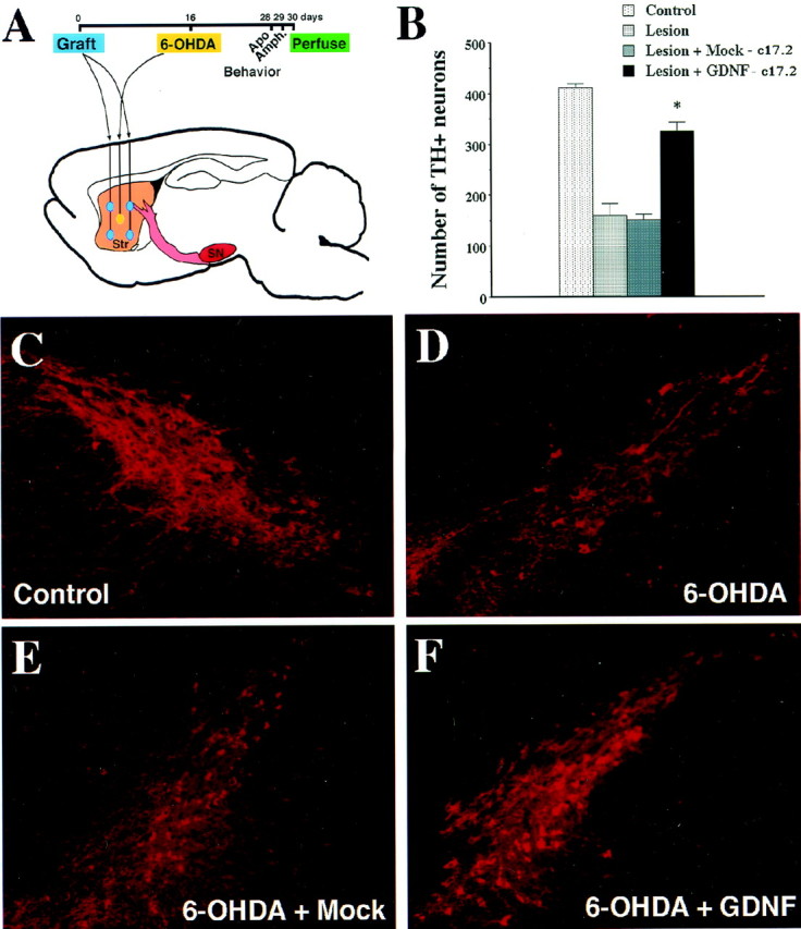
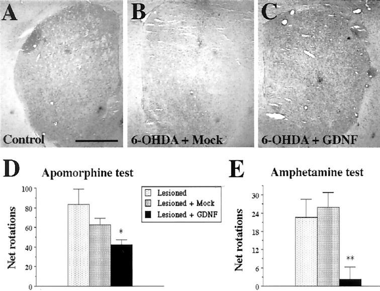
Neural stem cells (NSCs) have been proposed as tools for treating neurodegeneration because of their capacity to give rise to cell types appropriate to the structure in which they are grafted. In the present work, we explore the ability of NSCs to stably express transgenes and locally deliver soluble molecules with neuroprotective activity, such as glial cell line-derived neurotrophic factor (GDNF). NSCs engineered to release GDNF engrafted well in the host striatum, integrated and gave rise to neurons, astrocytes, and oligodendrocytes, and maintained stable high levels of GDNF expression for at least 4 months. The therapeutic potential of intrastriatal GDNF-NSCs grafts was tested in a mouse 6-hydroxydopamine model of Parkinson's disease. We found that GDNF-NSCs prevented the degeneration of dopaminergic neurons in the substantia nigra and reduced behavioral impairment in these animals. Thus, our results demonstrate that NSCs efficiently express therapeutic levels of GDNF in vivo, suggesting a use for NSCs engineered to release neuroprotective molecules in the treatment of neurodegenerative disorders, including Parkinson's disease.


Figures

Fig. 1.
Fig. 1.
Establishment of a GDNF-overexpressing neural stem cell line. A, Schematic representation of the pCAGGS-GIB expression vector that was used for transfection of the c17.2 cells. A rat GDNF cDNA and a mini-intron IRES-bleo resistance gene fragment were cloned after the β-actin promoter and a CMV-immediate early (CMV-IE) enhancer, in the pCAGGS vector.B and C show two representative experiments, in which GDNF mRNA was analyzed by RPA in control and GDNF-transfected cell lines. IVS, Synthetic intron.B, GDNF mRNA expression was determined in the parental c17.2 cell line (P) and five clones (5, 10, 12,16, 36) transfected with the pCAGGS-GIB construct. Yeast tRNA (Y) was used as a negative control. Clone 36, the highest expressor, was named GDNF-c17.2 and further characterized. C, GDNF mRNA expression was also examined in proliferating and differentiated (in N2 medium for 1 week) MT-c17.2 control cells (M) and the GDNF-c17.2 cells (G). Note that the difference in GDNF mRNA expression between the MT-c17.2 and the GDNF-c17.2 cell lines is even greater after differentiation than while proliferating.D, ELISA analysis of supernatants from proliferating cells in vitro showed that the control cells (Control; c17.2 or MT-c17.2) released <1 ng of GDNF per 106 cells in 1 d, whereas the GDNF-c17.2 cell line produced ∼100 ng of GDNF per 106 cells in 1 d (n = 3–4; *p < 0.0001; unpaired Student's t test).
Fig. 2.
Fig. 2.
GDNF-c17.2 and MT-c17.2 grafted cells survive the grafting procedure well, but only GDNF-c17.2 cells express high levels of GDNF in vivo. A–C, MT-c17.2 and GDNF-c17.2 grafted cells in the striatum 4.5 d after grafting.A, X-Gal histochemistry (LacZ) confirmed the presence of MT-c17.2 grafted cells. Note the migration out of the graft by some of the cells as soon as 4.5 d. B, No GDNF immunoreactivity was detected in MT-c17.2 grafts on adjacent slides. C, GDNF immunohistochemistry showed many GDNF-positive cells in the GDNF-c17.2 grafts, which also were found migrating away from the graft site at this time point.D, The presence of cells in the grafted striatum was also verified at later time points (1 and 10 months) by PCR against thelacZ vector. Cells could be detected in the GDNF or mock grafted striatum (+) but not in the contralateral striatum (−). The signal was similar at 1 month (as shown in the Mock Graft) and at 10 months (shown in the GDNF Graft). Proliferating c17.2 cells (c17.2 vitro) were used as positive control, and cells not transfected withlacZ expression vectors were used as negative control.E, Increased levels of GDNF mRNA expression could also be detected by RPA in the striatum 15 d after grafting of the GDNF-c17.2 cells (G) but not after grafting the MT-c17.2 cells (M). The levels of expression of GDNF in the nongrafted striatum (−) were higher than in the control grafted striatum (M) because the MT-c17.2 cells express less GDNF than the intact adult striatum. GDNF expression in the GDNF-c17.2 grafted striatum was more than five times higher than in the MT-c17.2 grafted striatum. Yeast tRNA was used as negative control. Values correspond to one representative experiment (n = 2). Scale bar (in A):A–C, 250 μm.
Fig. 3.
Fig. 3.
GDNF-c17.2 and MT-c17.2 cells engraft well and disperse within the striatum by 1 month after grafting.A–D, ISH showed the presence of cells expressing very high levels of GDNF mRNA in the GDNF grafted striatum (B, D) but not in the contralateral side (A, C). Note the abundance and dispersion of the signal in the grafted striatum (B). At higher magnification and bright field (C, D), it is possible to observe one endogenous GDNF-expressing cell (arrowhead in C) and many grafted cells with high levels of GDNF mRNA expression (D). E, G, The presence of MT-c17.2 cells was verified by β-Gal immunohistochemistry against thelacZ product (α-Lac) at 30 d after grafting. β-Gal-immunoreactive cells (magnified in G) were detected in the ipsilateral striatum to the graft (E). F, H, GDNF-c17.2 cells were also detected 1 month after grafting by GDNF immunohistochemistry.F, GDNF-c17.2 cells (magnified inH) displayed a similar distribution to cells expressing high levels of GDNF mRNA. Scale bars: (inA) A, B, E, F, 500 μm; (inC) C, D, 100 μm; (in G)G, H, 100 μm.
Fig. 4.
Fig. 4.
GDNF-c17.2 and MT-c17.2 engrafted for at least 4 months, the longest time point analyzed morphologically.A, B, GDNF-c17.2 cells engrafted well in the adult striatum and were detected by GDNF immunohistochemistry in the ipsilateral striatum of all nude mice but not in the contralateral striatum (A). MT-c17.2 cells showed a similar pattern of engraftment and were readily detected by β-Gal immunohistochemistry against the lacZ product (α-Lac) 4 months after grafting in the ipsilateral striatum (D) but not in the contralateral side (C). Scale bar (in A):A–D, 500 μm. E, F, At 4 months after grafting, we also found that GDNF-c17.2 cells differentiated and did not express the neural stem cell marker nestin (F). Instead, nestin was found to be abundantly expressed by most of the grafted cells by 4.5 d (E). Scale bar (in E): E,F, 100 mm.
Fig. 5.
Fig. 5.
Double immunohistochemistry showing that MT-c17.2 (A–C) and GDNF-c17.2 cells (D–F) behave as multipotent NSCs after intrastriatal grafting in the adult CD-1 mice and give rise to neurons (A, D), astrocytes (B, E), and oligodendrocytes (C,F). A–C, Double immunohistochemistry with anti-β-Gal antibodies (LacZ, in green), and antibodies against NeuN (A), GFAP (B), and CNPase (C), all in red, showed that MT-c17.2 can give rise to all three neuronal lineages in vivo. D–F, Double immunohistochemistry with anti-GDNF antibodies (in green) and antibodies against NeuN (D), GFAP (E), and CNPase (F), all in red, showed that GDNF-c17.2 can give rise to all three neuronal lineages. Note that most GDNF-c17.2 cells stained with anti-CNPase antibodies, suggesting that they mainly become oligodendrocytes (see Table 2). Scale bar (in A): A–F, 25 μm.
Fig. 6.
Fig. 6.
Intrastriatal grafting of the GDNF-c17.2 cells results in the retrograde labeling of substantia nigra dopaminergic neurons. A–D, Adjacent sections from the ipsilateral (A, C) or contralateral (B, D) substantia nigra to intrastriatal GDNF-c17.2 grafts were processed for immunohistochemistry with antibodies against TH (A, B) and GDNF (C, D) 1 month after grafting. GDNF immunoreactivity over background level was detected in the ipsilateral substantia nigra but not in the contralateral side.E–G, Double immunohistochemistry for TH (E) and GDNF (F) revealed that GDNF was contained within dopaminergic neurons in the substantia nigra pars compacta (G), suggesting that GDNF was retrogradely transported by substantia nigra dopaminergic neurons from the ipsilateral striatum. Scale bars: (in D)A–D, 250 μm; (in E)E–G, 50 μm.
Fig. 7.
Fig. 7.
GDNF-c17.2 grafts protected substantia nigra dopaminergic neurons in a 6-OHDA model of Parkinson's disease. A, Schematic drawing of the positions at which the cells were grafted and the 6-OHDA injection was performed. The contralateral side was left intact. The grafting was performed at day 0, the 6-OHDA injection at day 16, and perfusion at day 30. Apomorphine- and amphetamine-induced circling behavior were studied at days 28 and 29, respectively. B, Quantification of the number of substantia nigra TH-positive neurons in the indicated experimental conditions. Values represent the mean ± SEM (n = 5–7) of the number of TH-positive cells counted in serial sections through the substantia nigra. *p < 0.0001 versus lesioned substantia nigra grafted or not with the MT-c17.2 cell line as determined by one-way ANOVA (significant effect of treatment, p< 0.0001; F(3,32) = 101.1).C–F, TH immunohistochemistry showed that grafting of the MT-c17.2 cells did not prevent the loss of dopamine neurons (compare D and E with C). Instead, GDNF-c17.2 cells (F) prevented the loss of dopamine neurons in the substantia nigra.
Fig. 8.
Fig. 8.
GDNF-c17.2 grafted cells prevented the 6-OHDA-induced reduction of TH staining in the striatum and behavioral abnormalities associated with unilateral 6-OHDA lesions.A–C, TH immunostaining in sections through the intact striatum (A), the 6-OHDA lesioned and MT-c17.2 grafted striatum (B), and the 6-OHDA lesioned and GDNF-c17.2 grafted striatum (C).D, Total net apomorphine-induced rotations contralateral to the lesioned side during 5 min at 10 min after administration. *p < 0.01 versus lesioned substantia nigra as determined by one-way ANOVA (significant effect of treatment,p < 0.03; F(2,15)= 4.564). E, Total net amphetamine-induced rotations ipsilateral to the lesioned side during 3 min at 15, 30, and 45 min after administration. **p < 0.01 versus lesioned substantia nigra grafted or not with the MT-c17.2 cell line as determined by one-way ANOVA (significant effect of treatment,p < 0.006; F(2,14)= 7.549). Scale bar (in A): A–C, 1 mm.

Similar articles

Cited by

References

    1. Åkerud P, Alberch J, Eketjäll S, Wagner J, Arenas E. Differential effects of GDNF and Neurturin on developing and adult substantia nigra dopaminergic neurons. J Neurochem. 1999;73:70–78. - PubMed
    1. Arenas E, Trupp M, Åkerud P, Ibáñez CF. GDNF prevents degeneration and promotes the phenotype of brain noradrenergic neurons in vivo. Neuron. 1995;15:1465–1473. - PubMed
    1. Barneoud P, Parmentier S, Mazadier J, Miquet JM, Boireau A, Dubedat P, Blanchard J-C. Effects of complete and partial lesions of the dopaminergic mesotelencephalic system on skilled forelimb use in the rat. Neuroscience. 1995;67:837–848. - PubMed
    1. Beck K, Valverde J, Alexi T, Poulsen K, Moffat B, Vandlen R, Rosenthal A, Hefti F. Mesencephalic dopaminergic neurons protected by GDNF from axotomy-induced degeneration in the adult brain. Nature. 1995;373:339–341. - PubMed
    1. Benedetti S, Pirola B, Pollo B, Magrassi L, Bruzzone MG, Rigamonti D, Galli R, Selleri S, Di Meco F, De Fraja C, Vescovi A, Cattaneo E, Finocchiaro G. Gene therapy of experimental brain tumors using neural progenitor cells. Nat Med. 2000;6:447–450. - PubMed

Publication types

MeSH terms