. 2001 Oct 15;21(20):8164-73.
doi: 10.1523/JNEUROSCI.21-20-08164.2001.

Selective immunolesions of cholinergic neurons in mice: effects on neuroanatomy, neurochemistry, and behavior

Affiliations

Selective immunolesions of cholinergic neurons in mice: effects on neuroanatomy, neurochemistry, and behavior

J Berger-Sweeney et al. J Neurosci. .

Abstract

The ability to selectively lesion mouse basal forebrain cholinergic neurons would permit experimental examination of interactions between cholinergic functional loss and genetic factors associated with neurodegenerative disease. We developed a selective toxin for mouse basal forebrain cholinergic neurons by conjugating saporin (SAP), a ribosome-inactivating protein, to a rat monoclonal antibody against the mouse p75 nerve growth factor (NGF) receptor (anti-murine-p75). The toxin proved effective and selective in vitro and in vivo. Intracerebroventricular injections of anti-murine-p75-SAP produced a dose-dependent loss of choline acetyltransferase (ChAT) activity in the hippocampus and neocortex without affecting glutamic acid decarboxylase (GAD) activity. Hippocampal ChAT depletions induced by the immunotoxin were consistently greater than neocortical depletions. Immunohistochemical analysis revealed a dose-dependent loss of cholinergic neurons in the medial septum (MS) but no marked loss of cholinergic neurons in the nucleus basalis magnocellularis after intracerebroventricular injection of the toxin. No loss of noncholinergic neurons in the MS was apparent, nor could we detect loss of noncholinergic cerebellar Purkinje cells, which also express p75. Behavioral analysis suggested a spatial learning deficit in anti-murine-p75-SAP-lesioned mice, based on a correlation between a loss of hippocampal ChAT activity and impairment in Morris water maze performance. Our results indicate that we have developed a specific cholinergic immunotoxin for mice. They also suggest possible functional differences in the mouse and rat cholinergic systems, which may be of particular significance in attempts to develop animal models of human diseases, such as Alzheimer's disease, which are associated with impaired cholinergic function.


Figures
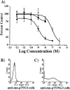
Fig. 1.
Fig. 1.
A, Cytotoxicity of anti-murine-p75-SAP to NG3 cells that express murine p75 and C6 glioma cells that do not. ●, Anti-murine-p75-SAP versus NG3 cells; ○, SAP versus NG3 cells; ▪, anti-murine-p75 versus C6 cells.B, FACS analysis of rat C6 glioma cells with anti-murine-p75-SAP and FITC-labeled goat anti-rat IgG. This profile was identical to the profile of FITC-labeled secondary antibody alone.C, Labeling of NG3 anti-murine-p75 antibody and FITC-labeled goat anti-rat secondary antibody. Data show the binding of the antibody to the surface of cells that express murine p75.
Fig. 2.
Fig. 2.
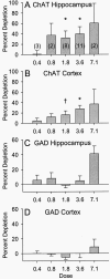
The percentage of depletion of ChAT (A, B) and GAD (C, D) after different doses (ranging from 0.4–7.1 μg/μl) of intracerebroventricular injections of anti-murine-p75-SAP. Data are percentage of depletion ± SEM. Anti-murine-p75-SAP injections result in a dose-dependent decrease in ChAT activity in the hippocampus (A) and neocortex (B). In contrast, there is not a dose-dependent loss of GAD activity in the hippocampus (C) or neocortex (D).
Fig. 3.
Fig. 3.
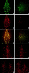
Immunohistochemistry for ChAT (A–C, G–I) and calbindin (D–F) after intracerebroventricular injection of saline (A, D, G), 1.78 μg of anti-murine-p75-SAP (B, E, H), or 3.55 μg of anti-murine-p75-saporin (C, F, I). ChAT-positive neurons in the MS (A–C) are lost dose-dependently, whereas calbindin-positive neurons in the MS (D–F) are still present even after the highest dose of toxin. In contrast, there is no apparent loss of ChAT-positive neurons in the nBM at any dose of toxin (G–I).
Fig. 4.
Fig. 4.
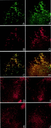
Immunofluorescence staining in the MS for ChAT (A, F), p75 (B, G), and double-labeling of ChAT/p75 (C, H) after intracerebroventricular injections of saline (A–C) or anti-murine-p75-SAP (F–H). Immunofluorescence staining for noncholinergic neurons using calbindin (D, I) and parvalbumin (E, J) after intracerebroventricular injections of saline (D, E) or anti-murine-p75-SAP (I, J) is shown. ChAT-positive and p75-positive neurons in the MS are lost after immunotoxin injections, whereas there is no apparent loss of noncholinergic calbindin and parvalbumin staining in the septum.
Fig. 5.
Fig. 5.
Immunofluorescence staining in the nBM for ChAT (A, F), p75 (B, G), and double-labeling of ChAT/p75 (C, H) after intracerebroventricular injections of saline (A–C) or anti-murine-p75-SAP (F–H). Immunofluorescence staining for noncholinergic neurons using calbindin (D, I) and parvalbumin (E, J) after intracerebroventricular injections of saline (D, E) or anti-murine-p75-SAP (I, J) is shown. The immunotoxin injections do not have a dramatic effect on any of the markers in the nBM.
Fig. 6.
Fig. 6.
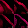
Immunofluorescence staining in the cerebellum of p75 (A, C) and calbindin (B, D) after intracerebroventricular injections of saline (A, B) and immunotoxin (C, D). The immunotoxin injections do not have a dramatic effect on any of these markers in the cerebellum.
Fig. 7.
Fig. 7.
Swim maze performance in the saline-injected control mice and immunotoxin-injected mice. A, Time to reach the hidden platform on training trials was measured over three blocks in a 1 d swim maze task (spatial swim time). Control mice performed significantly better than lesioned mice on all three blocks of trials. B, Saporin-lesioned mice were also impaired in learning to swim to a visible platform, as measured by time to reach the visible platform on four training trials (cued swim time). However, the main effect on swim time on spatial training trials remained when swim time on cued trials was partialled out as a covariate.C, Correlation between proximity to platform on the spatial probe trial and hippocampal ChAT depletion. Better spatial performance was correlated significantly with higher hippocampal ChAT activity.

Similar articles

Cited by

References

    1. Arters J, Hohmann CF, Mills J, Olaghere O, Berger-Sweeney J. Sexually dimorphic responses to neonatal basal forebrain lesions in mice. I. Behavior and neurochemistry. J Neurobiol. 1998;37:582–594. - PubMed
    1. Baxter MG, Chiba AA. Cognitive functions of the basal forebrain. Curr Opin Neurobiol. 1999;9:178–183. - PubMed
    1. Berger-Sweeney J, Berger UV, Sharma M, Paul CA. Effects of carbon dioxide-induced anesthesia on cholinergic parameters in rat brain. Lab Anim Sci. 1994a;44:369–371. - PubMed
    1. Berger-Sweeney J, Heckers S, Mesulam MM, Wiley RG, Lappi DA, Sharma M. Differential effects on spatial navigation of immunotoxin-induced cholinergic lesions of the medial septal area and nucleus basalis magnocellularis. J Neurosci. 1994b;14:4507–4519. - PMC - PubMed
    1. Bornemann K, Staufenbiel M. Transgenic mouse models of Alzheimer's disease. Ann NY Acad Sci. 2000;908:260–266. - PubMed

Publication types

MeSH terms