. 2001 Oct 15;21(20):8247-61.
doi: 10.1523/JNEUROSCI.21-20-08247.2001.

The enterins: a novel family of neuropeptides isolated from the enteric nervous system and CNS of Aplysia

Affiliations

The enterins: a novel family of neuropeptides isolated from the enteric nervous system and CNS of Aplysia

Y Furukawa et al. J Neurosci. .

Abstract

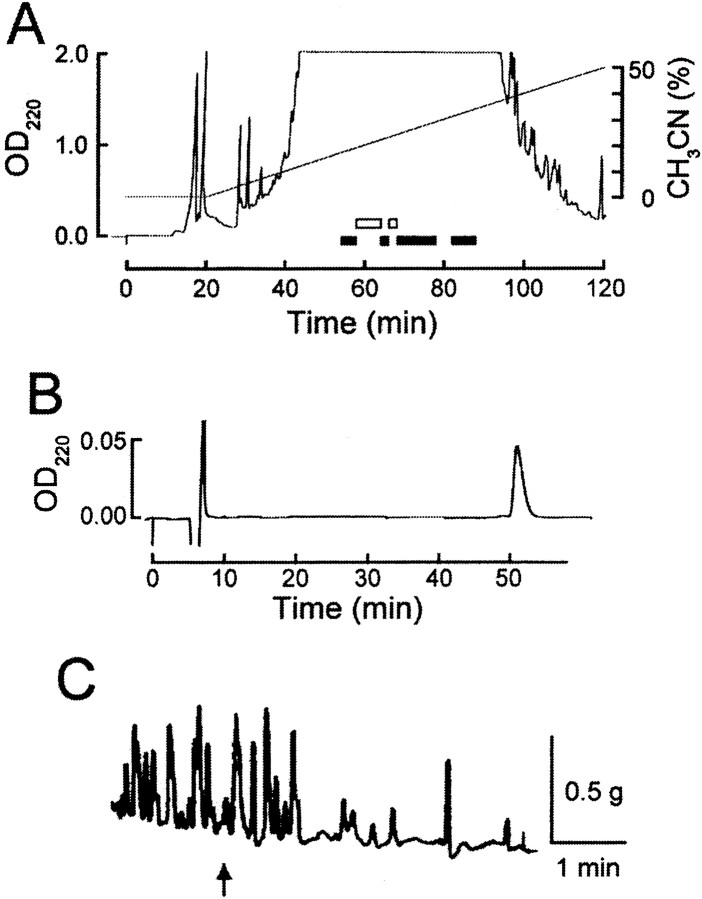
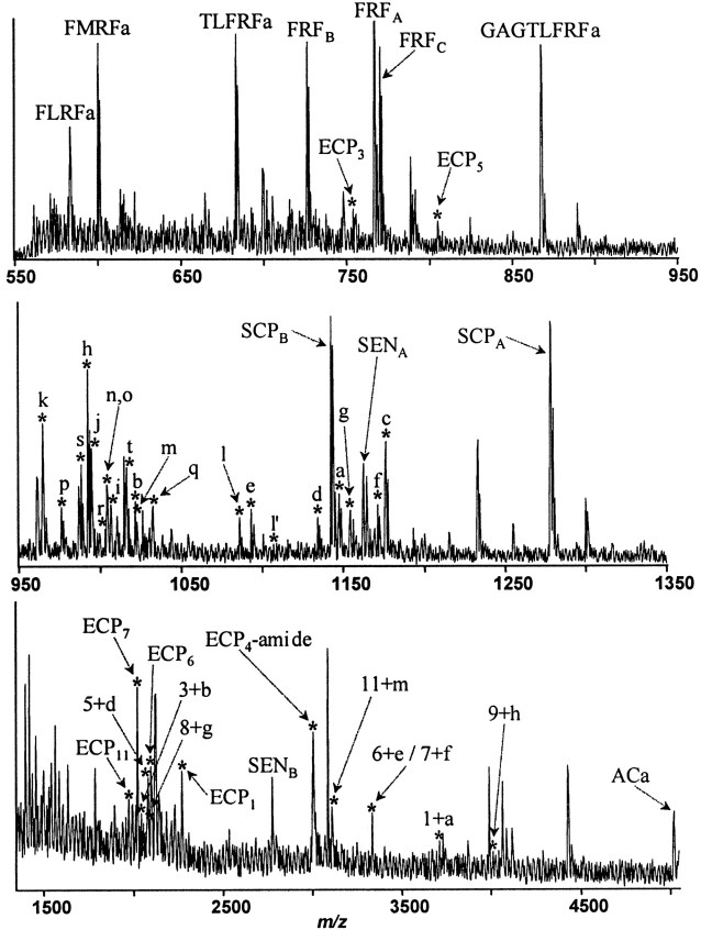
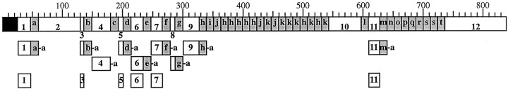
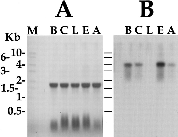
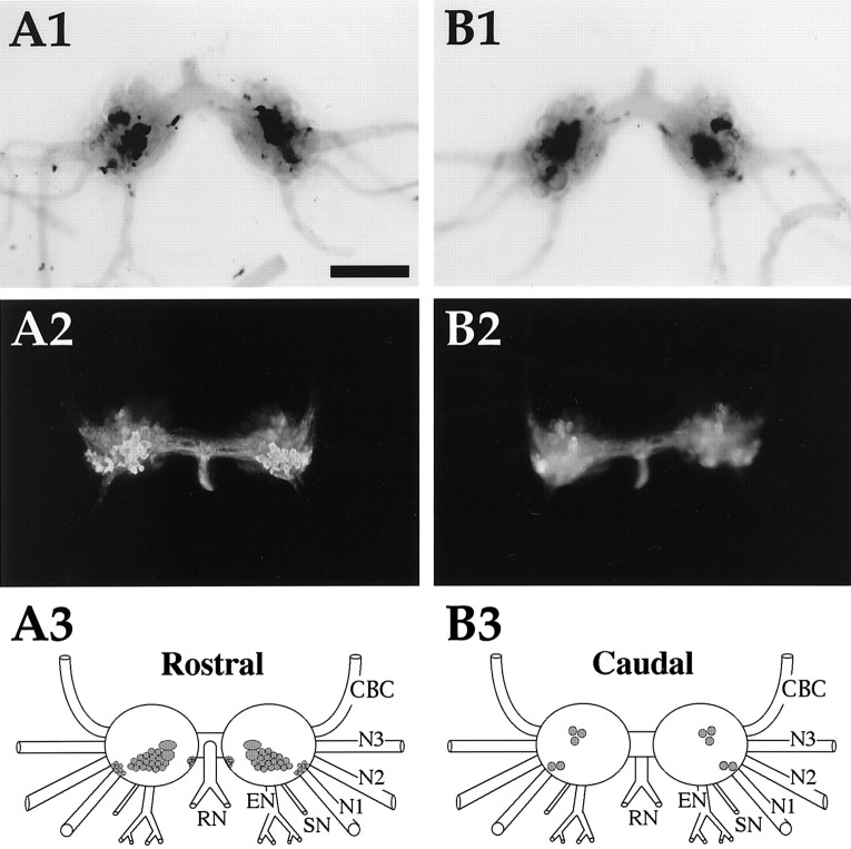
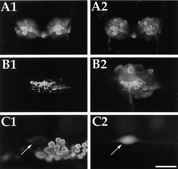
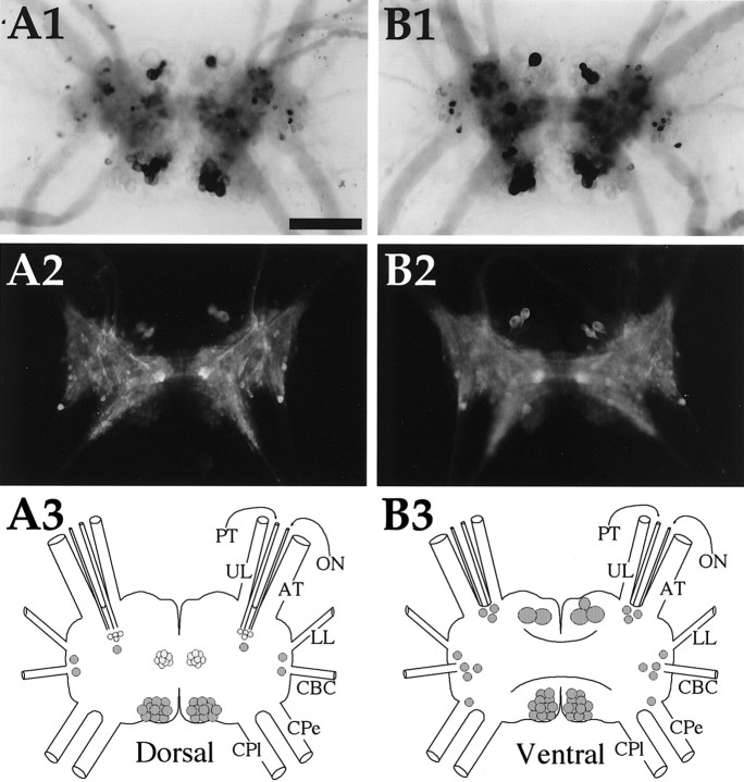
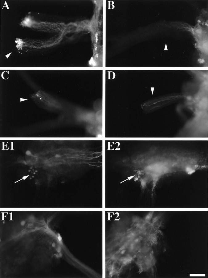
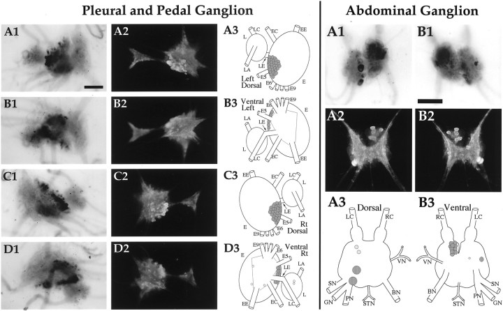
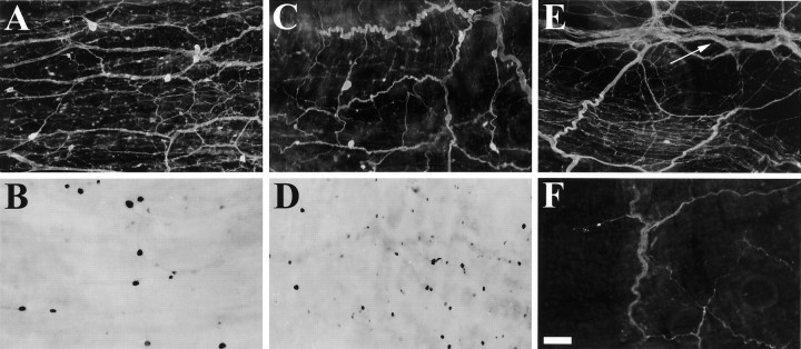
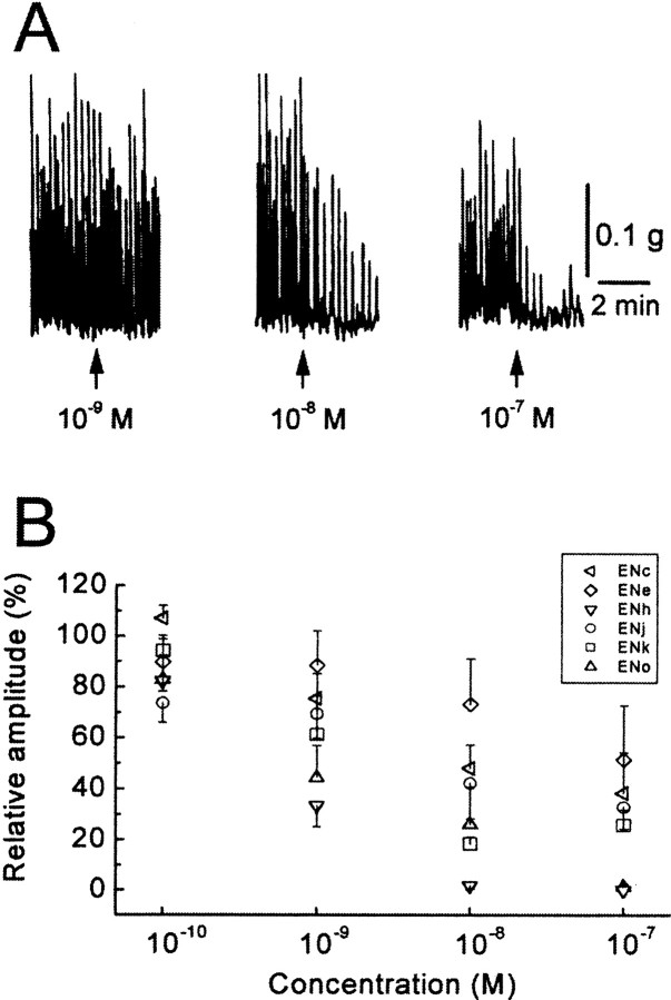
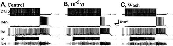
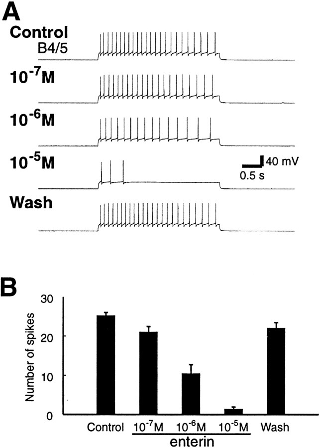
To identify neuropeptides that have a broad spectrum of actions on the feeding system of Aplysia, we searched for bioactive peptides that are present in both the gut and the CNS. We identified a family of structurally related nonapeptides and decapeptides (enterins) that are present in the gut and CNS of Aplysia, and most of which share the HSFVamide sequence at the C terminus. The structure of the enterin precursor deduced from cDNA cloning predicts 35 copies of 20 different enterins. Northern analysis, in situ hybridization, and immunocytochemistry show that the enterins are abundantly present in the CNS and the gut of Aplysia. Using matrix-assisted laser desorption/ionization time-of-flight mass spectrometry we characterized the enterin-precursor processing, demonstrated that all of the precursor-predicted enterins are present, and determined post-translational modifications of various enterins. Enterin-positive neuronal somata and processes were found in the gut, and enterins inhibited contractions of the gut. In the CNS, the cerebral and buccal ganglia, which control feeding, contained the enterins. Enterin was also present in the nerve that connects these two ganglia. Enterins reduced the firing of interneurons B4/5 during feeding motor programs. Such enterin-induced reduction of firing also occurred when excitability of B4/5 was tested directly. Because reduction of B4/5 activity corresponds to a switch from egestive to ingestive behaviors, enterin may contribute to such program switching. Furthermore, because enterins are present throughout the nervous system, they may also play a regulatory role in nonfeeding behaviors of Aplysia.


Figures

Fig. 1.
Fig. 1.
Purification of enterins from gut extracts ofA. kurodai. A, The second step of HPLC fractionation by a reverse-phase column. Retained material was eluted with a 200 min linear gradient of 0–100% of acetonitrile (CH3CN) in 0.1% TFA. As seen in this figure, most retained materials come out within 100 min. Fractions shown to contain bioactive substances are indicated by either black (inhibitory action) or white (excitatory action)bars. The enterins were purified from the fractions shown by a third black bar. B, The final purification step by RP-HPLC. A retained substance was eluted isocratically with 21.5% CH3CN/0.1% TFA.C, Biological activity of the purified substance in the triturating stomach of Aplysia. At anarrow, a small aliquot containing the 1/100 of purified substance shown in B was applied to the preparation.
Fig. 2.
Fig. 2.
Sequence of the predicted enterin precursor protein. Open reading frame of the cDNA encoding the precursor of APGYSHSFVamide is shown. Amino acids are numbered atright, and predicted amidated enterins areunderlined. The enterin precursor predicts 35 copies of 20 different enterins. Monobasic and dibasic cleavage sites are shown in bold, and the predicted signal sequence cleavage site between G(25) and T(26) is shown in lowercase letters.
Fig. 3.
Fig. 3.
MALDI TOF mass spectrum (550–5000 Da range) from isolated buccal neurons of the rostral sensory cluster. Detection of the mass spectral peaks predicted by the putative amidated peptides on the enterin precursor confirm that they are in fact processed from the precursor. Assigned peaks are labeled with corresponding peptides. Peptides derived from five precursors, FMRFamide, FRFamide, SCP, sensorin, and enterin are also detected. Peaks corresponding to the products of the enterin precursor are shown with anasterisk. Lowercase letters signify the corresponding enterins, and numbers signify the corresponding enterin-connecting peptides (ECP). Peaks labeled with a letter plus numbercorrespond to amidated enterins still connected to their preceding ECP. Unamidated enterins were not detected. Enterins with a Gln at the N terminus (ENn, ENo, ENq, ENs, ENt) were detected only as a pyroglutamine form of the peptide. The enterin with a Glu at the N terminus (Enl) was detected as both E (labeled l′) and pE (labeled l) forms of the peptide. Peaks with masses corresponding to all of the 20 fully processed enterins were detected. Two of the enterins, ENn and ENo, are isomers and could not be distinguished on the basis of molecular mass. In addition, some ECP and ECP-enterin peptides were detected. One of the ECPs, ECP4, was detected as an amidated form (ECP4-amide).
Fig. 4.
Fig. 4.
Summary of the enterin precursor structure and the enterin precursor-derived peptides. Schematic representation of the location of the enterins and enterin-connecting peptides on the precursor is shown with the amino acids numbered at thetop. The black box represents the signal peptide, shaded boxes represent enterins, andwhite boxes represent ECPs. Lowercase letters identify corresponding enterins (ENa to ENt), and thenumbers identify corresponding ECPs (ECP1 through ECP12). Although all of the enterins were detected by MALDI-TOF MS as fully processed peptides, this is not shown on the schematic for the sake of simplicity. Processed peptides, other than the enterins, that were detected by MALDI-TOF MS are shown on the schematic below their origin on the precursor. A lowercase -a denotes that the amidated peptide was detected. Many enterins that were still connected to their preceding ECP were detected. Many of the ECPs were also detected, and ECP4 was detected as an amidated form.
Fig. 5.
Fig. 5.
Northern blot of ganglionic distribution of enterin mRNA. A, Methylene blue-stained Northern blot of total RNA shows equal loading in all lanes. Aplysiaribosomal RNA runs as a single 18 sec band. B, Hybridization of the same blot with an enterin coding sequence cDNA probe. Kb, Kilobase; M, RNA size marker;B, buccal ganglion; C, cerebral ganglion;L, pleural ganglion; E, pedal ganglion;A, abdominal ganglion.
Fig. 6.
Fig. 6.
Enterin in the buccal ganglion. A1,In situ hybridization of rostral surface.A2, Immunocytochemistry of rostral surface. Immunoreactive axons are present in the CBC. A3, Drawing of the enterin-positive neurons on the rostral surface of buccal ganglia. B1, In situ hybridization of caudal surface. B2, Immunocytochemistry of caudal surface. B3, Drawing of the enterin-positive neurons on the caudal surface of buccal ganglia. CBC, Cerebrobuccal connective; N1, nerve 1 (B4); N2, nerve 2 (B5); N3, nerve 3 (B6); SN, salivary nerve (B3); EN, esophageal nerve (B2);RN, radula nerve (B1). Neurons drawn in darker shades of gray stain more intensely. Scale bar (shown in A1 for all panels), 500 μm.
Fig. 7.
Fig. 7.
Enterin in the radula mechanoafferent sensory neurons of the buccal ganglion. A, A buccal ganglion from a juvenile Aplysia (10 gm) double labeled with rat antibody to enterin (A1, rhodamine red) and rabbit antibody to SCP (A2, fluorescein). B, A buccal hemiganglion from an adult Aplysia (200 gm). Enterin immunostaining (rhodamine red) is shown in B1, and SCP immunostaining (fluorescein) is shown in B2. C, A buccal hemiganglion from an adult animal immunostained with enterin (C1) in which B21 (arrows) was electrophysiologically identified and injected with carboxyfluorescein (C2). Scale bar (shown in C2): A, B, 500 μm; C, 100 μm.
Fig. 8.
Fig. 8.
Enterin in the cerebral ganglion.A1, In situ hybridization of dorsal surface. A2, Immunocytochemistry of dorsal surface.A3, Drawing of the enterin neurons on the dorsal cerebral ganglion. B1, In situhybridization of ventral surface. B2, Immunocytochemistry of ventral surface. B3, Drawing of the enterin neurons on the ventral cerebral ganglion.UL, Upper labial nerve; PT, posterior tentacular nerve; ON, optic nerve; AT, anterior tentacular nerve; LL, lower labial nerve;CBC, cerebrobuccal connective; Cpe, cerebropedal connective; CPl, cerebropleural connective. Neurons drawn in darker shades of graystain more intensely. Scale bar (shown in A1 for all panels), 500 μm.
Fig. 9.
Fig. 9.
Enterin immunostaining in backfills of the CBC.A, Radula nerve (arrowhead) of the buccal ganglion (right) shows accumulation of enterin immunoreactivity, especially at the cut end. This suggests that the source of these axons is in the buccal ganglion. B, Esophageal nerve (arrowhead) of the buccal ganglion (right) shows depletion of enterin immunoreactivity from the cut end of the nerve (left). Some residual enterin immunoreactivity is observed in axons of this nerve nearer the buccal ganglion. C, CBC (arrowhead) on the side of the buccal ganglion (right) shows some accumulation of enterin immunoreactivity at the cut end of the nerve (left). D, CBC (arrowhead) on the side of the cerebral ganglion (right) also shows some accumulation of enterin immunoreactivity at the cut end of the nerve (left). E1, CBC backfill of the buccal ganglion (caudal surface of the contralateral hemiganglion) shows a cluster of a few small backfilled neurons (arrow) near the esophageal nerve and nerve 1.E2, Enterin immunostaining of the same field as in E1 shows that some of the backfilled neurons are enterin immunopositive (arrow). F1, CBC backfill of the cerebral ganglion (dorsal surface) shows several backfilled neurons in the ipsilateral E cluster. F2, Enterin immunostaining of the same field as in F1 shows that the backfilled neurons are not enterin immunopositive. Scale bar (shown in F2), 200 μm for all panels.
Fig. 10.
Fig. 10.
Enterin in the pleural, pedal, and abdominal ganglia. Pleural and Pedal Ganglion: A1,In situ hybridization of left ganglion pair dorsal surface. A2, Immunocytochemistry of the left ganglion pair dorsal surface. A3, Drawing of the enterin neurons on the dorsal surface of the left ganglion pair. B1,In situ hybridization of left ganglion pair ventral surface. B2, Immunocytochemistry of the left ganglion pair ventral surface. B3, Drawing of the enterin neurons on the ventral surface of the left ganglion pair. C1,In situ hybridization of right ganglion pair dorsal surface. C2, Immunocytochemistry of the right ganglion pair dorsal surface. C3, Drawing of the enterin neurons on the dorsal surface of the right ganglion pair. D1,In situ hybridization of right ganglion pair ventral surface. D2, Immunocytochemistry of the right ganglion pair ventral surface. D3, Drawing of the enterin neurons on the ventral surface of the right ganglion pair. L, Pleural ganglion; E, pedal ganglion; LE, pleuropedal connective; EE, pedal commissure;EC, cerebropedal connective; LC, cerebropleural connective; LA, pleuroabdominal connective; E5, posterior tegumentary nerve (P5);E6, anterior parapodial nerve (P6); E9, posterior pedal nerve (P9). Not all nerves are drawn for simplicity.Abdominal ganglion: A1, In situ hybridization of dorsal surface. A2, Immunocytochemistry of dorsal surface. A3, Drawing of the enterin neurons on the dorsal abdominal ganglion.B1, In situ hybridization of ventral surface. B2, Immunocytochemistry of ventral surface.B3, Drawing of the enterin neurons on the ventral abdominal ganglion. LC, Left pleuroabdominal connective;RC, right pleuroabdominal connective; VN, vulvar nerve; BN, branchial nerve; STN, spermathecal nerve; PN, pericardial nerve;GN, genital nerve; SN, siphon nerve. Neurons drawn in darker shades of graystain more intensely. Scale bars, 500 μm.
Fig. 11.
Fig. 11.
Enterin in the digestive tract. A, Esophagus immunostaining. B, Esophagus in situ hybridization. C, Crop immunostaining.D, Crop in situ hybridization.E, Stomatogastric ring (arrow) immunostaining. The crop is below and triturating stomach is above thearrow. F, Filter chamber immunostaining. All panels are from 10–15 gm Aplysia. Scale bar (shown in F), 200 μm for all panels.
Fig. 12.
Fig. 12.
Inhibitory actions of enterins on theAplysia triturating stomach. A, Effects of ENh on spontaneous contractions of the triturating stomach.B, Concentration–response relationships of six enterins. Each symbol shows a mean of three to four preparations, and upward or downward bar indicates SE of the mean. The potency of four nonapeptides that have a consensus sequence of XPGYSHSFVamide appeared to be almost identical. One-way ANOVA showed that there was no statistically significant difference between the enterins tested except at 107m (1010m, F(3,12) = 1.50,p > 0.26; 109m,F(3,12) = 1.91, p> 0.18; 108m,F(3,12) = 2.69, p> 0.09; 107m,F(3,12) = 10.00, p< 0.002). At 107m, ENo and ENh seemed to be more potent than ENk or ENj (Tukey–Kramer test; p < 0.05). Two tested decapeptides, ENc and ENe, may be slightly less than the nonapeptides. If the results of the two decapeptides are included in the analysis, there is a statistically significant difference in their potency (1010m,F(5,17) = 2.81, p< 0.05; 109m,F(5,17) = 2.88, p< 0.05; 108m,F(5,17) = 4.74, p< 0.01; 107m,F(5,17) = 3.73, p< 0.05). At these concentrations, ENe was less potent than ENh (Tukey–Kramer test; p < 0.01 at 108m,p < 0.05 at 107m).
Fig. 13.
Fig. 13.
Enterin reduces spike activity of multifunctional neurons B4/5 during feeding motor program elicited by CBI-2. Single cycles of feeding motor programs, comprising a protraction–retraction sequence, were elicited every 2 min by CBI-2 stimulation at 9 Hz. CBI-2 stimulation was terminated after the end of the protraction phase. Protraction phase was monitored by activity in the I2 nerve (I2). Retraction phase was monitored by sustained depolarization in B4/5 that occurs after termination of I2 nerve activity. The activity of radula closing motor neuron B8, also shown in radula nerve (RN), occurred during the retraction phase. This suggests that the motor program was ingestive. Superfusion of 10 μm ENk had no significant effect on the duration of protraction and retraction but reduced the B4/5 firing rate (Control = 77 spikes;105M = 24 spikes; Wash = 46 spikes).
Fig. 14.
Fig. 14.
Enterin reduces the excitability of the multifunctional neurons B4/5. A, Sample recording from B4/5. A 3 sec depolarizing pulse was applied to induce regular firing of B4/5 at ∼8 Hz every half minute. The B4/5 firing was reduced when ENk was applied, and the ENk-induced reduction of B4/5 firing was concentration-dependent. B, Group data (mean ± SEM; n = 4) showing the inhibitory effects of ENk on B4/5 excitability.

Similar articles

Cited by

References

    1. Bateman A, Solomon S, Bennett HP. Post-translational modification of bovine pro-opiomelanocortin. Tyrosine sulfation and pyroglutamate formation, a mass spectrometric study. J Biol Chem. 1990;265:22130–22136. - PubMed
    1. Brezden BL, Yeoman MS, Gardner DR, Benjamin PR. FMRFamide-activated Ca2+ channels in Lymnaea heart cells are modulated by “SEEPLY,” a neuropeptide encoded on the same gene. J Neurophysiol. 1999;81:1818–1826. - PubMed
    1. Brezina V, Weiss KR. Analyzing the functional consequences of transmitter complexity. Trends Neurosci. 1997;20:538–543. - PubMed
    1. Brunet JF, Shapiro E, Foster SA, Kandel ER, Iino Y. Identification of a peptide specific for Aplysia sensory neurons by PCR-based differential screening. Science. 1991;252:856–859. - PubMed
    1. Busby WH, Quackenbush GE, Humm J, Youngblood WW, Kizer JS. An enzyme(s) that converts glutaminyl-peptides into pyroglutamyl-peptides. Presence in pituitary, brain, adrenal medulla, and lymphocytes. J Biol Chem. 1987;262:8532–8536. - PubMed

Publication types

MeSH terms

Associated data

LinkOut - more resources