

Experimental induction of the two-host life cycle of Sarcocystis cruzi between dogs and Korean native calves
- PMID: 11590912
- PMCID: PMC2721071
- DOI: 10.3347/kjp.2001.39.3.227
Experimental induction of the two-host life cycle of Sarcocystis cruzi between dogs and Korean native calves
Abstract
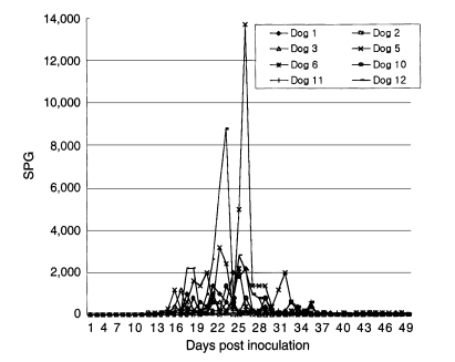
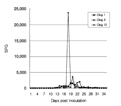
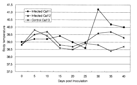
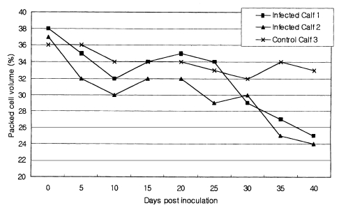
Eight dogs were experimentally infected with Sarcocystis by oral inoculation of cardiac muscle from naturally infected cattle. The infected dogs commenced discharging of sporocysts in the feces after 10 to 12 days of inoculation, and continued until 20 and 35 days after inoculation. Three dogs were reinfected with cardiac muscle from the naturally infected cattle. Sporocysts reappeared in the feces on 12 to 13 days after reinfection. Sarcocystis sporocysts collected from the experimentally infected dogs were fed to each of the two 30-day-old Korean native calves. The infected calves remained clinically normal, except for the high fever (> or = 40 degrees C) and decreased hematocrit values on day 30 to 40 post inoculation. Muscular cysts of Sarcocystis were found from infected calves on day 40 post inoculation. Proliferative forms of Sarcocystis were also observed in the muscle of infected calves. These results suggest that the Sarcocystis cruzi found in Korean native cattle has a 2-host life cycle with dogs as the definitive host and Korean native calves as the intermediate host.
Figures
References
-
- Carrigan MJ. An outbreak of sarcocystosis in dairy cattle. Aust Vet J. 1986;63:22–24. - PubMed
-
- Dubey JP. A review of Sarcocystis of domestic animals and of other coccidia of cats and dogs. J Am Vet Med Assoc. 1976;169:1061–1078. - PubMed
-
- Dubey JP, Speer CA, Epling GP. Sarcocystosis in newborn calves fed Sarcocystis cruzi sporocysts from coyotes. Am J Vet Res. 1982;43:2147–2164. - PubMed
-
- Fayer R. Production of Sarcocystis cruzi sporocysts by dogs fed experimentally infected and naturally infected beef. J Parasitol. 1977;63:1072–1075. - PubMed
-
- Fayer R, Johnson AJ, Lunde M. Abortion and other signs of disease in cows experimentally infected with Sarcocystis fusiformis from dogs. J Infect Dis. 1976;134:624–628. - PubMed
MeSH terms
LinkOut - more resources
Full Text Sources
