. 2001:2:18.
doi: 10.1186/1471-2121-2-18. Epub 2001 Aug 20.

Cell adhesion and signaling on the fibronectin 1st type III repeat; requisite roles for cell surface proteoglycans and integrins

Affiliations

Cell adhesion and signaling on the fibronectin 1st type III repeat; requisite roles for cell surface proteoglycans and integrins

K O Mercurius et al. BMC Cell Biol. 2001.

Abstract

Background: The first type III repeat of fibronectin is known to be involved in fibronectin matrix assembly, and recombinant proteins from this type III repeat can inhibit cell proliferation, tumor metastasis and angiogenesis. We have analyzed the way rat aortic smooth muscle cells (RASMCs) interact with a recombinant protein encompassing a C-terminal portion of the first type III repeat of fibronectin (protein III1-C).

Results: Cells are able to adhere to and spread on III1-C coated on a dish. Both beta1 integrins and cell surface heparan sulfate proteoglycans serve as receptors for III1-C. For example, cell attachment to III1-C is partially inhibited by agents that block beta1 integrins or by heparin. Complete inhibition of cell attachment is seen only when integrin blocking agents are combined with heparin. Affinity chromatography revealed the binding of proteins that likely represent the integrin beta1 and alpha5 submits to a III1-C column. Cell adhesion to III1-C results in robust ERK1/2 activation that is blocked by integrin-blocking agents. In addition, cell adhesion to III1-C and ERK1/2 activation by III1-C are both inhibited by heparan sulfate but not by chondroitin sulfate. Moreover, heparitinase treatment, but not chondroitinase treatment of RASMCs results in reduced cell adhesion and ERK1/2 activation. Affinity chromatography experiments demonstrated that 35SO4-labeled cell surface heparan sulfate proteoglycans bound specifically to III1-C.

Conclusions: The results suggest that the 1st type III repeat of fibronectin contains a previously unrecognized cell adhesion domain that stimulates robust ERK1/2 activation in RASMCs. Cells interact with this domain through cell surface heparan sulfate proteoglycans and integrins, and both classes of receptors are required for optimal cell adhesion and ERK1/2 activation.


Figures

Figure 1
Figure 1
Effect of III1-C on cell spreading.Panel A. RASMCs were plated onto either uncoated dishes (C and 11C panels) or dishes coated with 20 μg/ml fibronectin (FN, FN+C and FN+11C panels) in the presence of either no recombinant protein in the media (FN panel), or 25 μM III1-C (FN+C and C panels), or 25 μM III 11-C in the media (FN+11C and 11C panels). After 30 min at 37°C the cells were fixed, stained with coomassie blue and photographed under a light microscope. Bar equals 100 μm. Panel B. Quantitation of cell spreading over time in the culture conditions described for Panel A. Each data point is the average of four samples; similar results were obtained from four independent experiments.
Figure 2
Figure 2
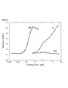
Cell attachment to III1-C. Fibronectin (■), III1-C (□) or III 11-C (●) at various concentrations were coated onto plastic wells. RASMCs were seeded onto the coated wells and the cells were allowed to adhere for 30 min at 37°C. The cells were fixed, stained and the amount of cell attachment was measured. Each sample was performed in triplicate and the experiment was done three times with comparable results each time.
Figure 3
Figure 3
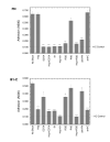
Blocking of cell attachment to III1-C RASMCs were seeded onto wells coated with either 20 μg/ml FN (top panel), or 25 μM III1-C (bottom panel) in the absence or presence of various inhibitors. The inhibitors were 100 μg/ml heparin (Hep), 10 mM EDTA (EDTA), 50 μg/ml anti-β1 blocking mAb Ha2/5 (pi), 0.5 mg/ml GRGDSP peptide (RGD), 0.5 mg/ml GRADSP peptide (RAD), 100 μg/ml anti-FN polyclonal antibody (anti-FN), and 60 μg/ml anti-IIIl-C polyclonal antibody (anti-C). After 30 min at 37°C the cells were fixed, stained with crystal violet and the amount of cell attachment was measured. The dashed line indicates the amount of signal seen on the negative control protein III 11-C, and represents the background A600 reading in these experiments. Statistical analysis was done by Student's T-test (2-tailed); * p < .05, ** p < .005, *** p < .0005. Each sample was done in quadruplicate and the experiment was performed three times with similar results each time.
Figure 4
Figure 4
Affinity chromatography of 125I surface labeled proteins on III1-C Sepharose RASMCs were surface labeled with 125 I, lysed in octylglucoside lysis buffer, and these cell lysates were passed over affinity columns containing either FN (FN), III1-C (C), or III 11-C (11C). The columns were washed and bound proteins were eluted with 10 mM EDTA in lysis buffer. Samples from the final wash before elution (W lanes), and from the elution (E lanes) fractions of each column were analyzed by SDS-PAGE and phosphorimager analysis. Numbers to the left of the panel indicate the migration of molecular mass markers (kDa). Asterisks mark the positions of bands that bound to and were eluted from the FN and III1-C columns.
Figure 5
Figure 5
Activation of ERK by cell adhesion to III1-C. RASMCs were collected into DMEM + 0.5% BSA and either kept in suspension in the absence (-) or presence of 2 ng/ml PDGF (PDGF), or were seeded onto dishes precoated with either 20 μg/ml FN (FN), 25 μM III1-C (C), or 25 μM III 11-C (11C). Note that the only cells that received growth factor are the cells in the PDGF lane, which received the growth factor 10 min before lysis. After 5, 15, 30, 45 or 60 min at 37°C the cells were lysed in sample buffer and analyzed by inununoblotting with anti-phospho-p44/42 MAPK antibodies (Phos-ERK panel). The blot was then stripped and reprobed with anti-ERK antibodies (ERK panel) to analyze protein loading. This experiment was done more than 3 times with similar results each time.
Figure 6
Figure 6
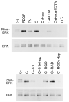
Inhibition of III1-C mediated ERK activation by various integrin inhibitors. RASMCs were collected in DMEM+0.5% BSA and were then either left in suspension in the absence (-) or presence of PDGF (PDGF), or plated onto dishes precoated with either fibronectin (FN), III1-C (C and C+... lanes), or III 11-C (11C), as described in the legend to Fig. 5. Some samples of cells plated onto III1-C also contained either 100 μg/ml heparin (C+Hep), 10 mM EDTA (C+EDTA), 50 μg/ml anti-β1 blocking mAb Ha2/5 (pi), 0.5 mg/ml GRGDSP peptide (RGD), 0.5 mg/ml GRADSP peptide (RAD), or combinations of heparin plus integrin inhibitors, as indicated. After 30 min at 37°C cells were lysed in SDS-PAGE sample buffer and samples were analyzed by immunoblotting with anti-phospho-p44/42 MAPK antibodies (Phos-ERK panels). Blots were then stripped and reprobed with anti-ERK antibodies (ERK panels) to analyze protein loading. The experiment was done 3 times with similar results each time.
Figure 7
Figure 7
Inhibition of III1-C mediated ERK activation by glycosaminoglycans. RASMCs were collected into DMEM + 0.5% BSA and either kept in suspension (-) or cells were seeded onto dishes precoated with 25 μM III1-C (all other lanes). Cells seeded onto III1-C contained either no glycosaminoglycan in the medium (C) or 80 μg/ml heparin (C+Hep), hyaluronic acid (C+HA), heparan sulfate (C+HS), dermatan sulfate (C+DS), chondroitin-4-sulfate (C+C4S), or chondroitin-6-sulfate (C+C6S). After 30 min at 37°C cells were processed and analyzed by immunoblotting with anti-phospho-p44/42 MAPK antibodies (Phos-ERK panel). The blot was subsequently stripped and reprobed with anti-ERK antibodies (ERK panel) as described in the legend to Fig. 5. The experiment was done 3 times with similar results each time.
Figure 8
Figure 8
Inhibition of III1-C mediated ERK activation by heparitinase. RASMCs were collected in DMEM+0.5% BSA and were then treated in suspension with either no GAGase ((-), PDGF, FN, C, Hep, and 11C samples) or with 0.1 u/ml of heparitinase (H'ase), or 0.1 u/ml of chondroitinase ABC (C'ase) for 1 hr at 37°C. Cells were then either left in suspension in the absence (-) or presence of PDGF (PDGF) for 10 min, or cells were plated onto dishes precoated with either fibronectin (FN), III1-C (C, Hep, H'ase, and C'ase lanes), or III 11-C (11C). One sample of cells plated onto III1-C contained 100 μg/ml heparin (Hep) in the medium. Cell samples were analyzed by immunoblotting with anti-phospho-p44/42 MAPK antibodies (Phos-ERK panel) followed by anti-ERK antibodies (ERK panel) as described in the legend to Fig. 5. The experiment was performed four times with similar results each time.
Figure 9
Figure 9
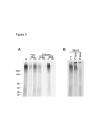
Affinity chromatography of 35 SO4-labeled proteoglycans on III1-C Sepharose. Panel A. RASMCs were labeled with 35SO4, which preferentially labels the glycosaminoglycan chains of proteoglycans. Cells were lysed in NP40 buffer and lysates were applied to either III1-C Sepharose (C lanes) or III 11-C Sepharose (11C lanes) columns. The flow through fractions were collected (Flow Thru lanes), the columns were washed, and the bound material was eluted by first applying 8 M urea elution buffer (Urea lanes), washing the columns with PBS, then collecting the Sepharose beads and boiling them in SDS-PAGE sample buffer (SDS lanes). The lane labeled St shows a sample of the starting material. All samples were separated on SDS-PAGE gels then detected by phosphorimager analysis. Numbers to the left of the panel indicate the migration of molecular mass markers (kDa). Panel B. Samples of the starting material were treated with either no enzyme ((-)), or with 0.03 u/ml chondroitinase ABC (C'ase), or 0.03 u/ml heparitinase (H'ase), then analyzed by SDS-PAGE and phosphorimager. The experiment was performed twice with similar results both times.
Figure 10
Figure 10
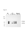
Affinity chromatography HSPGs on III1-C Sepharose. RASMCs were collected by trypsinization and lysed in NP40 buffer. The lysate was then treated either without (-) or with (+) 0.1 u/ml heparitinase for 1 hr at 37°C. Heparitinase treated samples were then applied to either III1-C Sepharose (lanes marked C) or III 11-C Sepharose (lanes marked 11C). One sample was applied to a III1-C Sepharose column in the presence of 0.5 mg/ml heparin (lanes marked C•H). The flow through fractions were collected (FT lanes), the columns were washed extensively, then the Sepharose beads were collected and boiled in SDS-PAGE sample buffer (Bound lanes). Samples were analyzed by immunoblotting with the 3G10 antibody. Antibody 3G10 recognizes the uronate stubs that remain associated with core proteins after heparitinase digestion. Thus, after heparitinase digestion 3G10 shows the sizes of HSPG core proteins. Note that the major HSPGs have core protein sizes of 46 kDa and 70 kDa, and both of these HSPGs bind to the III1-C column but not the III 11-C column. The experiment was performed twice with similar results both times.

Similar articles

Cited by

References

    1. Hynes RO. Fibronectins New York: Springer-Verlag; 1990.
    1. Morla A, Zhang Z, Ruoslahti E. Superfibronectin is a functionally distinct form of fibronectin. Nature. 1994;367:193–196. doi: 10.1038/367193a0. - DOI - PubMed
    1. Bourdoulous S, Orend G, MacKenna DA, Pasqualini R, Ruoslahti E. Fibronectin matrix regulates activation of RHO and CDC42 GTPases and cell cycle progression. J Cell Biol. 1998;143:267–276. doi: 10.1083/jcb.143.1.267. - DOI - PMC - PubMed
    1. Mercurius KO, Morla AO. Inhibition of vascular smooth muscle cell growth by inhibition of fibronectin matrix assembly. Circ Res. 1998;82:548–556. - PubMed
    1. Saulnier R, Bhardwaj B, Klassen J, Leopold D, Rahimi N, Tremblay E, Mosher D, Elliott B. Fibronectin fibrils and growth factors stimulate anchorage-independent growth of a murine mammary carcinoma. Exp Cell Res. 1996;222:360–369. doi: 10.1006/excr.1996.0045. - DOI - PubMed

Publication types

MeSH terms

LinkOut - more resources