. 2001 Nov;183(21):6234-43.
doi: 10.1128/JB.183.21.6234-6243.2001.

Identification of the partitioning site within the repABC-type replicon of the composite Paracoccus versutus plasmid pTAV1

Affiliations

Identification of the partitioning site within the repABC-type replicon of the composite Paracoccus versutus plasmid pTAV1

D Bartosik et al. J Bacteriol. 2001 Nov.

Abstract

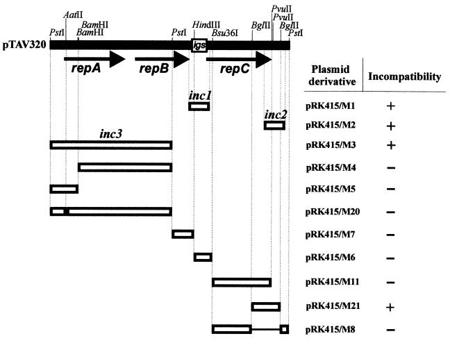
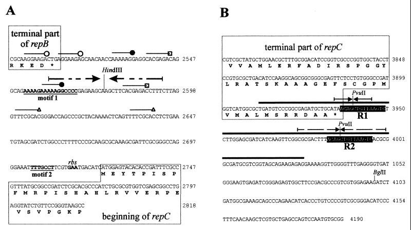
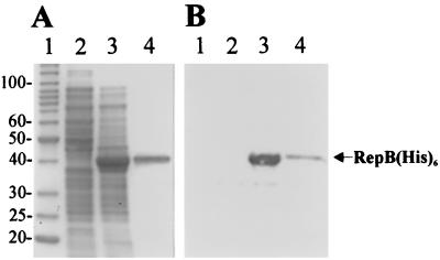
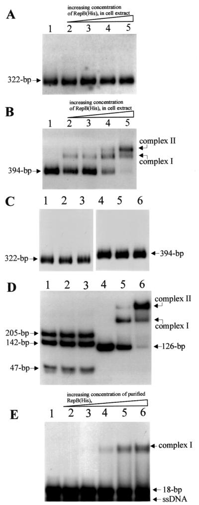
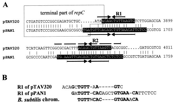
The replicator region of composite plasmid pTAV1 of Paracoccus versutus (included in mini-replicon pTAV320) belongs to the family of repABC replicons commonly found in plasmids harbored by Agrobacterium and Rhizobium spp. The repABC replicons encode three genes clustered in an operon, which are involved in partitioning (repA and repB) and replication (repC). In order to localize the partitioning site of pTAV320, the two identified incompatibility determinants of this mini-replicon (inc1, located in the intergenic sequence between repB and repC; and inc2, situated downstream of the repC gene) were PCR amplified and used together with purified RepB fusion protein (homologous to the type B partitioning proteins binding to the partitioning sites) in an electrophoretic mobility shift assay. The protein bound only inc2, forming two complexes in a protein concentration-dependent manner. The inc2 region contains two long (14-bp) repeated sequences (R1 and R2). Disruption of these sequences completely eliminates RepB binding ability. R1 and R2 have sequence similarities with analogous repeats of another repABC replicon of plasmid pPAN1 of Paracoccus pantotrophus DSM 82.5 and with centromeric sequences of the Bacillus subtilis chromosome. Excess RepB protein resulted in destabilization of the inc2-containing plasmid in Escherichia coli. On the other hand, the inc2 region could stabilize another unstable replicon in P. versutus when RepA and RepB were delivered in trans, proving that this region has centromere-like activity. Thus, it was demonstrated that repA, repB, and inc2 constitute a functional system for active partitioning of pTAV320.


Figures

FIG. 1
FIG. 1
Identification of incompatibility determinants (inc) of pTAV320. Only the restriction sites used to construct recombined plasmids are shown on the pTAV320 restriction map. The transcriptional orientations of the three open reading frames are indicated by arrows. The conserved intergenic sequence between repB and repC (igs) is enclosed in a box. The open boxes represent DNA restriction or PCR-amplified fragments of pTAV320 that were cloned into the pRK415 vector and tested for the presence of the inc regions; the designations of the pRK415 derivatives are on the right. For details concerning construction see Table 1. The lines joining the boxes indicate regions of pTAV320 removed by deletion. The smallest cis-acting regions of pTAV320 carrying identified incompatibility determinants are designated inc1 and inc2. A plus sign indicates incompatibility and a minus sign indicates compatibility of pTAV320 with derivatives of pRK415 introduced into P. versutus UW225(pTAV320).
FIG. 2
FIG. 2
Nucleotide sequences of the inc1 (A) and inc2 (B) regions of mini-replicon pTAV320. Several short repeated sequences identified in inc1 are indicated by different lines. Motif 1 and motif 2, the two motifs of inc1 sequences that are visibly conserved in the igs of repABC replicons, are indicated by boldface type and underlined. Two direct repeats of inc2, R1 and R2, are indicated by a black background. The region of inc2 amplified with primers INC10 and INC11 and used for EMSA is indicated by a thick line over the sequence. The palindromic sequences of the inc regions are indicated by arrows. The terminal fragments of the repB and repC genes are enclosed in boxes. The amino acid sequences of the proteins are given below the nucleotide sequences. The HindIII, PvuII, and BglII restriction sites important for the analysis are indicated. A presumptive ribosome binding site (rbs) is indicated. Coordinate numbers for the pTAV320 sequence are indicated on the right (accession number U60522).
FIG. 3
FIG. 3
(A) Coomassie blue-stained SDS-PAGE gel of overexpressed (in E. coli M15) and purified RepB(His)6 protein. The gel was loaded with the following samples: protein molecular weight standards (lane 1) (the molecular masses of the proteins [in kilodaltons] are indicated on the left); crude cell lysate of strain M15(pQE30/repB) (lane 2); crude cell lysate of strain M15(pQE30/repB) after IPTG induction (lane 3); purified RepB(His)6 protein (lane 4). (B) Western transfer of proteins visualized on the gel in panel A and immunodetection of RepB(His)6 with Penta · His antibody.
FIG. 4
FIG. 4
Autoradiograph of EMSA results, showing the binding ability of RepB(His)6 protein [present in crude protein extract of RepB(His)6-overproducing E. coli M15 or purified] in 32P-labeled inc1 and inc2 regions of pTAV320. (A and B) inc1 fragment (A) and inc2 fragment (B) incubated with different concentrations (2.5, 5, 10, and 20 μg/ml in lanes 2 to 5, respectively) of M15(pQE30/repB) crude protein extract. Lane 1 contained labeled fragment with no protein added. (C) inc1 fragment (lanes 1 to 3) and inc2 fragment (lanes 4 to 6) incubated (i) with M15(pQE30) crude protein extract (20 μg/ml) (lanes 2 and 5), (ii) with purified IHF protein (3 μg/ml) (lanes 3 and 6), and (iii) without protein (lanes 1 and 4). (D) The inc2 fragment was digested with PvuII (lanes 1 to 3) and incubated with 2.5 and 5 μg of purified RepB(His)6 protein per ml (lanes 2 and 3, respectively). No protein was added to the reaction mixture in lane 1. The 126-bp PCR-amplified fragment of the inc2 region (Fig. 2B) (lanes 4 to 6) was incubated with 2.5 and 5 μg of purified RepB(His)6 protein per ml (lanes 5 and 6, respectively). No protein was added to the reaction mixture in lane 4. (E) An 18-bp oligonucleotide containing a single R1/R2 sequence was incubated with 0.5, 1, 2.5, 5, and 10 μg of purified RepB(His)6 protein per ml (lanes 2 to 6, respectively). No protein was added to the reaction mixture in lane 1. The positions of the protein-DNA complexes and the sizes of the unbound probes are indicated on the right and left. ssDNA, single-stranded DNA.
FIG. 5
FIG. 5
Plasmid stability of pABW3 (Kmr) and pABW3/BGL coexisting in trans with compatible pRK415 (Tcr) or pRK415/AB (carrying repA and repB genes of pTAV320) in P. versutus UW225 cells. Symbols: ▾, UW225 carrying plasmids pABW3 and pRK415; ○, UW225 carrying plasmids pABW3/BGL and pRK415; □, UW225 carrying plasmids pABW3 and pRK415/AB; ●, UW225 carrying plasmids pABW3/BGL and pRK415/AB. The data are averages based on three independent experiments.
FIG. 6
FIG. 6
(A) Alignment (BESTFIT; GCG software) of nucleotide sequences of the corresponding regions of P. versutus pTAV320 and the repABC replicator region of plasmid pPAN1 from P. pantotrophus DSM 82.5. The R1 and R2 repeated sequences are shown with a black background. The palindromic sequences are indicated by arrows. Terminal parts of the repC genes are enclosed in a box. Coordinates of the pTAV320 and pPAN1 sequences are indicated on the right. (B) Manual alignment of nucleotide sequences of R1 of pTAV320 and R1 of pPAN1 repeats and consensus centromeric sequence of the B. subtilis chromosome (chrom.) (22). Nucleotides common to two sequences at any position are indicated by boldface type. Dashes indicate gaps introduced to maximize the alignment.

Similar articles

Cited by

References

    1. Austin S, Abeles A L. Partitioning of unit-copy miniplasmids to daughter cells. II. The partition region of miniplasmid P1 encodes an essential protein and a centromere-like site at which it acts. J Mol Biol. 1983;169:373–387. - PubMed
    1. Austin S, Nordström K. Partition-mediated incompatibility of bacterial plasmids. Cell. 1990;60:351–354. - PubMed
    1. Bartosik D, Baj J, Wlodarczyk M. Construction and preliminary characterization of mini-derivatives of large (107-kb) cryptic plasmid of the sulphur bacterium Thiobacillus versutus. FEMS Microbiol Lett. 1995;129:169–174.
    1. Bartosik D, Bialkowska A, Baj J, Wlodarczyk M. Construction of mobilizable cloning vectors derived from pBGS18 and their application for analysis of replicator region of a pTAV202 mini-derivative of Paracoccus versutus pTAV1 plasmid. Acta Microbiol Pol. 1997;46:379–383. - PubMed
    1. Bartosik D, Baj J, Wlodarczyk M. Molecular and functional analysis of pTAV320-repABC type replicon of a composite pTAV1 plasmid of Paracoccus versutus. Microbiology. 1998;144:3149–3157. - PubMed

Publication types

LinkOut - more resources