. 2001 Oct 23;98(22):12677-82.
doi: 10.1073/pnas.221449198. Epub 2001 Oct 9.

A model for gene therapy of human hereditary lymphedema

Affiliations

A model for gene therapy of human hereditary lymphedema

M J Karkkainen et al. Proc Natl Acad Sci U S A. .

Abstract

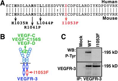
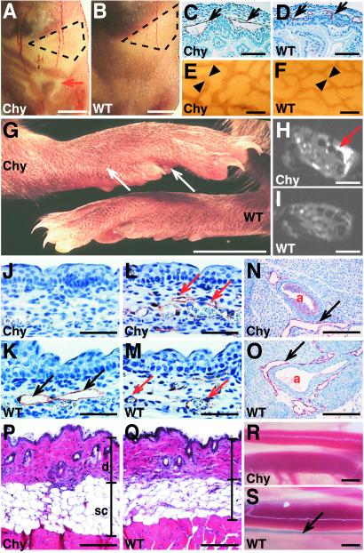
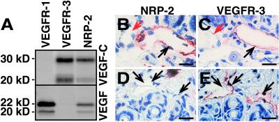
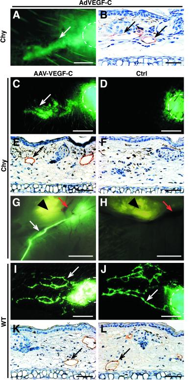
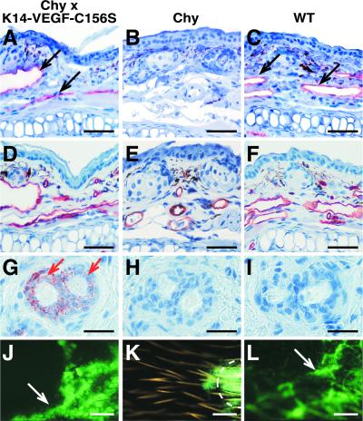
Primary human lymphedema (Milroy's disease), characterized by a chronic and disfiguring swelling of the extremities, is associated with heterozygous inactivating missense mutations of the gene encoding vascular endothelial growth factor C/D receptor (VEGFR-3). Here, we describe a mouse model and a possible treatment for primary lymphedema. Like the human patients, the lymphedema (Chy) mice have an inactivating Vegfr3 mutation in their germ line, and swelling of the limbs because of hypoplastic cutaneous, but not visceral, lymphatic vessels. Neuropilin (NRP)-2 bound VEGF-C and was expressed in the visceral, but not in the cutaneous, lymphatic endothelia, suggesting that it may participate in the pathogenesis of lymphedema. By using virus-mediated VEGF-C gene therapy, we were able to generate functional lymphatic vessels in the lymphedema mice. Our results suggest that growth factor gene therapy is applicable to human lymphedema and provide a paradigm for other diseases associated with mutant receptors.


Figures

Figure 1
Figure 1
The VEGFR-3(I1053F) mutant is tyrosine kinase inactive. (A) The alignment of the human and mouse VEGFR-3 amino acid sequences showing the lymphedema-linked human mutations (bold) and the I1053F mutation found in the Chy mice (red). (B) Localization of the I1053F mutation within the catalytic domain of the ligand-bound VEGFR-3 heterodimer. (C) VEGFR-3(I1053F) is kinase inactive. We transfected cells with Mock, WT, or I1053F VEGFR-3 expression vectors and analyzed VEGFR-3 by immunoprecipitation and Western blotting of the cell lysates using phosphotyrosine antibodies (Upper). We also confirmed the expression of similar amounts of VEGFR-3 (Lower).
Figure 2
Figure 2
Defective lymphatic vessels in the Chy mice. (A and B) The Chy mice were recognized by the accumulation of chyous fluid into the abdomen (red arrow). WT littermate is shown as a comparison. The stomach is marked with a dashed line. (CF) The subserosal lymphatic vessels are enlarged in the Chy mouse intestine as detected by VEGFR-3 IHC (C and D), and by VEGFR-3 wholemount staining (E and F). (G) The feet of the Chy mice are swollen (arrows), when compared with a WT littermate. (H and I) MRI of the mouse feet shows the prominent hyperintensity in the Chy mouse foot, which is absent in a WT mouse. (J and K) The lymphatic endothelium (black arrows) of E15.5 skin was visualized by VEGFR-3 IHC in the Chy and WT mice. Note the absence of the lymphatic vessels in the Chy mice. (L and M) PECAM-1 IHC reveals no differences in the blood vasculature (red arrows). (N and O) VEGFR-3 IHC indicates that the lymphatics surrounding the aorta (a) in the Chy mice are similar to those of the WT mice. (P and Q) The hematoxylin-eosin staining of the back skin shows that the dermis (d) and s.c. adipose tissue (sc) are thickened in the Chy mice when compared with the WT littermate. (R and S) The transport of the Evans blue dye into the collecting lymphatic vessels (arrow) was visualized in the WT but not in the Chy mice after intradermal injection of the dye. [Bars = 5 mm (A and B), 90 μm (C and D), 200 μm (E and F), 5 mm (G), 100 μm (H and I), 70 μm (JM), 200 μm (N and O), 210 μm (P and Q), and 1.5 mm (R and S).]
Figure 3
Figure 3
NRP-2 binds VEGF-C and is differentially expressed in the visceral organs and in the skin. (A) Labeled VEGF-C is precipitated by VEGFR-3-IgG and by NRP-2-IgG, but not by VEGFR-1-IgG fusion protein. (B and C) The intestinal VEGFR-3-positive lymphatic vessels (C; black arrow) stain also for NRP-2 (B), whereas the blood vessels are not stained (red arrows). (D and E) The VEGFR-3-positive vessels in the skin (E) are not stained by the NRP-2 antibodies (D). (Bars = 35 μm.)
Figure 4
Figure 4
Analysis of the AAV-VEGF-C expression. (A) AAV-produced VEGF-C polypeptides were metabolically labeled and subjected to immunoprecipitation with VEGF-C antibodies (IP) or to binding assay with soluble VEGFR-3 and NRP-2 IgG fusion proteins. AAV-EGFP-infected cells were used as a negative control and VEGF-C produced in 293T cells as a positive control (p-C). (B) Northern blotting of total RNA from AAV-VEGF-C-infected and control ears of a Chy mouse.
Figure 5
Figure 5
Gene therapy by using viral VEGF-C overexpression. (A and B) Adenovirally induced VEGF-C overexpression in a Chy mouse ear induces formation of functional lymphatic vessels, as analyzed by fluorescent microlymphography (A) or by VEGFR-3 IHC (B). The dye depot is marked with a dashed line. (C and D) Microlymphography of the lymphatic vessels 7 weeks after AAV infection reveals functional lymphatic vessels (white arrows) in the AAV-VEGF-C-infected Chy ear (C) when compared with the control ear (D). (E and F) VEGFR-3 IHC shows that the AAV-VEGF-C-infected Chy ears contain VEGFR-3-positive vessels (E, black arrows), whereas no staining is detected in the AAV-EGFP-infected ears (F). (G and H) The fluorescent dextran was collected by the draining lymphatic vessel (white arrow) in the AAV-VEGF-C-treated Chy ear (G), unlike in the noninfected ear of the same mouse (H). Red arrows mark the blood vessels, and an arrowhead marks the cartilage of the ear. (I and J) In WT mice, there are no major changes in lymphatic vasculature after AAV-VEGF-C infection (I), when compared with the control ears (J). (K and L) The same is also confirmed by VEGFR-3 IHC. [Bars = 200 μm (A), 150 μm (B, E, F, K, and L), 300 μm (C, D, I, and J), and 5 mm (G and H).]
Figure 6
Figure 6
Lymphatic vessel growth in the Chy × K14-VEGF-C156S mice. (AC) Staining by using VEGFR-3 antibodies shows lymphatic vessels (black arrows) in the ear of the Chy × K14-VEGF-C156S mice, in comparison with the aplastic lymphatic vessels in the Chy mouse ear (B), or with the WT littermate (C). (DF) PECAM IHC confirms that the blood vasculature is normal in all mice. (GI) VEGF-C IHC shows the VEGF-C156S transgene expression in the basal cells of the hair follicles (red arrows). (JL) Fluorescent microlymphography shows the functional lymphatic capillary network in the Chy × K14-VEGF-C156S ear (J), resembling that of the WT mouse (L). The FITC-dextran is not collected into the lymphatic vessels in the Chy mouse ear (K). [Bars = 70 μm (AF), 25 μm (GI), and 600 μm (JL).]

References

    1. Witte M H, Way D L, Witte C L, Bernas M. In: Regulation of Angiogenesis. Goldberg I D, Rosen E M, editors. Basel: Birkhäuser; 1997. pp. 65–112.
    1. Ferrell R E, Levinson K L, Esman J H, Kimak M A, Lawrence E C, Barmada M M, Finegold D N. Hum Mol Genet. 1998;7:2073–2078. - PubMed
    1. Witte M H, Erickson R, Bernas M, Andrade M, Reiser F, Conlon W, Hoyme H E, Witte C L. Lymphology. 1998;31:145–155. - PubMed
    1. Evans A L, Brice G, Sotirova V, Mortimer P, Beninson J, Burnand K, Rosbotham J, Child A, Sarfarazi M. Am J Hum Genet. 1999;64:547–555. - PMC - PubMed
    1. Karkkainen M J, Ferrell R E, Lawrence E C, Kimak M A, Levinson K L, McTigue M A, Alitalo K, Finegold D N. Nat Genet. 2000;25:153–159. - PubMed

Publication types

MeSH terms

Substances