. 2001 Oct 23;98(22):12754-9.
doi: 10.1073/pnas.221450098. Epub 2001 Oct 9.

Brain-specific expression of an exogenous gene after i.v. administration

Affiliations

Brain-specific expression of an exogenous gene after i.v. administration

N Shi et al. Proc Natl Acad Sci U S A. .

Abstract

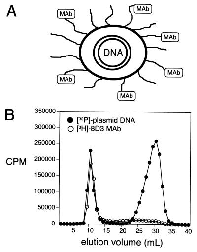
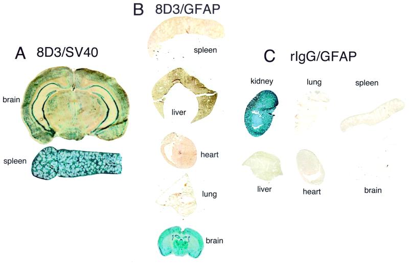
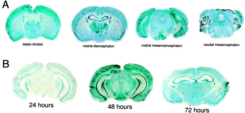
The treatment of brain diseases with gene therapy requires the gene to be expressed throughout the central nervous system, and this is possible by using gene targeting technology that delivers the gene across the blood-brain barrier after i.v. administration of a nonviral formulation of the gene. The plasmid DNA is targeted to brain with pegylated immunoliposomes (PILs) using a targeting ligand such as a peptidomimetic mAb, which binds to a transporting receptor on the blood-brain barrier. The present studies adapt the PIL gene targeting technology to the mouse by using the rat 8D3 mAb to the mouse transferrin receptor. Tissue-specific expression in brain and peripheral organs of different exogenous genes (beta-galactosidase, luciferase) is examined at 1-3 days after i.v. injection in adult mice of the exogenous gene packaged in the interior of 8D3-PIL. The expression plasmid is driven either by a broadly expressed promoter, simian virus 40, or by a brain-specific promoter taken from the 5' flanking sequence of the human glial fibrillary acidic protein (GFAP) gene. The transgene is expressed in both brain and peripheral tissues when the simian virus 40 promoter is used, but the expression of the exogenous gene is confined to the brain when the transgene is under the influence of the brain-specific GFAP promoter. Confocal microscopy colocalizes immunoreactive bacterial beta-galactosidase with immunoreactive GFAP in brain astrocytes. These studies indicate that tissue-specific gene expression in brain is possible after the i.v. administration of a nonviral vector with the combined use of gene targeting technology and tissue-specific gene promoters.


Figures

Figure 1
Figure 1
(A) A double-stranded supercoiled plasmid DNA is encapsulated in the interior of 75- to 85-nm liposomes, which are pegylated with PEG2000, and 1–2% of the PEG strands are conjugated with a targeting ligand, such as a mAb (1, 2). (B) Elution of 8D3 PILs through a Sepharose CL-4B gel filtration column allowed for separation of the PILs from unconjugated mAb and from exteriorized nuclease-digested DNA. The comigration of the interiorized [32P]-DNA and the [3H]8D3 mAb demonstrated the plasmid DNA and the targeting mAb were incorporated in the same structure.
Figure 2
Figure 2
(A) β-Galactosidase histochemistry was performed on organs removed 2 days after an i.v. injection of PILs carrying the pSV-β-galactosidase plasmid, driven by the SV40 promoter, and conjugated with the 8D3 rat mAb. (B) 5-Bromo-4-chloro-3-indoyl-β-d-galactoside histochemistry of organs removed from mice 2 days after a single i.v. injection of 8D3 PILs carrying the GFAP/β-galactosidase plasmid. (C) β-Galactosidase gene expression in mouse organs removed 48 h after a single i.v. injection of the GFAP/β-galactosidase plasmid incorporated in PILs that were targeted with nonspecific rat IgG (rIgG). All organs were negative except for kidney. The histochemical product in kidney is derived from endogenous mammalian β-galactosidase-like activity in that organ. Kidney removed from noninjected mice yielded similar β-galactosidase reaction product.
Figure 3
Figure 3
(A) β-Galactosidase histochemistry in mouse brain removed 48 h after a single i.v. injection of the GFAP/β-galactosidase plasmid encapsulated in the interior of 8D3-targeted PILs. Coronal sections at different levels of the brain are shown. (B) β-Galactosidase histochemistry is shown for brain removed from mice at 24, 48, and 72 h after a single i.v. injection of the GFAP/β-galactosidase plasmid encapsulated in the interior of 8D3 PILs.
Figure 4
Figure 4
Confocal microscopy of mouse brain removed 48 h after a single i.v. injection of the GFAP/β-galactosidase plasmid encapsulated in the interior of 8D3 PILs. (A, D, and G) The confocal microscopy of immunoreactive β-galactosidase, stained with a fluorescein-labeled secondary antibody. (B, E, and H) Immunoreactive GFAP, stained with a rhodamine-labeled secondary antibody. (C, F, and I) The respective superimposed images showing colocalization of the β-galactosidase and the GFAP in astrocyte cell bodies (C) and astrocyte processes (F and I). The yellow/orange images in are the result of the superimposition of the green (β-galactosidase) and red (GFAP) images. No staining of brain was obtained with either mouse IgG/fluorescein-conjugated goat anti-mouse IgG (J) or goat IgG/rhodamine-conjugated donkey anti-goat IgG (K). (Scale bar: 10 μm.)

References

    1. Shi N, Pardridge W M. Proc Natl Acad Sci USA. 2000;97:7567–7572. . (First Published June 6, 2000; 10.1073/pnas.130187497) - PMC - PubMed
    1. Shi N, Boado R J, Pardridge W M. Pharm Res. 2001;18:1091–1095. - PubMed
    1. Brenner M, Kisseberth W C, Su Y, Besnard F, Messing A. J Neurosci. 1994;14:1030–1037. - PMC - PubMed
    1. Segovia J, Vergara P, Brenner M. Neurosci Lett. 1998;242:172–176. - PubMed
    1. Lee H J, Engelhardt B, Lesley J, Bickel U, Pardridge W M. J Pharmacol Exp Ther. 2000;292:1048–1052. - PubMed

Publication types