. 2001 Oct 9;98(21):11857-62.
doi: 10.1073/pnas.191250198.

Hierarchical self-assembly of chiral rod-like molecules as a model for peptide beta -sheet tapes, ribbons, fibrils, and fibers

Affiliations

Hierarchical self-assembly of chiral rod-like molecules as a model for peptide beta -sheet tapes, ribbons, fibrils, and fibers

A Aggeli et al. Proc Natl Acad Sci U S A. .

Abstract

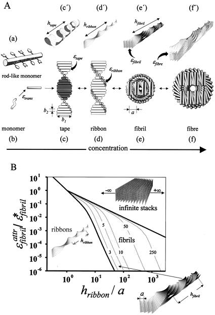
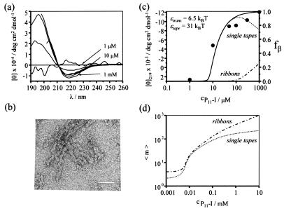
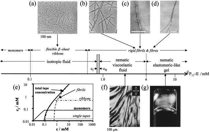
A generic statistical mechanical model is presented for the self-assembly of chiral rod-like units, such as beta-sheet-forming peptides, into helical tapes, which with increasing concentration associate into twisted ribbons (double tapes), fibrils (twisted stacks of ribbons), and fibers (entwined fibrils). The finite fibril width and helicity is shown to stem from a competition between the free energy gain from attraction between ribbons and the penalty because of elastic distortion of the intrinsically twisted ribbons on incorporation into a growing fibril. Fibers are stabilized similarly. The behavior of two rationally designed 11-aa residue peptides, P(11)-I and P(11)-II, is illustrative of the proposed scheme. P(11)-I and P(11)-II are designed to adopt the beta-strand conformation and to self-assemble in one dimension to form antiparallel beta-sheet tapes, ribbons, fibrils, and fibers in well-defined solution conditions. The energetic parameters governing self-assembly have been estimated from the experimental data using the model. The 8-nm-wide fibrils consist of eight tapes, are extremely robust (scission energy approximately 200 k(B)T), and sufficiently rigid (persistence length l(fibril) approximately 20-70 microm) to form nematic solutions at peptide concentration c approximately 0.9 mM (volume fraction approximately 0.0009 vol/vol), which convert to self-supporting nematic gels at c > 4 mM. More generally, these observations provide a new insight into the generic self-assembling properties of beta-sheet-forming peptides and shed new light on the factors governing the structures and stability of pathological amyloid fibrils in vivo. The model also provides a prescription of routes to novel macromolecules based on a variety of self-assembling chiral units, and protocols for extraction of the associated energy changes.


Figures

Figure 1
Figure 1
Model of hierarchical self-assembly of chiral rod-like units. (A) Local arrangements (cf) and the corresponding global equilibrium conformations (c′–f′) for the hierarchical self-assembling structures formed in solutions of chiral molecules (a), which have complementary donor and acceptor groups, shown by arrows, via which they interact and align to form tapes (c). The black and the white surfaces of the rod (a) are reflected in the sides of the helical tape (c), which is chosen to curl toward the black side (c′). The outer sides of the twisted ribbon (d), of the fibril (e), and of the fiber (f) are all white. One of the fibrils in the fiber (f′) is drawn with darker shade for clarity. (e and f) The front views of the edges of fibrils and fibers, respectively. Geometrical sizes (the numbers in parentheses show the values of the corresponding geometric sizes for P11-I and P11-II peptides, based on x-ray diffraction data and molecular modeling): interrod separation in a tape b2 (b2 = 0.47 nm); tape width, equal to the length of a rod, b1 (b1 = 4 nm); interribbon distance in the fibril, α (α = 1.6–2 nm for P11-I, and α = 2–2.4 nm for P11-II). (B) Phase diagram of a solution of twisted ribbons that form fibrils. The scaled variables are as follows: relative helix pitch of isolated ribbons hribbon/α, and relative side-by-side attraction energy between ribbons ɛformula image*fibril*fibril ≡ (2π2b22) ktwist; see the text and Fig. 1 Ad and Ae′ for notations]. The areas divided by the thick lines reveal the conditions where ribbons, fibrils, and infinite stacks of completely untwisted ribbons are stable. The dotted lines are lines of stability for fibrils containing p ribbons (p are written on the lines); kbend/ktwist = 0.1.
Figure 2
Figure 2
Self-assembly of P11-I. (a) Far-UV CD spectra as a function of peptide concentration. The solutions were prepared by mixing the dry peptide with the required volume of water adjusted to pH = 2 with phosphoric acid. Data were collected with 1-month-old solutions stored at 20°C. For interpretation of the CD spectra, see the text and legend of Fig. 4 a and b. (b) Negatively stained TEM image of single “curly” tapes, reminiscent of Fig. 1Ac′; the scale bar = 50 nm. (c) Plot of the β-sheet fraction in solution (black circles) as a function of total peptide concentration, based on the CD data (the mean residue ellipticity [θ] at 219 nm is taken as a linear function of the β-sheet fraction in solution). The solid line is the fit of the data with the single tape theory. The best-fit values of the energetic parameters ɛtrans and ɛtape, which were chosen to comply with the concentration dependence of the CD data and with the observed lengths of tapes at c = 5 mM, are shown in the panel. (d) Theoretical concentration dependence of the average number 〈m〉 of peptides per single tape (dotted line) and in ribbons (dash-dot line), based on the energetic parameters derived from the fit (c). Minimum number of peptides in tapes is two, and in ribbons is four. The predicted lengths of tapes and ribbons are in agreement with the observed lengths in the TEM pictures for the same peptide concentration.
Figure 3
Figure 3
Aggregate structures and liquid crystalline phase behavior observed in solutions of P11-II in water with increasing c (log scale). The electron micrographs (a) of ribbons (c = 0.2 mM), and (b) of fibrils (c = 6.2 mM) were obtained with a 4-month-old solution after platinum rotary shadowing. The observed micrometer-long contour length may be limited by multiple ruptures of the fibrils during preparation of the samples for TEM imaging. Higher resolution TEM images of ribbons were also obtained by using negatively stained samples. Micrographs c (c = 6.2 mM) and d (c = 6.2 mM) were obtained with a 1-month-old solution after uranyl acetate negative staining. CD and Fourier transfer IR (FTIR) have confirmed that the fibrils are made of β-sheet structures. X-ray diffraction data have also shown arcs corresponding to 0.47 nm periodicity, consistent with the expected interstrand distance in a β-sheet (unpublished data). The TEM micrographs show the principle aggregate structures whose populations cj = fjc (fj is the fraction of peptides incorporated in the jth structure) change with peptide concentration, as depicted in e. The curves in e were calculated with the generalized model described in the text (see also Fig. 4d). The aggregation behavior of the peptide, probed by using time-resolved fluorescence anisotropy and CD of filtered solutions, is fully consistent with the expectations of the model. The polarizing optical micrograph (f) shows the thick thread-like texture observed for a solution with c = 3.7 mM in a 0.2-mm pathlength microslide. (g) A self-supporting birefringent gel (c = 6.2 mM) in an inverted 10-mm o.d. glass tube, viewed between crossed polarizers. The scale bars in a, b, c, and d = 100 nm, in f = 100 μm.

References

    1. Artsaenko O, Kettig B, Fiedler K, Conrad K, During K. Mol Breeding. 1998;4:313–319.
    1. Urry D. Trend Biotechnol. 1999;17:249–257. - PubMed
    1. Panitch A, Yamaoka T, Fournier M J, Mason T L, Tirrell D A. Macromolecules. 1999;32:1701–1703.
    1. Krejchi M T, Atkins E D T, Waddon A J, Fournier J, Mason T L, Tirrell D A. Science. 1994;265:1427–1432. - PubMed
    1. Zhang S G, Altman M. React Funct Polym. 1999;41:91–102.

Publication types

LinkOut - more resources