. 2001 Oct 9;98(21):11967-72.
doi: 10.1073/pnas.211439798.

Crystal structure of the central region of bovine fibrinogen (E5 fragment) at 1.4-A resolution

Affiliations

Crystal structure of the central region of bovine fibrinogen (E5 fragment) at 1.4-A resolution

J Madrazo et al. Proc Natl Acad Sci U S A. .

Abstract

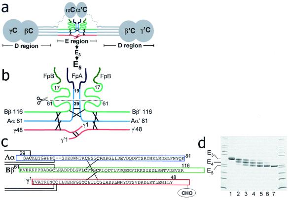
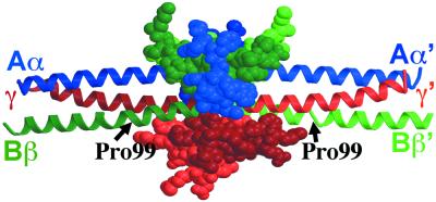
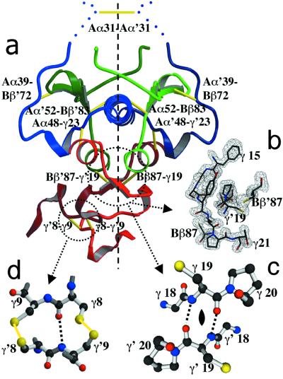
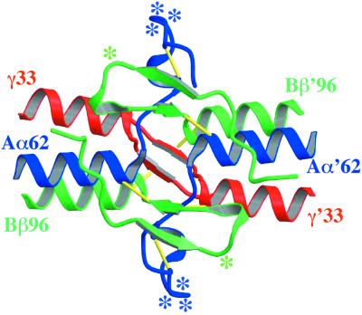
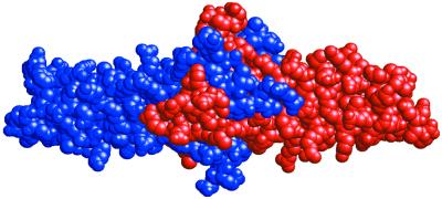
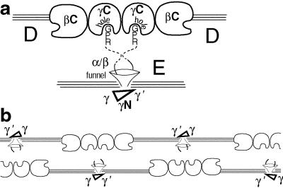
The high-resolution crystal structure of the N-terminal central region of bovine fibrinogen (a 35-kDa E(5) fragment) reveals a remarkable dimeric design. The two halves of the molecule bond together at the center in an extensive molecular "handshake" by using both disulfide linkages and noncovalent contacts. On one face of the fragment, the Aalpha and Bbeta chains from the two monomers form a funnel-shaped domain with an unusual hydrophobic cavity; here, on each of the two outer sides there appears to be a binding site for thrombin. On the opposite face, the N-terminal gamma chains fold into a separate domain. Despite the chemical identity of the two halves of fibrinogen, an unusual pair of adjacent disulfide bonds locally constrain the two gamma chains to adopt different conformations. The striking asymmetry of this domain may promote the known supercoiling of the protofibrils in fibrin. This information on the detailed topology of the E(5) fragment permits the construction of a more detailed model than previously possible for the critical trimolecular junction of the protofibril in fibrin.


Figures

Figure 1
Figure 1
(a) Schematic diagram of the intact fibrinogen dimer highlighting (in color) the central location of the E region. Here, the N-terminal regions of the Aα (blue), Bβ (green), and γ (red) chains from the two halves of the molecule are covalently linked by 11 disulfide bonds (black lines). The C-terminal regions of the chains form globular domains (depicted by circles). Coiled coils are depicted by parallel lines, and disordered segments are dotted. (b) Schematic diagram of the central E region showing the N-terminal portions of the Aα and Bβ chains cleaved sequentially by plasmin and chymotrypsin to generate E5 (below scissors) (see Methods). This fragment is missing both the fibrinopeptides (FpA and FpB, shaded dark) and the thrombin-exposed polymerization knobs of fibrin (α19–22 and β17–20) but includes all of the central region's disulfide bonds. (c) Amino acid sequence of one half of the E5 dimer indicates that its generation from E3 (whose approximate length is represented by the solid rectangles) is due to the removal of several residues from the N termini of the Aα chains and the C termini of the Bβ and γ chains, including the γAsn-52-linked carbohydrates (CHO) (see Methods). (d) Time course of the chymotryptic digestion of the 45-kDa bovine fibrinogen fragment E3 reveals that an intermediate 40-kDa “E4 ” fragment is produced before the appearance of the 35-kDa E5 product. The outer lanes are molecular mass markers; lanes 1–6 are the E3 fragment before and 1, 2, 4, 6, and 10 h after addition of chymotrypsin; lane 7 is purified E5. (See digestion conditions in supporting information, www.pnas.org.)
Figure 2
Figure 2
The two sets of Aα (blue), Bβ (green), and γ (red) chains form four domains in fragment E5. Each of the coiled-coil domains (ribbons) consists of chains from the same half molecule and are slightly bent at the location of proline Bβ99. The funnel-shaped domain (green and blue space-filling model), composed of the Aα and Bβ chains, and the γN domain (red space filling model) both include chains from the two molecular halves. One monomer is shaded darker than the other.
Figure 3
Figure 3
Locations and conformational effects of disulfide bonds in E5. (a) Cross section of most of the fragment (C-terminal portions of the coiled-coil domains are omitted) viewed along the long axis of the molecule. This view shows that the disulfide bonds (yellow), which brace the N-terminal end of each coiled-coil domain, are located in the interior of the structure relative to the disulfide bonds that connect the two molecular halves. The color coding is the same as in Fig. 2. The N-terminal portion of the γN domain (Bottom), which includes the two disulfide bonds between residues 8 and 9 of opposite γ chains (see d), is located to the side of the fragment's 2-fold dimeric axis (dashed line). Residues 29–34 of the two Aα chains (Top), including the disulfide bond that links them at residue 31, are poorly ordered in the fragment, and their locations (bold dotted lines) are approximate. (b and c) Magnified views of the symmetrical C-terminal portion of the γN domain. b includes residues γ15–21 and γ′19–21, as well as residues Bβ87 and Bβ′87, to which the γ and γ′ chains, respectively, are disulfide linked; also shown is the corresponding 1.6-Å-resolution electron density map produced by using 2FoFc coefficients and phases calculated from an E5 model omitting these residues. High resolution is required to distinguish the closely interacting halves of the fragment. c shows the short antiparallel β-sheet in this region; this structure is compatible with the 2-fold axis of the dimer (oval symbol), because identical residues (γ19 and γ′19) in the sheet are located directly opposite each other. Note, for example, the identical chemical environments of the carbonyl oxygens of residues γ19 and γ′19. (The side chains of residues γ18 and γ′18 are omitted for clarity.) (d) Atomic model of the disulfide bonds in the N-terminal portion of the γN domain, which locally orient the two γ chains in an antiparallel but asymmetric manner. The covalent linkage of residue 8 from one chain with residue 9 of the other prevents the register between the chains necessary for 2-fold symmetry. As a result, the hydrogen-bonding pattern is different for the two chains. Note here the different chemical environments of the carbonyl oxygens of residues γ8 and γ′8. The conformations of the N-terminal 14 residues of the two γ chains differ from one another.
Figure 4
Figure 4
Fragment E5 has an unusual funnel-shaped domain with an apolar cavity. The relatively large rims and walls of this cavity are formed by the Aα (blue) and Bβ (green) chains; the small floor is formed by the C-terminal portion of the γN domain (red). Possible binding sites for thrombin (stars) are located predominantly on the rims and exterior sides of the walls of this domain. In this view, the γN domain and the C-terminal portions of the coiled coils are omitted for clarity.
Figure 5
Figure 5
The two halves of fragment E5 (shown in red and blue) form an intertwined dimer and contain many stabilizing contacts within a relatively small region. The convoluted nature of the dimeric interface results primarily from the pairs of N-terminal Bβ and γ chains (best seen in Figs. 4 and 2, respectively). These chains appear as if they had exchanged, in evolution, identical elements between the two halves of the molecule, and the structure is reminiscent of “3-D domain-swapped” dimers (see text and ref. 29).
Figure 6
Figure 6
Schematic model of the basic protofibrillar unit of polymerized fibrin (3, 15, 32) (which consists of two half-staggered filaments of end-to-end bonded fibrin molecules), now taking into account the domain structure of bovine E5. (a) In this model of the DDE interface, the γC domain receptor pockets (holes) for the N-terminal Aα knobs and the funnel-shaped domain face each other. The γN domain is located on the exterior of the protofibril (see text). [Note that, for simplicity, the offset between γC domains at the D–D interface (32) is not shown, and the distance between the two filaments is relatively arbitrary.] (b) If the γN domain does not influence the formation of individual protofibrils (as its location in this protofibril model suggests), then fibrin molecules may not pack regularly with respect to the asymmetry in this domain. The exterior of the protofibril would then not display a regular repeat, a feature that could affect subsequent lateral associations between protofibrils. (Additional nonuniformity may also arise from the offset at the D–D interface.)

References

    1. Doolittle R F, Everse S J, Spraggon G. FASEB J. 1996;10:1464–1470. - PubMed
    1. Stryer L, Cohen C, Langridge R. Nature (London) 1963;197:793–794. - PubMed
    1. Weisel J W, Phillips G N, Jr, Cohen C. In: Molecular Biology of Fibrinogen and Fibrin. Mosesson M W, Doolittle R F, editors. New York: NY Acad. Sci.; 1983. pp. 367–379. - PubMed
    1. Hantgan R, McDonagh J, Hermans J. In: Molecular Biology of Fibrinogen and Fibrin. Mosesson M W, Doolittle R F, editors. New York: NY Acad. Sci.; 1983. pp. 344–366.
    1. Weisel J W, Veklich Y, Gorkun O. J Mol Biol. 1993;232:285–297. - PubMed

Publication types

Substances

LinkOut - more resources