. 2001 Oct 9;98(21):12003-8.
doi: 10.1073/pnas.211229198. Epub 2001 Oct 2.

Functional cloning and characterization of a UDP- glucuronic acid decarboxylase: the pathogenic fungus Cryptococcus neoformans elucidates UDP-xylose synthesis

Affiliations

Functional cloning and characterization of a UDP- glucuronic acid decarboxylase: the pathogenic fungus Cryptococcus neoformans elucidates UDP-xylose synthesis

M Bar-Peled et al. Proc Natl Acad Sci U S A. .

Abstract

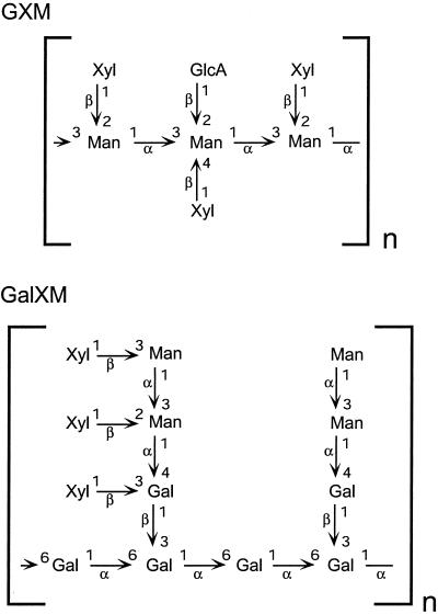
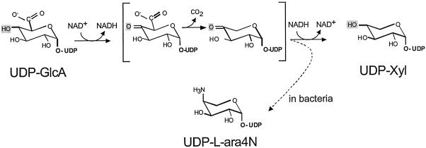
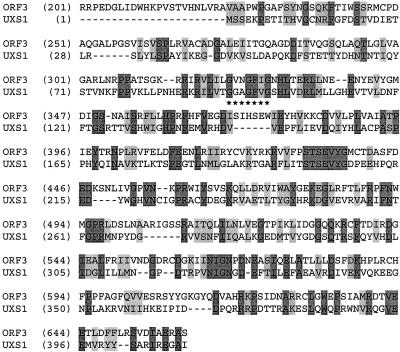
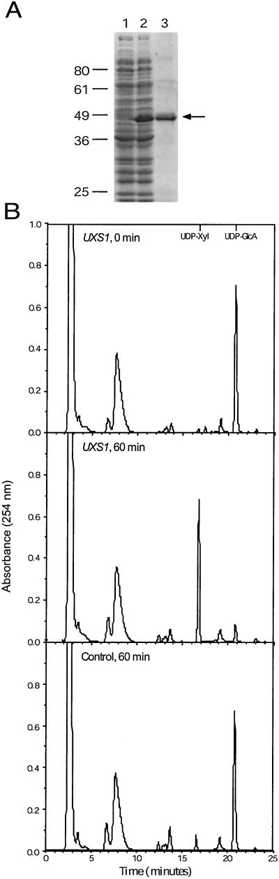
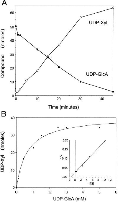
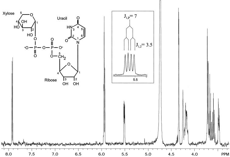
UDP-xylose is a sugar donor required for the synthesis of diverse and important glycan structures in animals, plants, fungi, and bacteria. Xylose-containing glycans are particularly abundant in plants and in the polysaccharide capsule that is the major virulence factor of the pathogenic fungus Cryptococcus neoformans. Biosynthesis of UDP-xylose is mediated by UDP-glucuronic acid decarboxylase, which converts UDP-glucuronic acid to UDP-xylose. Although this enzymatic activity was described over 40 years ago it has never been fully purified, and the gene encoding it has not been identified. We used homology to a bacterial gene, hypothesized to encode a related function, to identify a cryptococcal sequence as putatively encoding a UDP-glucuronic acid decarboxylase. A soluble 47-kDa protein derived from bacteria expressing the C. neoformans gene catalyzed conversion of UDP-glucuronic acid to UDP-xylose, as confirmed by NMR analysis. NADH, UDP, and UDP-xylose inhibit the activity. Close homologs of the cryptococcal gene, which we termed UXS1, appear in genome sequence data from organisms ranging from bacteria to humans.


Figures

Figure 1
Figure 1
Structures of cryptococcal capsular polysaccharides. The structures of C. neoformans glucuronoxylomannan (GXM) (serotype B) and galactoxylomannan (GalXM) are shown. Not shown are the 6-O-acetyl groups that modify up to 10% of mannose residues in GXM. Component monosaccharides are d-pyranoses.
Figure 2
Figure 2
Model for the mechanism of UDP-GlcA decarboxylase, based on refs. and . The compounds in brackets are 4-keto intermediates. Depending on the system the same intermediates could give rise to end products of UDP-Xyl or UDP-aminoarabinose (dashed arrow, in bacteria). The substituent at the 4-position is shaded in each structure.
Figure 3
Figure 3
Comparison of C. neoformans Uxs1 to S. typhimurium Orf3. Identical amino acids are shaded dark gray and similar residues are shaded light gray. The NAD+ binding motif of Uxs1 is underscored with * and is aligned with a similar motif in the bacterial sequence that falls in a well-conserved region. The N-terminal 200 residues of Orf3 are not shown.
Figure 4
Figure 4
Expression and assay of Uxs1. (A) SDS/PAGE analysis of bacterial proteins stained with Coomassie blue. Lane 1, soluble protein from bacteria containing vector alone; lane 2, soluble protein from bacteria expressing Uxs1; lane 3, Uxs1 protein purified by nickel chromatography. The migration positions of standards (in kDa) are shown at left, and the arrow indicates Uxs1. (B) HPLC profiles of standard assays performed for the times indicated using soluble protein from bacteria carrying either a plasmid containing UXS1 or a control plasmid. As indicated at the top the UDP-xylose standard elutes just before 17 min (at 0.21 M KH2PO4) and UDP-GlcA just before 21 min (at 0.28 M KH2PO4). The large peak near 8 min is NAD+.
Figure 5
Figure 5
Characterization of product formation. (A) Time course. Standard Uxs1 reactions (Experimental Procedures) were carried out for the indicated times, and the amounts of reactant and product were analyzed. ●, UDP-GlcA; ○, UDP-Xyl. (B) Effect of UDP-GlcA concentration. Reactions (15 min) were performed without added NAD+ and in the presence of the indicated concentrations of UDP-GlcA. (Inset) A reciprocal plot of the data.
Figure 6
Figure 6
Product analysis. The one-dimensional proton NMR spectrum of the product of the Uxs1 assay is shown. (Left Inset) The structure of UDP-Xyl. (Boxed Inset) An expanded region of the axis just above 5.5 ppm to show the xylose H1 peak splitting.

Similar articles

Cited by

References

    1. Chang Y C, Kwon-Chung K J. Mol Cell Biol. 1994;14:4912–4919. - PMC - PubMed
    1. Casadevall A, Perfect J R. Cryptococcus neoformans. Washington, DC: Am. Soc. Microbiol.; 1998.
    1. Buchanan K L, Murphy J W. Emerg Infect Dis. 1998;4:71–83. - PMC - PubMed
    1. Rodrigues M L, Alviano C S, Travassos L R. Microbes Infection. 1999;1:293–301. - PubMed
    1. Cherniak R, Sundstrom J B. Infect Immun. 1994;62:1507–1512. - PMC - PubMed

Publication types

MeSH terms

Associated data

LinkOut - more resources