. 2001 Oct 9;98(21):12056-61.
doi: 10.1073/pnas.221269198. Epub 2001 Oct 2.

Proteomic method identifies proteins nitrated in vivo during inflammatory challenge

Affiliations

Proteomic method identifies proteins nitrated in vivo during inflammatory challenge

K S Aulak et al. Proc Natl Acad Sci U S A. .

Abstract

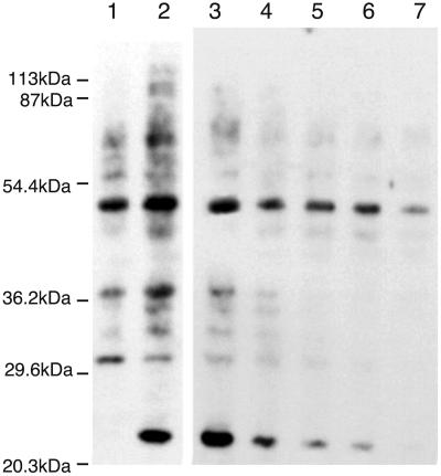
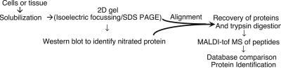
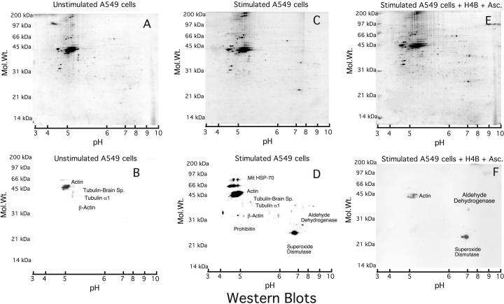
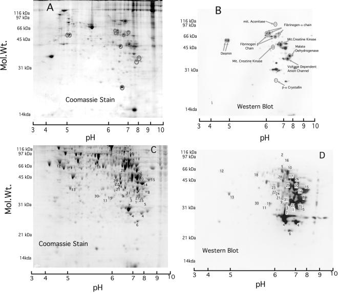
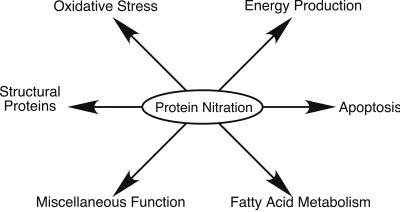
Inflammation in asthma, sepsis, transplant rejection, and many neurodegenerative diseases associates an up-regulation of NO synthesis with increased protein nitration at tyrosine. Nitration can cause protein dysfunction and is implicated in pathogenesis, but few proteins that appear nitrated in vivo have been identified. To understand how this modification impacts physiology and disease, we used a proteomic approach toward targets of protein nitration in both in vivo and cell culture inflammatory disease models. This approach identified more than 40 nitrotyrosine-immunopositive proteins, including 30 not previously identified, that became modified as a consequence of the inflammatory response. These targets include proteins involved in oxidative stress, apoptosis, ATP production, and other metabolic functions. Our approach provides a means toward obtaining a comprehensive view of the nitroproteome and promises to broaden understanding of how NO regulates cellular processes.


Figures

Figure 1
Figure 1
Western blot analysis of the effects of aminoguanidine on anti-nitrotyrosine immunopositive proteins in A549 cells. A549 cells were uninduced (lane 1) or stimulated with cytokines (lanes 2–7: IFN-γ 1,000 units/ml; tumor necrosis factor-α 10 units/ml; IL1-β 10 units/ml). Cytokine-stimulated cells were incubated with aminoguanidine at the time of induction (0, .5, 1, 1.5, 2, and 3 mM in lanes 2–7, respectively). After 3 days, the cells were lysed immediately by using SDS/PAGE loading buffer and 20 μg of protein extracts loaded onto a 12% polyacrylamide gel. Nitrated proteins then were identified by Western blotting using an anti-nitrotyrosine monoclonal antibody.
Figure 2
Figure 2
Overview of the proteomic approach to analyze and identify proteins by using a high throughput system (see Materials and Methods for details).
Figure 3
Figure 3
Anti-nitrotyrosine immunopositive proteins in A549 cells. Cell lysates (300 μg) were run onto two-dimensional gels, and immunoreactive proteins were identified. A, B, and C are the two-dimensional acrylamide gels stained with gel code blue, and C, D, and E are the corresponding Western blots. Stimulation was carried out by using cytokines (C and D: IFN-γ, 1,000 units/ml; tumor necrosis factor-α 10 units/ml; IL1-β 10 units/ml) or with cytokines plus 100 μM H4B in 300 μM ascorbate (E and F). Western blots are exposed for 3 (B and D) and 15 (F) sec. On the 3-sec exposure of F, the intensity of manganese superoxide dismutase (MnSOD) and actin was much lower compared with D (data not shown). Identified proteins are indicated on the Western blots. Gels D and E show similar expression of all of the proteins that were detected by immunoreactivity; therefore the change in signal intensity is caused by decreased protein nitration.
Figure 4
Figure 4
Anti-nitrotyrosine immunopositive proteins in lung and liver of rats induced with LPS. Rats were induced with 10 mg/kg LPS and killed after 18 h. A (lung lysate, 400 μg) and C (liver lysate, 500 μg) are two-dimensional acrylamide gels stained with gel code blue, and B and D are the corresponding Western blots probed with an anti-nitrotyrosine monoclonal antibody. Identified proteins are listed on lung Western blot (B) but numbered on the liver Western blot (D) with the key in Table 1.
Figure 5
Figure 5
Functional classification of anti-nitrotyrosine immunoreactive proteins.

References

    1. Beckman J S, Koppenol W H. Am J Physiol. 1996;271:C1424–C1437. - PubMed
    1. Halliwell B, Zhao K, Whiteman M. Free Radical Res. 1999;31:651–669. - PubMed
    1. Ischiropoulos H. Arch Biochem Biophys. 1998;356:1–11. - PubMed
    1. Gole M D, Souza J M, Choi I, Hertkorn C, Malcolm S, Foust R F, III, Finkel B, Lanken P N, Ischiropoulos H. Am J Physiol. 2000;278:L961–L967. - PubMed
    1. Kamisaki Y, Wada K, Bian K, Balabanli B, Davis K, Martin E, Behbod F, Lee Y C, Murad F. Proc Natl Acad Sci USA. 1998;95:11584–11589. - PMC - PubMed

Publication types

LinkOut - more resources