. 2001 Oct 9;98(21):12078-83.
doi: 10.1073/pnas.211427098. Epub 2001 Oct 2.

Functional communication between endogenous BRCA1 and its partner, BARD1, during Xenopus laevis development

Affiliations

Functional communication between endogenous BRCA1 and its partner, BARD1, during Xenopus laevis development

V Joukov et al. Proc Natl Acad Sci U S A. .

Abstract

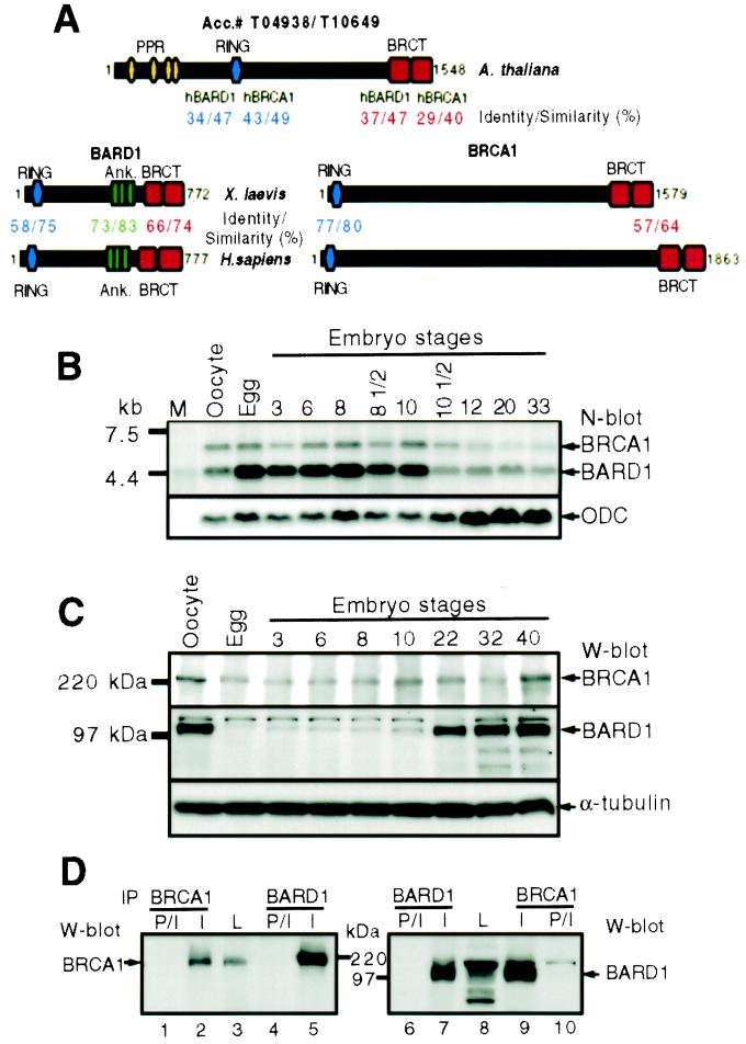
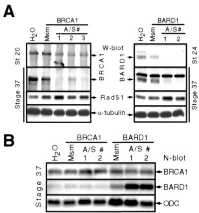
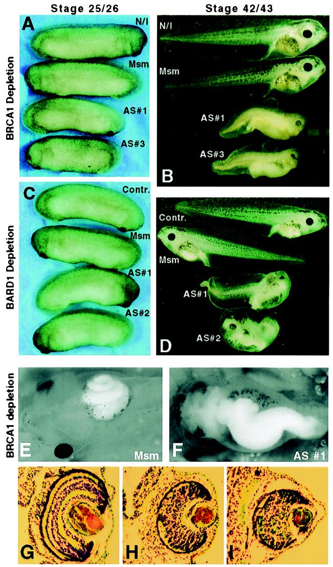
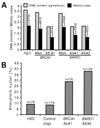
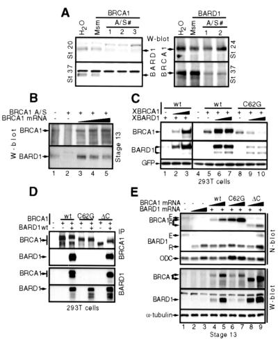
The breast and ovarian susceptibility protein 1 (BRCA1) heterodimerizes with its structural relative, the BRCA1-associated RING domain protein (BARD1), which may have tumor suppressing function in its own right. Both proteins have evolved from a common evolutionary ancestor, and both exist in Xenopus laevis where, similar to their mammalian homologs, they form functional heterodimers. Depleting frog embryos of either BARD1 or BRCA1 led to similar and widely defective developmental phenotypes as well as depletion of the other polypeptide due to its decreased stability. Thus, each protein, in part, controls the abundance, stability, and function of the other, and these effects are heterodimerization-dependent. The interdependent nature of BRCA1 and BARD1 function supports the view that BARD1/BRCA1 heterodimers play a major role in breast and ovarian cancer suppression.


Figures

Figure 1
Figure 1
BRCA1 and BARD1 Orthologs of A. thaliana and X. laevis. (A) Domain structure of frog and human BRCA1 and BARD1, and the putative Arabidopsis BRCA1/BARD1 ortholog. PPR, Pentatrico Peptide Repeat; RING, RING finger domain; Ank, Ankyrin repeat; BRCT, BRCA1 C- terminal motif. The first and the last residues of each protein are noted. The sequence identity/similarity between certain segments of human BRCA1 and BARD1 (the RING, ankyrin repeats, and 2xBRCT motifs) and their frog and Arabidopsis orthologs are indicated. (B) Electophoretically fractionated total RNA from embryos at the indicated stages was hybridized to specific, radioactively labeled xBRCA1-, xBARD1-, and Xenopus ornithine decarboxylase (ODC) probes. Bands were detected by autoradiography. mRNA transcripts and positions of RNA markers [the Radiolabeled RNA Ladder System (GIBCO), lane M] are indicated. (C) Total cell extracts of ten oocytes/eggs/embryos per sample were subjected to IP with xBRCA1- and the xBARD1-specific Abs. Immunoprecipitates were fractionated by SDS/PAGE and gels immunoblotted with xBRCA1- and xBARD1-specific Abs. Lysate from ½ of an embryo was immunoblotted with the α-tubulin Ab as a loading control. (D) BRCA1 and BARD1 complex formation in Xenopus embryos. Lysates of stage-12 embryos were IPd with xBRCA1- or xBARD1-specific immune (I) Ig, or preimmune (P/I) Ig. Lysates (L) and immunoprecipitates were analyzed by Western blotting using xBRCA1 (Left) or xBARD1 (Right) Ab. Combined lysates from two embryos were loaded in lanes 3 and 8. Precipitates from combined lysates of five embryos were loaded in lanes 1, 2, 6, and 7; precipitates from combined lysates of 20 embryos were loaded in lanes 4, 5, 9, and 10, respectively.
Figure 2
Figure 2
Depletion of BRCA1 and BARD1 with specific antisense MOs. (A) Embryos were injected with 40 ng of alternative BRCA1-specific (BRCA1A/S #1, #2, and #3) or BARD1- specific (BARD1 A/S #1 and #2) antisense MOs. The control embryos were injected with water (H2O) or with 40 ng of MOs that were each identical to the BRCA1- or BARD1-specific antisense MOs #1 but contained four dispersed nucleotide Msm. Lysates of the indicated embryos were analyzed by Western blotting with Rad51- and α-tubulin-specific Ab or by IP and Western blotting with xBRCA1- and xBARD1-specific Abs. (B) Total RNA from stage-37 embryos, which were treated as in A, was analyzed by Northern blot hybridization, as described for Fig. 1B.
Figure 3
Figure 3
BRCA1- and BARD1-depleted Xenopus embryos. (A and B) Embryos were either left uninjected (N/I) or injected with MOs: xBRCA1 Msm (30 ng), xBRCA1 AS#1 (30 ng), and xBRCA1 AS#3 (60 ng); (C and D) xBARD1 Msm (30 ng), xBARD1 AS#1 (30 ng), and xBARD1 AS#2 (70 ng) or with the control MO (Contr., 70 ng). Representative embryos at stages 25/26 (Left) and 42/43 (Right) are shown. Note the progressive phenotypic changes in the antisense-treated embryos. (E and F) The ventral view of representative stage-45 embryos, injected with 20 ng of the xBRCA1 Msm (E) or xBRCA1 AS#1 (F). Note the poor segregation of the alimentary canal and impaired coiling of the intestinal tube in the embryo even at this relatively low dose of antisense MO (F). Similar defects were observed in the BARD1-depleted embryos (data not shown). (GI) The eye of a stage-42 embryo, injected with 25 ng of the BRCA1 Msm (G), xBRCA1 AS#1 (H), or xBARD1 AS#1 (I). Note the lack of retinal layers and the absence of degeneration of nuclei in the central lens fibers in the antisense MO-treated embryos (H and I). Similar defects were observed in embryos injected with xBRCA1 AS#3 and xBARD1 AS#2. The eye structures of the noninjected and the mismatch MO-injected embryos were histologically indistinguishable from one another (data not shown).
Figure 4
Figure 4
Deficient proliferation and chromosomal instability in BRCA1- and BARD1-deficient embryos. (A) Embryos were injected with water or with 30 ng each of the MOs: xBRCA1 Msm, xBRCA1 AS#1, xBARD1 Msm, and xBARD1 AS#1, or with 60 ng of xBARD1 AS#2. The DNA content and the mitotic index were analyzed at stage 37. (B) Embryos were injected with xBRCA1 AS#1 (25 ng), xBARD1 AS#2 (60 ng), or a control oligonucleotide (60 ng). The chromosome number was analyzed in the stage-37–40 embryos. Aneuploid chromosome numbers ranged from 32 to 104 per cell in the antisense-treated embryos. A euploid, somatic X. laevis cell contains 36 chromosomes (38).
Figure 5
Figure 5
Reciprocal control of BRCA1 and BARD1 abundance. (A) Cell lysates from the MO-treated embryos were analyzed by standardized Western blotting for BARD1 and BRCA1 abundance, respectively, at the indicated stages. Identical samples of embryo cell lysates were analyzed in this experiment and in the experiment described in Fig. 2A. (B) BRCA1 and BARD1 protein in stage 13 embryos that had been injected with 40 ng of xBRCA1 AS#1 alone (lane 2) or coinjected with this reagent and with 200, 400, or 600 pg of xBRCA1 mRNA (lanes 3, 4, and 5, respectively). (C) 293 T cells were transfected with 3 μg of xBARD1 wt cDNA expression vector, alone (lane 1) or together with 3 μg or 9 μg of xBRCA1 wt cDNA expression vector (lanes 2 and 3, respectively), or with 3 μg of xBRCA1 wt cDNA (lane 5) or xBRCA1 C62G cDNA expression vector (lane 8), alone or together with 3 μg (lanes 6 and 9) or 9 μg (lanes 7 and 10) of xBARD1wt cDNA expression vector. The transfection mixtures each contained 5% of green fluorescent protein (GFP) cDNA, cloned in an expression vector. Each lane was analyzed by Western blotting with the indicated Ab. Anti-GFP Ab was used to detect GFP abundance in each lane. (D) 293 T cells were transfected with the indicated xBRCA1 cDNA expression vector, alone or together with the wt xBARD1 cDNA vector. The cell lysates were subjected to IP with the xBRCA1- or xBARD1-specific Ab (Upper and Lower, respectively), and each immunoprecipitate was immunoblotted with both of these Ab. (E) Xenopus embryos were injected with 300 and 600 pg of xBARD1 mRNA (lanes 2 and 3, respectively) or were coinjected with 300 pg of xBARD1 mRNA and 300 pg (lanes 4, 6, and 8) or 600 pg (lanes 5, 7, and 9) of the indicated xBRCA1 mRNA. At embryo-stage 13, total RNA was isolated from half of each injected embryo set and analyzed by Northern blot hybridization as described for Fig. 1B (Upper). The endogenous (E) and the recombinant (R) forms of the xBRCA1 and xBARD1 mRNAs are indicated by arrows. In extracts of the other half of each experimental embryo set, the abundance of xBRCA1 and xBARD1 were analyzed by IP/Western blotting as in Fig. 2A (Lower). α-tubulin was detected by direct Western blotting.

Similar articles

Cited by

References

    1. Miki Y, Swensen J, Shattuck-Eidens D, Futreal P A, Harshman K, Tavtigian S, Liu Q, Cochran C, Bennett L M, Ding W. Science. 1994;266:66–71. - PubMed
    1. Wu L C, Wang Z W, Tsan J T, Spillman M A, Phung A, Xu X L, Yang M C, Hwang L Y, Bowcock A M, Baer R. Nat Genet. 1996;14:430–440. - PubMed
    1. Hakem R, de la Pompa J L, Sirard C, Mo R, Woo M, Hakem A, Wakeham A, Potter J, Reitmair A, Billia F, Firpo E, Hui C C, Roberts J, Rossant J, Mak T W. Cell. 1996;85:1009–1023. - PubMed
    1. Liu C Y, Flesken-Nikitin A, Li S, Zeng Y, Lee W H. Genes Dev. 1996;10:1835–1843. - PubMed
    1. Ludwig T, Chapman D L, Papaioannou V E, Efstratiadis A. Genes Dev. 1997;11:1226–1241. - PubMed

Publication types

Associated data