. 2001 Oct 9;98(21):12168-73.
doi: 10.1073/pnas.221220998. Epub 2001 Oct 2.

Specific treatment of autoimmunity with recombinant invariant chains in which CLIP is replaced by self-epitopes

Affiliations

Specific treatment of autoimmunity with recombinant invariant chains in which CLIP is replaced by self-epitopes

F Bischof et al. Proc Natl Acad Sci U S A. .

Abstract

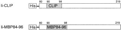
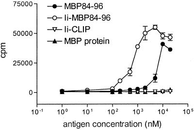
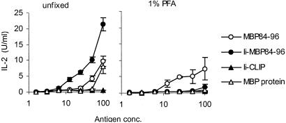
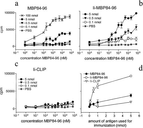
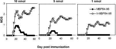
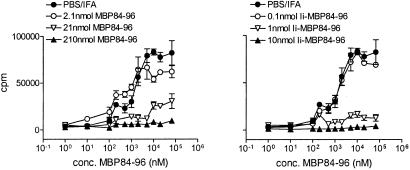
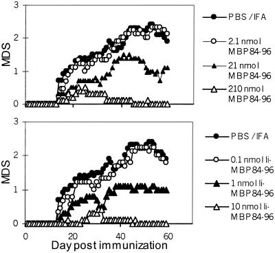
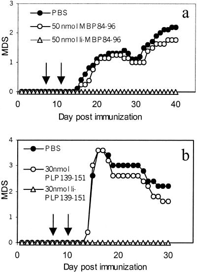
The invariant chain (Ii) binds to newly synthesized MHC class II molecules with the CLIP region of Ii occupying the peptide-binding groove. Here we demonstrate that recombinant Ii proteins with the CLIP region replaced by antigenic self-epitopes are highly efficient in activating and silencing specific T cells in vitro and in vivo. The Ii proteins require endogenous processing by antigen-presenting cells for efficient T cell activation. An Ii protein encompassing the epitope myelin basic protein amino acids 84-96 (Ii-MBP84-96) induced the model autoimmune disease experimental allergic encephalomyelitis (EAE) with a higher severity and earlier onset than the peptide. When applied in a tolerogenic manner, Ii-MBP84-96 abolished antigen-specific T cell proliferation and suppressed peptide-induced EAE more effectively than peptide alone. Importantly, i.v. administration of Ii proteins after EAE induction completely abrogated the disease, whereas peptides only marginally suppressed disease symptoms. Ii fusion proteins are thus more efficient than peptide in modulating CD4(+) T cell-mediated autoimmunity, documenting their superior qualities for therapeutic antigen delivery in vivo.


Figures

Figure 1
Figure 1
Design of the proteins. Depicted numbers indicate the amino acids of the murine Ii. The N-terminal hydrophobic region was replaced by a His6 tag. The CLIP core region at amino acids 90–98 was replaced by the epitope MBP84–96 to generate Ii-MBP84–96.
Figure 2
Figure 2
Lymph node cells of mice immunized with MBP84–96 proliferate in vitro when stimulated with Ii-MBP84–96 and MBP84–96 but not Ii-CLIP. Mice were immunized s.c. with 10 nmol MBP84–96 in CFA containing 200 μg M. tuberculosis. At day 10 postimmunization, single cell suspensions of draining lymph nodes were cultured along with the indicated concentrations of MBP84–96, Ii-MBP84–96, Ii-CLIP, or MBP. A total of 1 μCi/well [3H]thymidine was added after 96 h of culture, cells were harvested after an additional 12 h, and mean [3H]thymidine incorporation was measured by liquid scintillation counting. Background incorporation was less than 1,000 cpm and proliferation to the control antigen PPD, which is a component of the adjuvant, was >20,000 cpm (data not shown). Error bars represent SEM.
Figure 3
Figure 3
Ii-MBP84–96 requires endogenous antigen processing to stimulate MBP84–96-specific T cells. Spleen cells of naive mice were used unfixed or fixed with 1% paraformaldehyde and incubated along with titrated amounts of the indicated antigens. The MBP84–96-specific T cell hybridoma 6F11 was added for 36 h, and the IL-2 concentration in the supernatant was determined by proliferation of the IL-2-dependent cell line CTLL-2.
Figure 4
Figure 4
MBP84–96 and Ii-MBP84–96, but not Ii-CLIP, can prime MBP84–96-specific T cells in vivo. Mice were immunized s.c. with the indicated amount of MBP84–96 (a), Ii-MBP84–96 (b), or Ii-CLIP (c) in CFA containing 200 μg M. tuberculosis. Ten days later, single cell suspensions of draining lymph nodes were cultured in vitro along with the indicated concentrations of MBP84–96. After 96 h of culture, 1 μCi/well [3H]thymidine was added, cells were harvested after an additional 12 h, and proliferation was measured by liquid scintillation counting. Proliferation induced by the control antigen PPD was >40,000 cpm (data not shown) and background incorporation was less than 2,500 cpm in all cases. (d) The proliferation at the highest in vitro dose of MBP84–96 was plotted against the amount of antigen used for immunization.
Figure 5
Figure 5
Ii-MBP84–96 is more efficient in induction of EAE than MBP84–96. Groups of 10 mice were immunized on days 0 and 7 with 10, 5, or 1 nmol of either MBP84–96 or Ii-MBP84–96 in CFA containing 200 μg M. tuberculosis. On days 0 and 2, mice received in addition 200 ng pertussis toxin in PBS i.v. Clinical signs of EAE were assessed as described in Materials and Methods, and the mean disease score (MDS) was plotted against the day postimmunization. The incidence rates were determined as 10/10 in each of the groups immunized with 10, 5, and 1 nmol Ii-MBP84–96 and 8/10, 4/10, and 0/10 in the groups immunized with 10, 5, and 1 nmol MBP84–96, respectively.
Figure 6
Figure 6
Ii-MBP84–96 is more efficient than MBP84–96 in suppression of antigen-specific T cell responses. Mice received an i.p. injection of the indicated amount of MBP84–96, Ii-MBP84–96, or PBS emulsified in IFA at day 10 before s.c. immunization with 100 nmol MBP84–96 in CFA. Ten days postimmunization, proliferation of the draining lymph node cells cultured with MBP84–96 was determined as described in the legend to Fig. 4. Error bars indicate SEM. Background incorporation was between 1,450 and 5,400 cpm. The response to the control antigen PPD was >30,000 cpm in all cases.
Figure 7
Figure 7
Ii-MBP84–96 is more efficient than MBP84–96 in prevention of autoimmunity. Ten mice per group were injected i.p. with 100 μl of an emulsion containing the indicated amount of either MBP84–96, the fusion protein Ii-MBP84–96, or PBS in IFA. Ten days later, EAE was induced by s.c. injection of 130 nmol MBP84–96 emulsified in CFA at days 0 and 7 and i.v. injection of 200 ng pertussis toxin at days 0 and 2. EAE disease score was determined as described in Materials and Methods. The disease incidence for mice injected with PBS/IFA was 10/10, for mice injected with 2.1, 21, and 210 nmol MBP84–96, 9/9, 8/10, and 4/10 for mice that received 0.1, 1, and 10 nmol Ii-MBP84–96 8/8, 8/10, and 3/10, respectively.
Figure 8
Figure 8
Suppression of EAE by i.v. injection of recombinant Ii proteins. (a) EAE was induced by immunization with 130 nmol of MBP84–96 and i.v. injection of pertussis toxin as described in the legend to Fig. 7. At days 7 and 11 (arrows) mice received either 50 nmol MBP84–96, 50 nmol Ii-MBP84–96, or PBS i.v. Disease severity was scored as described in Materials and Methods. The incidence rates for the PBS- and MBP84–96-treated groups were 10/10 and for the Ii-MBP84–96-treated group 0/10. (b) EAE was induced by s.c. injection of 50 nmol PLP139–151 in CFA and i.v. injection of 200 ng pertussis toxin. Experimental groups received i.v. injections of PBS, 30 nmol PLP139–151, or 30 nmol Ii-PLP139–151, respectively. The incidence rates were 5/5 in the PBS- and peptide-treated groups and 0/5 in the Ii-PLP139–151- treated group. The results are representative of three independent experiments. MDS, mean disease score.

Similar articles

Cited by

References

    1. Ghosh P, Amaya M, Mellins E, Wiley D C. Nature (London) 1995;378:457–462. - PubMed
    1. Malcherek G, Gnau V, Jung G, Rammensee H G, Melms A. J Exp Med. 1995;181:527–536. - PMC - PubMed
    1. Avva R R, Cresswell P. Immunity. 1994;1:763–774. - PubMed
    1. Bijlmakers M J, Benaroch P, Ploegh H L. J Exp Med. 1994;180:623–629. - PMC - PubMed
    1. Freisewinkel I M, Schenck K, Koch N. Proc Natl Acad Sci USA. 1993;90:9703–9706. - PMC - PubMed

Publication types

MeSH terms