. 2001 Oct 9;98(21):12174-9.
doi: 10.1073/pnas.211191098. Epub 2001 Oct 2.

Cardiovascular, skeletal, and renal defects in mice with a targeted disruption of the Pkd1 gene

Affiliations

Cardiovascular, skeletal, and renal defects in mice with a targeted disruption of the Pkd1 gene

C Boulter et al. Proc Natl Acad Sci U S A. .

Abstract

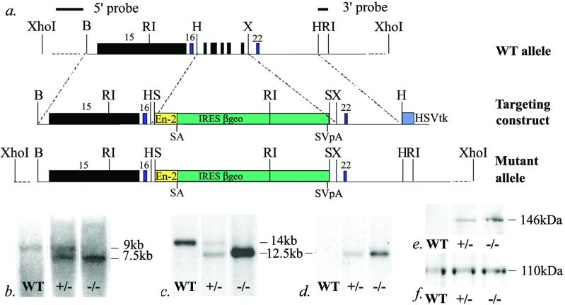
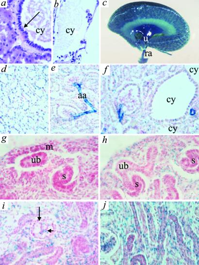
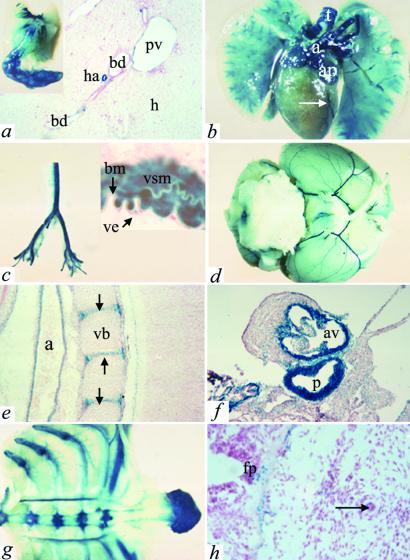
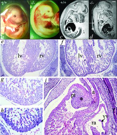
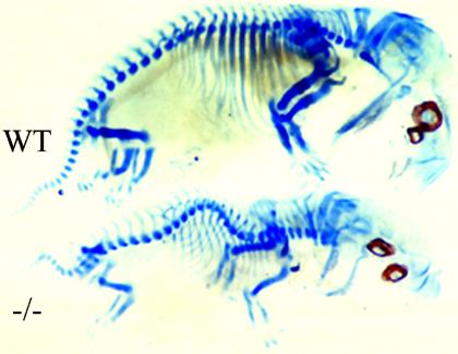
Autosomal dominant polycystic kidney disease (ADPKD) is characterized by cyst formation in the kidney, liver, and pancreas and is associated often with cardiovascular abnormalities such as hypertension, mitral valve prolapse, and intracranial aneurysms. It is caused by mutations in PKD1 or PKD2, encoding polycystin-1 and -2, which together form a cell surface nonselective cation ion channel. Pkd2-/- mice have cysts in the kidney and pancreas and defects in cardiac septation, whereas Pkd1(del34) -/- and Pkd1(L) -/- mice have cysts but no cardiac abnormalities, although vascular fragility was reported in the latter. Here we describe mice carrying a targeted mutation in Pkd1 (Pkd1(del17-21betageo)), which defines its expression pattern by using a lacZ reporter gene and may identify novel functions for polycystin-1. Although Pkd1(del17-21betageo) +/- adult mice develop renal and hepatic cysts, Pkd1(del17-21betageo) -/- embryos die at embryonic days 13.5-14.5 from a primary cardiovascular defect that includes double outflow right ventricle, disorganized myocardium, and abnormal atrio-ventricular septation. Skeletal development is also severely compromised. These abnormalities correlate with the major sites of Pkd1 expression. During nephrogenesis, Pkd1 is expressed in maturing tubular epithelial cells from embryonic day 15.5. This expression coincides with the onset of cyst formation in Pkd1(del34) -/-, Pkd1(L) -/-, and Pkd2-/- mice, supporting the hypothesis that polycystin-1 and polycystin-2 interact in vivo and that their failure to do so leads to abnormalities in tubule morphology and function.


Figures

Figure 1
Figure 1
Generation of a targeted disruption of Pkd1. (a) Pkd1 exons 17–21 were replaced with a lacZ-neomycinR fusion gene (βgeo) located downstream of an engrailed-2 gene donor intron (En-2) and splice acceptor site (SA), an IRES, and upstream of a simian virus 40 polyadenylation signal (SVpA). The positions of the 5′ and 3′ external probes are indicated. HSVtk, herpes simplex virus thymidine kinase gene; B, BamHI; RI, EcoRI; H, HindIII; X, XbaI; S, SalI. (b) A Southern blot of EcoRI-digested DNA from a cross of Pkd1del17–21βgeo +/− mice hybridized with an external 3′ probe showing the WT (+/+) (9 kb) and mutant (7.5 kb) alleles; this result was confirmed with the 5′-external probe (data not shown). (c) Northern analysis using a Pkd1 exon 15 probe demonstrated the ≈14-kb Pkd1 transcript in WT (+/+) and Pkd1del17–21βgeo +/− embryos and the predicted 12.5-kb mutant transcript in Pkd1del17–21βgeo +/− and −/− embryos; different intensities between RNA samples reflected different RNA loading are shown. (d) A neoR probe confirmed the presence of the 12.5-kb mutant transcript in Pkd1del17–21βgeo +/− and −/− embryos. (e) Independent translation of the βgeo gene was demonstrated by using an anti-β-galactosidase antibody to detect the predicted 146-kDa β-galactosidase-neomycin fusion protein. (f) Levels of polycystin-2 (110 kDa) were unaltered in Pkd1del17–21βgeo +/− and −/− E12.5 embryos compared with WT (+/+) littermates.
Figure 2
Figure 2
Renal cysts and Pkd1 expression in embryonic and adult kidneys of Pkd1del17–21βgeo +/− mice demonstrated by using X-Gal staining. Renal cysts (cy) in Pkd1del17–21βgeo +/− were lined with hyperplastic epithelial cells (a, arrowed) or cells with pyknotic apoptotic nuclei (b, ×60). (c) Whole-mount X-Gal staining of adult kidney. (d-f) Adult kidney frozen sections (×40). Expression is seen in all medullary collecting ducts (d), glomerular parietal, proximal and distal tubule epithelial cells (e), and in cystic epithelia (f). Strong staining of the afferent arterioles (aa) and other vascular structures was seen also. (g-j) Pkd1 expression in Pkd1del17–21βgeo +/− embryos during nephrogenesis (×60): g, E13.5; h and i, E15.5; j, E18.5. Expression is confined to mesenchyme (m) and maturing tubules. ub, ureteric bud; s, S-shaped body; long arrow, podocytes; short arrow, endothelium.
Figure 3
Figure 3
Expression of Pkd1 in nonrenal adult and embryonic tissues. (a) Adult liver [bd, bile ducts; ha, hepatic artery; pv, portal vein; h, hepatocytes (×10)]. (Inset) Whole-mount staining of gall bladder. (b) Adult heart. Pkd1 was expressed strongly in the aortic outflow tract (a), atrial appendages (ap), and major coronary vessels (arrow). The cartilage rings of the trachea (t) and bronchi and vessels in the lung are also positive. (c) Dissected adult aorta. (Inset) Frozen section adult aorta (×40). Pkd1 is expressed in vascular smooth muscle cells (vsm) and vascular endothelial cells (ve) separated by vascular basement membrane (bm). (d) Intracranial arteries. (e and f) Pkd1 expression in the embryonic cardiovascular system. (e) Aorta (a) at E14.5. The cartilage of the developing vertebral bodies (vb) and intervertebral discs (arrows) are also positive (×10). (f) E18.5 cardiac outflow tract (p, pulmonary artery; av, aortic valve), (×20). (g) Whole-mount staining of sternum and ribs. Note also strong staining in vessels. (h) E11.5 floor plate (fp) and notochord (arrow) are sites of Pkd1 expression (×40).
Figure 4
Figure 4
Abnormal cardiovascular development in Pkd1del17–21βgeo −/− embryos. (a–d) Pkd1del17–21βgeo −/− embryos at E13.5 (b and d) were edematous and hemorrhagic compared with littermate controls (a and c). Magnetic resonance imaging demonstrated the marked edema (*), subcutaneous hemorrhage (white arrow), pericardial effusions (long black arrow), and abnormal craniofacial development (short black arrow) of Pkd1del17–21βgeo −/− embryos (d) (scale bar, 1 mm). Hematoxylin/eosin staining of sagittal sections of E13.5 embryos showed hemorrhagic effusion and disorganization of the myocardial wall and ventricular septum in Pkd1del17–21βgeo −/− embryos (f and h) but not in WT littermates (e and g). lv, left ventricle; rv, right ventricle. e and f, ×10; g and h, ×40. (i) Transverse section showing the heart of a Pkd1del17–21βgeo −/− embryo at E13.5 showing a double outlet right ventricle (DORV) with the large dysplastic cushion mass forming the outlet septum (rv, right ventricle; ra, right atrium; pt, pulmonary trunk; a, aorta; vv, venous valves of the right atrium; *, outlet septum) (×10).
Figure 5
Figure 5
Abnormal skeletal development in Pkd1del17–21βgeo −/− mice. Cartilage staining of skeletal preparations from an E13.5 Pkd1del17–21βgeo −/− embryo and WT (+/+) littermate showing abnormalities in cranial, facial, axial, and long bone formation in the mutant.

Similar articles

Cited by

References

    1. Murcia N S, Sweeney W E, Jr, Avner E D. Kidney Int. 1999;55:1187–1197. - PubMed
    1. Calvet J P. Kidney Int. 1993;43:101–108. - PubMed
    1. Grantham J J. Am J Kidney Dis. 1996;28:788–803. - PubMed
    1. Richards W G, Sweeney W E, Yoder B K, Wilkinson J E, Woychik R P, Avner E D. J Clin Invest. 1998;101:935–939. - PMC - PubMed
    1. Carone F A, Butkowski R J, Nakamura S, Polenakovic M, Kanwar Y S. Kidney Int. 1994;46:1368–1374. - PubMed

Publication types

MeSH terms