. 2001 Oct 9;98(21):12180-5.
doi: 10.1073/pnas.201420298. Epub 2001 Oct 2.

Targeting tissue factor on tumor vascular endothelial cells and tumor cells for immunotherapy in mouse models of prostatic cancer

Affiliations

Targeting tissue factor on tumor vascular endothelial cells and tumor cells for immunotherapy in mouse models of prostatic cancer

Z Hu et al. Proc Natl Acad Sci U S A. .

Abstract

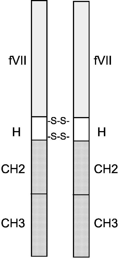
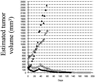
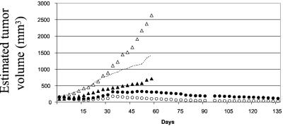
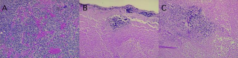
The efficacy and safety of an immunoconjugate (icon) molecule, composed of a mutated mouse factor VII (mfVII) targeting domain and the Fc effector domain of an IgG1 Ig (mfVII/Fc icon), was tested with a severe combined immunodeficient (SCID) mouse model of human prostatic cancer and an immunocompetent mouse model of mouse prostatic cancer. The SCID mice were first injected s.c. with a human prostatic tumor line, forming a skin tumor that produces a high blood titer of prostate-specific antigen and metastasizes to bone. The icon was encoded in a replication-incompetent adenoviral vector that was injected directly into the skin tumor. The tumor cells infected by the vector synthesize and secrete the icon into the blood, and the blood-borne icon binds with high affinity and specificity to mouse tissue factor expressed on endothelial cells lining the lumen of the tumor vasculature and to human tissue factor expressed on the tumor cells. The Fc domain of the icon activates a cytolytic immune attack against cells that bind the icon. The immunotherapy tests in SCID mice demonstrated that intratumoral injections of the adenoviral vector encoding the mfVII/human Fc icon resulted in long-term regression of the injected human prostatic tumor and also of a distant uninjected tumor, without associated toxicity to the mice. Comparable results were obtained with a SCID mouse model of human melanoma. At the end of the experiments the mice appeared to be free of viable tumor cells. This protocol also could be efficacious for treating cancer patients who have vascularized tumors.
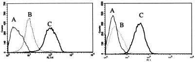

Figures

Figure 1
Figure 1
Diagram of a fVII/Fc icon. fVII, fVII with a Lys341 to Ala341 mutation. H, Hinge region of an IgG1 Ig. CH2 and CH3, Constant regions in the Fc domain of an IgG1 Ig.
Figure 2
Figure 2
Regression of a human prostatic tumor in SCID mice after intratumoral injections of an adenoviral vector encoding the mfVII/hFc icon. The first experiment involved two control mice (▵) and two treated mice (○), and a second experiment involved five control mice (▴) and six treated mice (●). The mice were injected s.c. in one flank with the human prostatic cancer line C4–2. When the tumors had grown to an estimated volume of about 350 mm3 for the first experiment and about 180 mm3 for the second experiment, intratumoral injections were started (day 0). The dose/injection was 1 × 10¹⁰ VP. The mice received during the next 20 days a total of seven injections for the first experiment and six injections for the second experiment either of the vector encoding the mfVII/hFc icon or the control vector. The tumors injected with the control vector grew rapidly, causing the mice to become seriously ill; all of the mice died between days 49 and 63. The tumors injected with the vector encoding the icon regressed initially, but started to grow again. The mice then received three injections for the first experiment and four injections for the second experiment from days 33 to 45, after which the tumors regressed and did not grow again for the duration of the experiments. At the end of the experiments only minute necrotic nodules of tumor tissue remained, similar to the tumor section shown in Fig. 5. Each point is an average value that varied about ±20% among the mice.
Figure 3
Figure 3
Regression of two human prostatic tumors in SCID mice after intratumoral injections of an adenoviral vector encoding the mfVII/hFc icon into one tumor. The mice were injected s.c. in both rear flanks with the human prostatic cancer line C4–2. The resulting skin tumor on one flank was injected with the vector encoding the icon (four mice) or with the control vector (four mice) on days 0, 3, 6, 9, 12, 15, 33, 36, 39, and 42, whereas the tumor on the other flank was left uninjected. The dose was 1 × 10¹⁰ VP per injection. The control mice were euthanized on days 53–57 and the icon-treated mice on day 138. ○, Tumors injected with the vector encoding the icon. ●, Uninjected tumors in the icon-treated mice. ▵, Tumors injected with the control vector. ▴, Uninjected tumors in the control mice; the broken line shows the curve after adjusting each point for the difference between the estimated volumes on day 0 for the injected tumors (169 mm3) and uninjected tumors (88 mm3). Each point is an average value that varied about ±25% among the mice.
Figure 4
Figure 4
Histology of the human prostatic tumors from the experiment described in Fig. 3. The tumors were excised during necropsy on day 138 for the icon-treated mice and on day 57 for the control mice. The sections were fixed in formaldehyde, embedded in paraffin, sectioned, and stained with hematoxylin and eosin. (A) Tumor injected with the control vector, showing dense vascularization and viable tumor cells. (B) Injected tumor from the icon-treated mouse. (C) Uninjected tumor from the icon-treated mouse. Note that B and C, and also the areas not shown here, do not contain viable tumor cells. (Magnification: ×25.)
Figure 5
Figure 5
Regression of two human melanoma tumors in SCID mice after intratumoral injections of an adenoviral vector encoding the mVII/hFc icon into one tumor. The mice were injected s.c. in both rear flanks with the human melanoma line TF2. The resulting skin tumor on one flank was injected with the vector encoding the icon (three mice) or with the control vector (three mice) on days 0, 3, 6, 9, 12, 15, 30, 36, 39, 42, and 45, whereas the tumor on the other flank was left uninjected. The dose was 6 × 10⁹ VP per injection. One control mouse was euthanized on day 53 and the other control mice on day 111. The icon-treated mice were euthanized on day 111. The histology of the melanoma tumors removed during necropsy was similar to the histology of the prostatic tumors shown in Fig. 4. ●, Tumors injected with the vector encoding the icon. ○, Uninjected tumors from the icon-treated mice. ▵, Tumors injected with the control vector. ▴, Uninjected tumors from the control mice. Each point is an average value that varied about ±20% among the mice.
Figure 6
Figure 6
Clearance of the mfVII/hFc icon from the plasma of SCID mice. (A) Five micrograms of icon protein was injected into the tail vein of two SCID mice on day 0, and blood samples were taken from the eyes on days 1, 3, 7, 14, 21, 32, 41, 47, and 51. The blood samples were mixed with buffered sodium citrate at a ratio of 1:9 (vol/vol), and the icon concentration was determined by fluorescence ELISA. (B) Five SCID mice were injected s.c. with 5 × 10⁵ human melanoma TF2 cells per mouse. The resulting skin tumors were injected with the vector encoding the mfVII/hFc icon on days 0, 3, 6, 9, 12, and 15. The dose was 6 × 10⁹ VP per injection. Blood samples were taken from the eyes on days 2, 17, 21, and 30, mixed with buffered sodium citrate at a ratio of 1:9 (vol/vol), and the icon concentration was determined by fluorescence ELISA.
Figure 7
Figure 7
Specific binding of the mfVII/hFc icon to the endothelium of a melanoma tumor in SCID mice. The mice were injected s.c. with the 6 × 10⁹ VP of human melanoma cells TF2, and the resulting skin tumor was injected with the vector encoding the mfVII/hFc icon on days 0, 3, 6, 9, 12, and 15. On the second day after the last injection, the mice were euthanized and immediately perfused with 4% formaldehyde in PBS for 2 min. Sections of the tumor, liver, and kidney were postfixed for 1 h, washed with PBS, infused with 10% DMSO, and stored in liquid nitrogen. Frozen sections cut 6 μm thick were placed on lysine-coated slides, rehydrated in PBS, blocked with 1% BSA in normal goat serum for 1 h, and incubated with goat anti-human Fc antibody followed by tetramethylrhodamine B isothiocyanate-conjugated anti-goat antibody. The sections were coated with Vectashield antifade (Vector Laboratories) and photographed with fluorescent confocal optics. (A) Tumor. (B) Liver. (C) Kidney. The sections were prepared and photographed by T. Ardito in P. McPhaedron's laboratory, Department of Pathology at Yale University. (Magnification: ×375.)
Figure 8
Figure 8
Effect of the mfVII/mFc icon on the growth of a mouse prostatic tumor in immunocompetent mice. The mouse prostatic tumor line RM-1 was injected s.c. in C57BL/6 mice, and when a skin tumor had grown to the size indicated on day 0 the mice were i.v. injected with the vector encoding the mfVII/mFc icon (five mice) or with the control vector (five mice). The injections were done on days 0, 4, 7, 10, 13, and 16 with a dose of 1 × 10¹⁰ VP per injection. ▵, Control vector. ○, Vector encoding the icon. Each point is an average value that varied by about ±20% among the mice.
Figure 9
Figure 9
FACS assays for binding of the mfVII/hFc and hfVII/hFc icons to human TF on human melanoma cells (TF2) (Left) and mouse TF on mouse melanoma cells (B16F10) (Right). (A) Control cells not exposed to an icon. (B) Cells exposed to the hfVII/hFc icon. (C) Cells exposed to the mfVII/hFc icon. The relative displacement of each curve is proportional to the number of icon molecules bound to the cells.

Similar articles

Cited by

References

    1. Folkman J. N Engl J Med. 1995;333:1757–1763. - PubMed
    1. Nemerson Y. Blood. 1988;71:1–8. - PubMed
    1. Drake T A, Morrissey J H, Edgington T S. Am J Pathol. 1989;134:1087–1097. - PMC - PubMed
    1. Flossel C, Luther T, Muller M, Albrecht S, Kasper M. Histochemistry. 1994;101:449–453. - PubMed
    1. Callander N S, Varki N, Rao L V. Cancer. 1992;70:1194–1201. - PubMed

Publication types

MeSH terms