

Shifts in cortical representations predict human discrimination improvement
- PMID: 11593042
- PMCID: PMC59801
- DOI: 10.1073/pnas.191176298
Shifts in cortical representations predict human discrimination improvement
Abstract
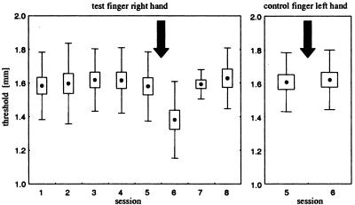
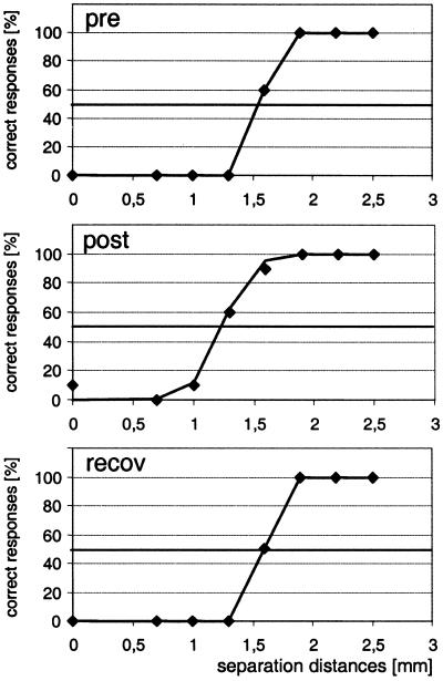
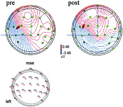
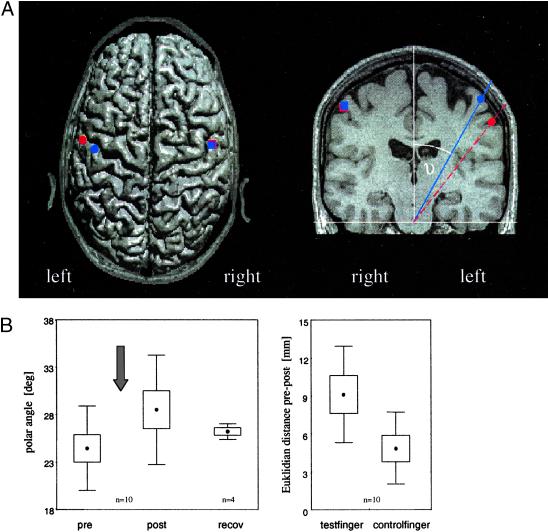
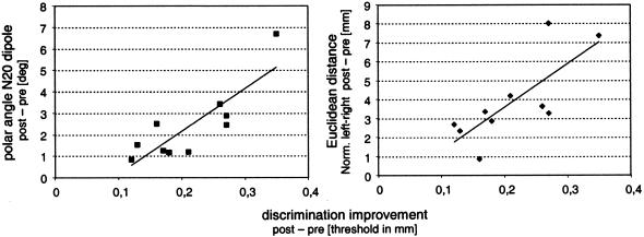
We report experiments combining assessment of spatial tactile discrimination behavior and measurements of somatosensory-evoked potentials in human subjects before and after short-term plastic changes to demonstrate a causal link between the degree of altered performance and reorganization. Plastic changes were induced by a Hebbian coactivation protocol of simultaneous pairing of tactile stimuli. As a result of coactivation, spatial discrimination thresholds were lowered; however, the amount of discrimination improvement was variable across subjects. Analysis of somatosensory-evoked potentials revealed a significant, but also variable shift in the localization of the N20-dipole of the index finger that was coactivated. The Euclidean distance between the dipole pre- and post-coactivation was significantly larger on the coactivated side (mean 9.13 +/- 3.4 mm) than on the control side (mean 4.90 +/- 2.7 mm, P = 0.008). Changes of polar angles indicated a lateral and inferior shift on the postcentral gyrus of the left hemisphere representing the coactivated index finger. To explore how far the variability of improvement was reflected in the degree of reorganization, we correlated the perceptual changes with the N20-dipole shifts. We found that the changes in discrimination abilities could be predicted from the changes in dipole localization. Little gain in spatial discrimination was associated with small changes in dipole shifts. In contrast, subjects who showed a large cortical reorganization also had lowest thresholds. All changes were highly selective as no transfer to the index finger of the opposite, non-coactivated hand was found. Our results indicate that human spatial discrimination performance is subject to improvement on a short time scale by a Hebbian stimulation protocol without invoking training, attention, or reinforcement. Plastic processes related to the improvement were localized in primary somatosensory cortex and were scaled with the degree of the individual perceptual improvement.
Figures
Similar articles
-
Sustained increase of somatosensory cortex excitability by tactile coactivation studied by paired median nerve stimulation in humans correlates with perceptual gain.J Physiol. 2007 Oct 15;584(Pt 2):463-71. doi: 10.1113/jphysiol.2007.140079. Epub 2007 Aug 16. J Physiol. 2007. PMID: 17702814 Free PMC article.
-
Pharmacological modulation of perceptual learning and associated cortical reorganization.Science. 2003 Jul 4;301(5629):91-4. doi: 10.1126/science.1085423. Science. 2003. PMID: 12843392 Clinical Trial.
-
Impaired tactile acuity in old age is accompanied by enlarged hand representations in somatosensory cortex.Cereb Cortex. 2009 Jul;19(7):1530-8. doi: 10.1093/cercor/bhn190. Epub 2008 Nov 13. Cereb Cortex. 2009. PMID: 19008462
-
Pharmacological suppression of plastic changes in human primary somatosensory cortex after motor learning.Exp Brain Res. 2003 Feb;148(4):525-32. doi: 10.1007/s00221-002-1324-1. Epub 2002 Dec 11. Exp Brain Res. 2003. PMID: 12582838 Clinical Trial.
-
Short-term functional plasticity of cortical and thalamic sensory representations and its implication for information processing.Adv Neurol. 1997;73:159-78. Adv Neurol. 1997. PMID: 8959213 Review.
Cited by
-
20 Hz Steady-State Response in Somatosensory Cortex During Induction of Tactile Perceptual Learning Through LTP-Like Sensory Stimulation.Front Hum Neurosci. 2020 Jun 30;14:257. doi: 10.3389/fnhum.2020.00257. eCollection 2020. Front Hum Neurosci. 2020. PMID: 32694988 Free PMC article.
-
Increased excitability of somatosensory cortex in aged humans is associated with impaired tactile acuity.J Neurosci. 2012 Feb 1;32(5):1811-6. doi: 10.1523/JNEUROSCI.2722-11.2012. J Neurosci. 2012. PMID: 22302820 Free PMC article.
-
Differential effects of synchronous and asynchronous multifinger coactivation on human tactile performance.BMC Neurosci. 2007 Jul 30;8:58. doi: 10.1186/1471-2202-8-58. BMC Neurosci. 2007. PMID: 17663778 Free PMC article.
-
Cortical excitability in human somatosensory and visual cortex: implications for plasticity and learning - a minireview.Front Hum Neurosci. 2023 Aug 17;17:1235487. doi: 10.3389/fnhum.2023.1235487. eCollection 2023. Front Hum Neurosci. 2023. PMID: 37662638 Free PMC article. Review.
-
Effects of combining 2 weeks of passive sensory stimulation with active hand motor training in healthy adults.PLoS One. 2014 Jan 9;9(1):e84402. doi: 10.1371/journal.pone.0084402. eCollection 2014. PLoS One. 2014. PMID: 24416229 Free PMC article. Clinical Trial.
References
-
- Cohen L G, Brasil N, Pascual-Leone A, Hallett M. Adv Neurol. 1993;63:187–200. - PubMed
-
- Pascual-Leone A, Torres F. Brain. 1993;116:39–52. - PubMed
-
- Elbert T, Pantev C, Wienbruch C, Rockstroh B, Taub E. Science. 1995;270:305–307. - PubMed
-
- Pantev C, Oostenveld R, Engelien A, Ross B, Roberts L E, Hoke M. Nature (London) 1998;392:811–814. - PubMed
-
- Sterr A, Muller M M, Elbert T, Rockstroh B, Pantev C, Taub E. Nature (London) 1998;391:134–135. - PubMed
Publication types
MeSH terms
LinkOut - more resources
Full Text Sources
Research Materials
