. 2001 Oct 9;98(21):12283-8.
doi: 10.1073/pnas.211086598. Epub 2001 Oct 2.

The in vivo role of p38 MAP kinases in cardiac remodeling and restrictive cardiomyopathy

Affiliations

The in vivo role of p38 MAP kinases in cardiac remodeling and restrictive cardiomyopathy

P Liao et al. Proc Natl Acad Sci U S A. .

Abstract

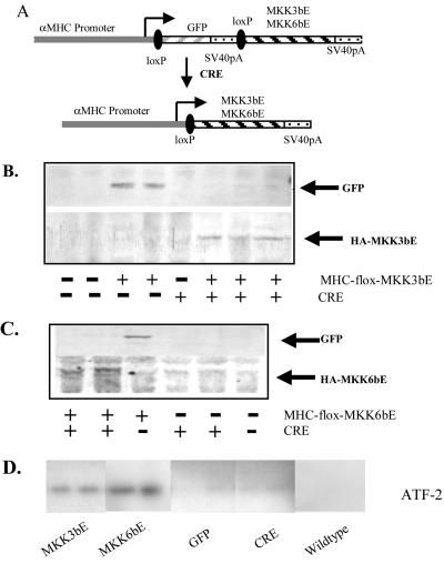
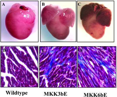
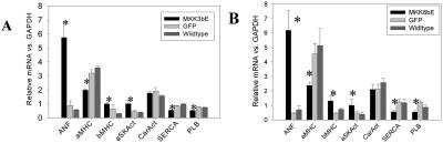
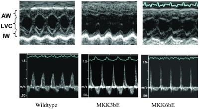
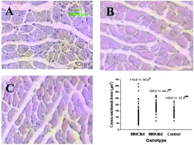
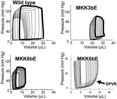
Stress-induced mitogen-activated protein kinase (MAP) p38 is activated in various forms of heart failure, yet its effects on the intact heart remain to be established. Targeted activation of p38 MAP kinase in ventricular myocytes was achieved in vivo by using a gene-switch transgenic strategy with activated mutants of upstream kinases MKK3bE and MKK6bE. Transgene expression resulted in significant induction of p38 kinase activity and premature death at 7-9 weeks. Both groups of transgenic hearts exhibited marked interstitial fibrosis and expression of fetal marker genes characteristic of cardiac failure, but no significant hypertrophy at the organ level. Echocardiographic and pressure-volume analyses revealed a similar extent of systolic contractile depression and restrictive diastolic abnormalities related to markedly increased passive chamber stiffness. However, MKK3bE-expressing hearts had increased end-systolic chamber volumes and a thinned ventricular wall, associated with heterogeneous myocyte atrophy, whereas MKK6bE hearts had reduced end-diastolic ventricular cavity size, a modest increase in myocyte size, and no significant myocyte atrophy. These data provide in vivo evidence for a negative inotropic and restrictive diastolic effect from p38 MAP kinase activation in ventricular myocytes and reveal specific roles of p38 pathway in the development of ventricular end-systolic remodeling.


Figures

Figure 1
Figure 1
Targeted expression of MKK3bE and MKK6bE in ventricular myocytes in vivo. (A) A schematic drawing of the transgene constructs used in the generation of floxed GFP/MKK3bE and floxed GFP/Mkk6bE transgenic mice (Upper) and resulting structure on cre/loxP-mediated DNA recombination in ventricular muscle cells after crossing with MLC-2v/Cre mice. (B and C) Representative Western blotting results for the expression of GFP, HA-MKK3bE/HA-MKK6bE recombinant proteins in mouse ventricles. The genotypes of the transgenic mice and their littermates are indicated at the bottom. −/− represents wild type; +/− and −/+ represent GFP-expressing MHC-flox-MKK3bE or MKK6bE- and CRE-expressing MLC-2v/cre single transgenic mice; +/+ represents MKK3bE- or MKK6bE-expressing double transgenic mice. (D) Representative autoradiograms of p38 kinase assays are presented. All samples were prepared from mice at similar age between 6 and 8 weeks.
Figure 2
Figure 2
Gross morphology of transgenic and wild-type control hearts (AC) with indicated genotypes taken under the same magnification using a stereo dissecting microscope. Noting the enlarged atria in MKK3bE- and MKK6bE-expressing hearts but no significant changes in external sizes of the ventricles. (DF) Trichrome staining of transgenic hearts with different genotypes as indicated. The fibrotic tissue is illustrated in dark blue. Noting the patchy staining pattern in both MKK3bE- and MKK6bE-expressing hearts, but absent in the wild-type control heart. (Magnifications: ×120.)
Figure 3
Figure 3
Relative expression levels of cardiac marker genes measured by a dot blot method. The values are means of 4–6 samples from each genotype group and error bars represents standard errors. ANF, atrial natriuretic factor; aMHC, α-MHC; bMHC, β-MHC; aSKAct, α-skeletal actin; CarAct, cardiac actin; SERCA, sarcoplasmic reticular calcium ATPase-2a; PLB, phospholamban. *, P < 0.05 compared with wild-type or GFP control littermates.
Figure 4
Figure 4
Echocardiographic evaluation of cardiac structure and function in wild-type and transgenic mice. (Upper) Representative two-dimensional guided M-mode images of the LV demonstrate minimally increased relative wall thickness and minimally reduced fractional shortening in MKK6bE transgenic mice (both statistically nonsignificant) and significantly reduced relative wall thickness and fractional shortening in MKK3bE transgenic mice compared to wild-type control littermates. Of note, the steeper slope of endocardial motion during early diastole indicates an increased rate of wall thinning and LV chamber expansion in transgenic mice. AW, anterior wall; IW, inferior wall; LVC, left ventricular cavity. (Lower) Trans-mitral Doppler recordings show increased velocity and shortened deceleration time of early left ventricular filling and decreased isovolumic relaxation time in transgenic mice consistent with “restrictive” filling pattern and impaired left ventricular compliance.
Figure 5
Figure 5
Representative hematoxylin/eosin cross sections of LVs from MKK3bE (A), MKK6bE (B), and wild-type control (C) hearts. The cross-sectional areas (D) from individual myofilaments were complied with mean value ± SD for each group labeled at the top. *, n = 198; P < 0.02 vs. control; P < 0.0001 vs. MKK6bE. **, n = 102; P < 0.001 vs. control. ***, n = 100. Note the heterogeneous myocyte filament sizes in both hematoxylin/eosin section (A) and area measurement (D) from MKK3bE heart.
Figure 6
Figure 6
Representative pressure-volume loops during basal (thick-line loop) and reduction of pre-load by vena cava occlusion (thin-line loops). MKK3bE-expressing hearts displayed a rightward shift of the end-systolic pressure-volume relation, whereas MKK6bE expressing hearts displayed a significantly leftward shift of the diastolic pressure-volume relation as compared with wild-type controls. (Lower Right) Reproduction of the diastolic relations on an expanded scale, demonstrating the marked increase in chamber passive stiffness measured in the MKK-expressing mutants. This example is from an MKK6bE animal, but very similar stiffening was measured in MKK3bE mice.

Similar articles

Cited by

References

    1. Cohn J N, Bristow M R, Chien K R, Colucci W S, Frazier O H, Leinwand L A, Lorell B H, Moss A J, Sonnenblick E H, Walsh R A, et al. Circulation. 1997;95:766–770. - PubMed
    1. Grossman W. Am J Med. 1980;69:576–584. - PubMed
    1. Chien K R, Grace A A. In: Heart Disease. Braunwald E, editor. Philadelphia: Saunders; 1997. pp. 1626–1649.
    1. Parmley W W. Clin Cardiol. 1992;15, Suppl. 1:I5–I12. - PubMed
    1. Chien K R. Cell. 1999;98:555–558. - PubMed

Publication types

MeSH terms

Substances