. 2001 Oct;13(10):2257-68.
doi: 10.1105/tpc.010185.

Plastid division is driven by a complex mechanism that involves differential transition of the bacterial and eukaryotic division rings

Affiliations

Plastid division is driven by a complex mechanism that involves differential transition of the bacterial and eukaryotic division rings

Miyagishima Sy et al. Plant Cell. 2001 Oct.

Abstract

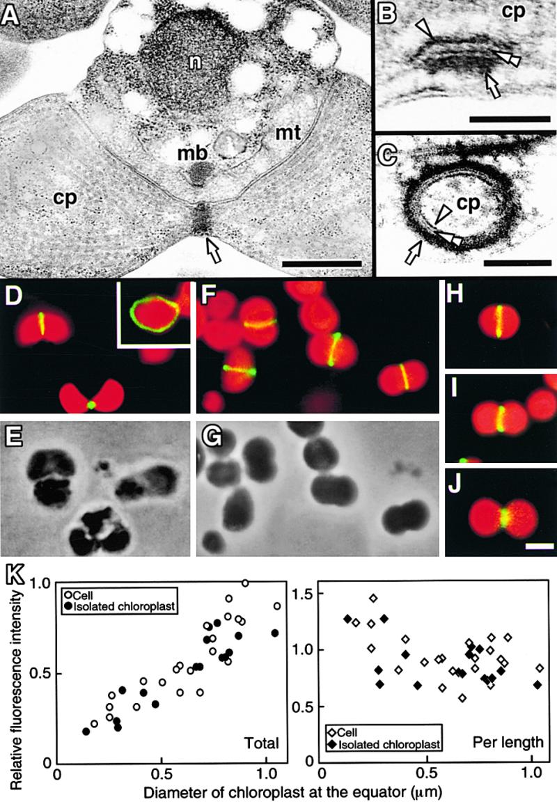
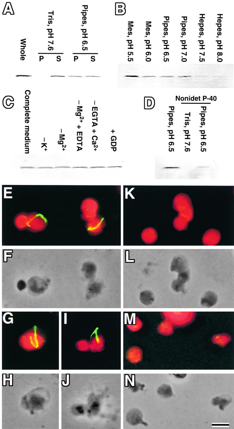
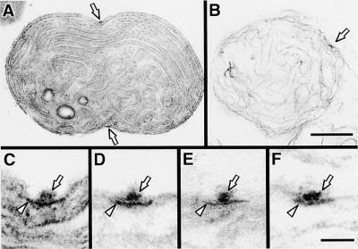
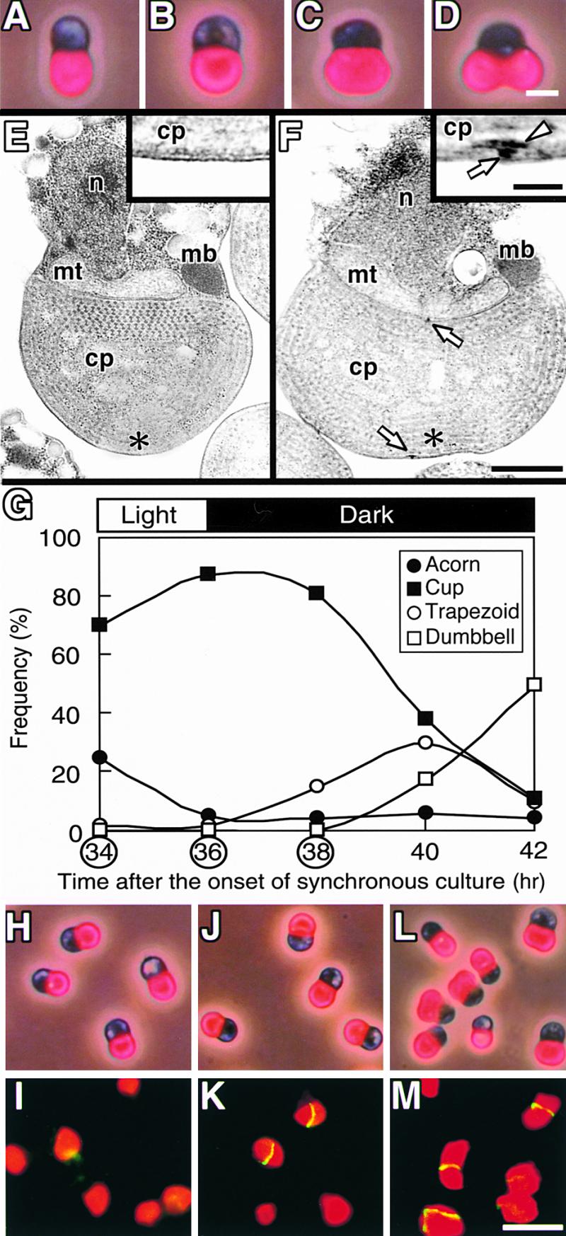
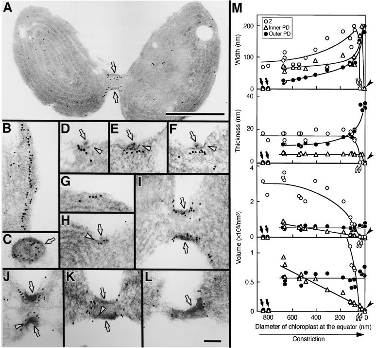
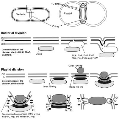
During plastid division, two structures have been detected at the division site in separate analyses. The plastid-dividing ring can be detected by transmission electron microscopy as two (or three) electron-dense rings: an outer ring on the cytosolic face of the outer envelope, occasionally a middle ring in the intermembrane space, and an inner ring on the stromal face of the inner envelope. The FtsZ ring, which plays a central role in bacterial division, also is involved in plastid division and is believed to have descended to plastids from cyanobacterial endosymbiosis. The relationship between the two structures is not known, although there is discussion regarding whether they are identical. Biochemical and immunocytochemical investigations, using synchronized chloroplasts of the red alga Cyanidioschyzon merolae, showed that the plastid FtsZ ring is distinct and separable from the plastid-dividing ring. The FtsZ ring localizes in stroma and faces the inner plastid-dividing ring at the far side from the inner envelope. The FtsZ ring and the inner and outer plastid-dividing rings form in that order before plastid division. The FtsZ ring disappears at the late stage of constriction before dissociation of the plastid-dividing ring, when the constriction is still in progress. Our results suggest that the FtsZ ring;-based system, which originated from a plastid ancestor, cyanobacteria, and the plastid-dividing ring;-based system, which probably originated from host eukaryotic cells, form a complex and are involved in plastid division by distinct modes.


Figures

Figure 1.
Figure 1.
Micrographs of the PD and Z Rings in C. merolae. (A) to (C) Electron micrographs of a dividing cell in which the PD ring is cut tangentially (A). Magnified cross-sectional (B) and circumferential (C) views of the PD ring show that it consists of three rings. (D) and (E) Immunofluorescence image of Z rings in dividing cells (D) and a phase-contrast image of the same field (E). The inset shows a circumferential image of the Z ring. Green fluorescence indicates the localization of FtsZ, and red indicates the autofluorescence of chlorophyll on the thylakoid. (F) and (G) Immunofluorescence image of Z rings in dividing chloroplasts isolated from a synchronous culture (F) and a phase-contrast image of the same field (G). (H) to (J) The Z ring in isolated chloroplasts at early (H), middle (I), and late stages of division (J). (K) Relative fluorescence intensity of fluorescein isothiocyanate labeling of the Z ring in whole cells (open circles and open diamonds) and isolated chloroplasts (closed circles and closed diamonds) plotted versus diameter of the chloroplast at the equator. The total intensity of the Z ring (left; open circles and closed circles) was converted to intensity per 1 μm (right; open diamonds and closed diamonds). Arrows, arrowheads, and double arrowheads indicate the outer, inner, and middle PD rings, respectively. cp, chloroplast; mb, microbody; mt, mitochondrion; n, nucleus. Bar in (A) = 500 nm; bar in (B) = 50 nm; bar in (C) = 100 nm; bar in (J) = 1 μm for (D) to (J).
Figure 2.
Figure 2.
Effect of Several Factors on the Stability of the Z Ring. (A) to (D) Isolated chloroplasts were burst by osmotic shock in hypotonic media and centrifuged, and FtsZ was detected by immunoblotting. (A) Sucrose-depleted chloroplast isolation medium (pH 7.6) and the same medium in which Tris (pH 7.6) was replaced with Pipes (pH 6.5) were used, and the pellet (P) and supernatant (S) were analyzed. Untreated isolated chloroplasts also were analyzed (whole). (B) Media buffered at various pH values were used, and the pellets were analyzed. (C) Chemicals were depleted from (−) or added to (+) medium containing Pipes (pH 6.5), KCl, MgCl2, and EGTA (complete medium), and the pellets were analyzed. (D) Media buffered with Pipes (pH 6.5) or Tris (pH 7.6) with or without 0.1% Nonidet P-40 were used, and the pellets were analyzed. (E) to (N) Immunofluorescence ([E], [G], [I], [K], and [M]) and phase contrast images ([F], [H], [J], [L], and [N]) of burst chloroplasts at pH 6.5 ([E] to [J]), pH 7.5 ([K] and [L]), and pH 5.5 ([M] and [N]) detecting the Z ring. (F), (H), (J), (L), and (N) are the same fields of view as (E), (G), (I), (K), and (M), respectively. Bar in (N) = 1 μm for (E) to (N).
Figure 3.
Figure 3.
Effect of pH on the Stability of the PD Ring. (A) and (B) Whole electron microscopic image of an isolated chloroplast before (A) and after (B) being burst in medium at pH 7.6. (C) to (F) Magnified cross-sectional image of the PD ring before (C) and after being burst in medium at pH 5.5 (D), 6.5 (E), or 7.5 (F). Arrows and arrowheads indicate the outer and inner PD rings, respectively. Bar in (B) = 500 nm for (A) and (B); bar in (F) = 50 nm for (C) to (F).
Figure 4.
Figure 4.
Timing of the Formation of the Z and PD Rings in Synchronous Culture. (A) to (D) Autofluorescence of chloroplast/phase–contrast images showing the transition of chloroplast shape in C. merolae in the order acorn (A), cup (B), trapezoid (C), and dumbbell (D). (E) and (F) Electron micrographs of cells with cup-shaped (E) or trapezoidal chloroplasts (F). Magnified images of the regions indicated by asterisks are shown in the insets. Arrows and the arrowhead indicate the outer and inner PD rings, respectively. cp, chloroplast; mb, microbody; mt, mitochondrion; n, nucleus. (G) Change in the frequency of cells containing acorn-shaped (closed circles), cup-shaped (closed squares), trapezoidal (open cir-
Figure 5.
Figure 5.
Immunoelectron Micrographs Showing the Localization and Change in Shape of the Z and PD Rings during Contraction. FtsZ was labeled with gold particles. (A) Whole image of an isolated dividing chloroplast. (B) and (C) Sections cut along the chloroplast division site in the early (B) and late (C) stages of division. (D) to (F) Sections cut perpendicular to the PD ring, in which the outer and inner PD rings are resolved into two distinct rings. (G) to (L) Sections showing sequential change in the PD ring and the label for the FtsZ protein. (M) Change in the width, thickness, and deduced volume of the region of gold particles labeling the Z ring (open circles), inner PD ring (open triangles), and outer PD ring (closed circles). The stage of division progresses from left to right. Because the diameter of the chloroplast at the equator decreases linearly with time (Miyagishima et al., 1999a), the x axis provides a relative time axis. Arrows and arrowheads in (A) to (L) indicate the outer and inner PD rings, respectively. In (M), closed arrows show points at which the Z ring has formed but the inner and outer PD rings have not yet formed, as in (G); open arrows show points at which the Z ring was absent, unlike the inner and outer PD rings, as in (K); closed arrowheads show points at which only the outer ring was present, as in (L). Bar in (A) = 500 nm; bar in (L) = 100 nm for (B) to (L).
Figure 6.
Figure 6.
Comparison of the Division Machinery between Bacteria and Plastids and a Model of the Machinery of Plastid Division. To illustrate the bacterial division machinery, the division complex characterized in E. coli is used. Of these proteins, MinC, MinD, MinE, FtsZ, and FtsI are found in the genome sequence of the cyanobacterium Synechocystis PCC6803. In bacterial division, MinC, MinD, and MinE determine the division site and FtsZ is first recruited at the expected division site. Other components of the division complex (ZipA, FtsA, FtsK, FtsQ, FtsL, FtsI, FtsN, and FtsW) are then recruited in a Z ring–dependent manner, and septum formation starts. In plastid division, at least MinD is involved in determining the division site. After the formation of the Z ring, the inner PD ring forms between the Z ring and the inner envelope. Finally, the outer PD ring (and the middle PD ring in C. merolae) forms and constriction starts. The Z and inner (and middle) PD rings decompose as they contract, whereas the outer PD ring contracts without loss of components. At the late stage of division, the Z and inner (and middle) PD rings disappear in that order. Finally, the outer PD ring pinches off the plastid and remains in the cytosol. CM, cytoplasmic membrane; IE, inner envelope; OE, outer envelope; OM, outer membrane; PG, peptide glycan.

Similar articles

Cited by

References

    1. Beech, P.L., and Gilson, P.R. (2000). FtsZ and organelle division in protists. Protist 151, 11–16. - PubMed
    1. Beech, P.L., Nheu, T., Schultz, T., Herbert, S., Lithgow, T., Gilson, P.R., and McFadden, G.I. (2000). Mitochondrial FtsZ in a chromophyte alga. Science 287, 1276–1279. - PubMed
    1. Bleazard, W., McCaffery, J.M., King, E.J., Bale, S., Mozdy, A., Tieu, Q., Nunnari, J., and Shaw, J.M. (1999). The dynamin-related GTPase Dnm1 regulates mitochondrial fission in yeast. Nat. Cell Biol. 1, 298–304. - PMC - PubMed
    1. Bramhill, D. (1997). Bacterial cell division. Annu. Rev. Cell Dev. Biol. 13, 395–424. - PubMed
    1. Bramhill, D., and Thompson, C.M. (1994). GTP-dependent polymerization of Escherichia coli FtsZ protein to form tubules. Proc. Natl. Acad. Sci. USA 91, 5813–5817. - PMC - PubMed

Publication types

MeSH terms