. 2001 Oct 11;413(6856):603-9.
doi: 10.1038/35098027.

A sperm ion channel required for sperm motility and male fertility

Affiliations

A sperm ion channel required for sperm motility and male fertility

D Ren et al. Nature. .

Abstract

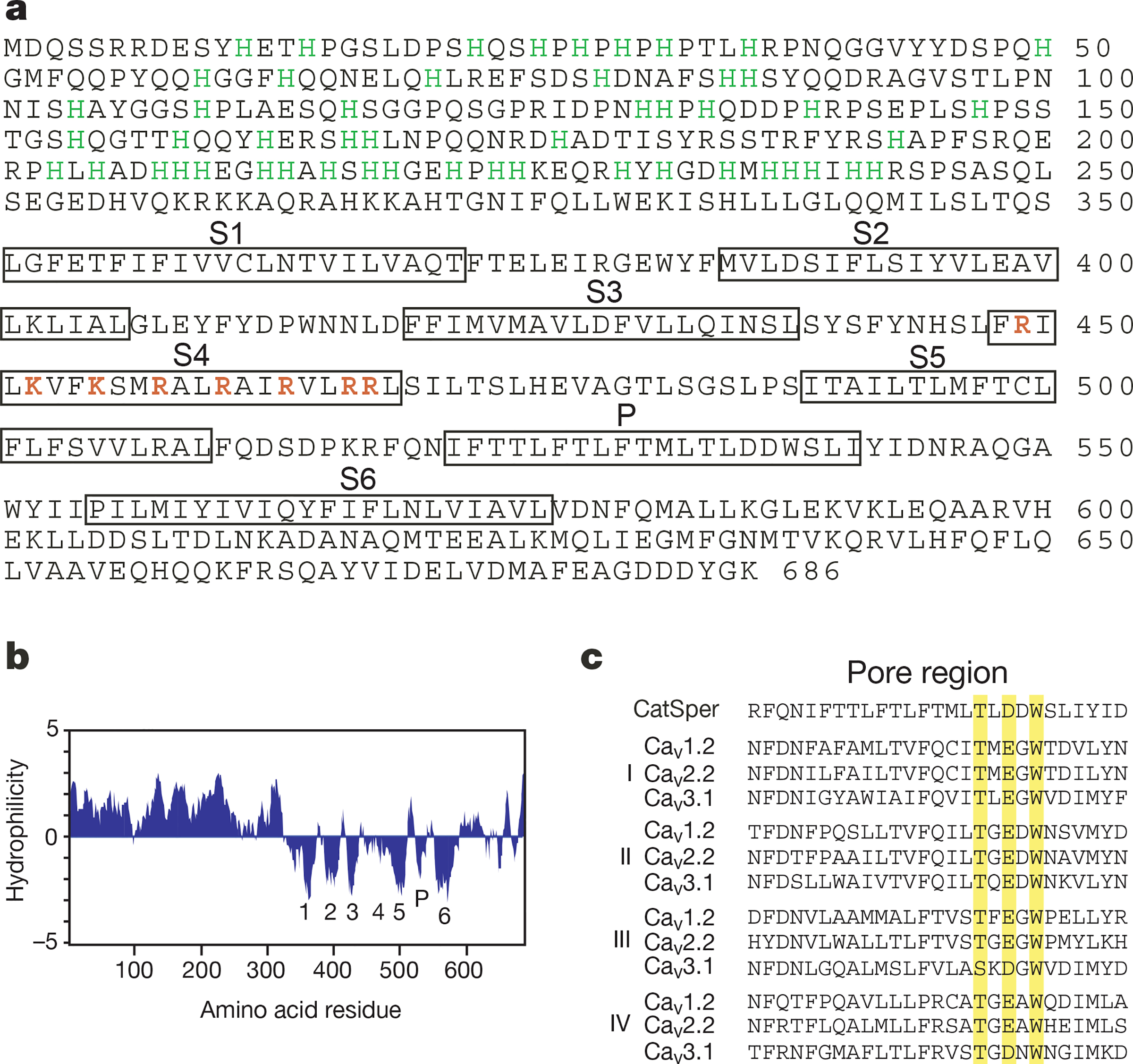
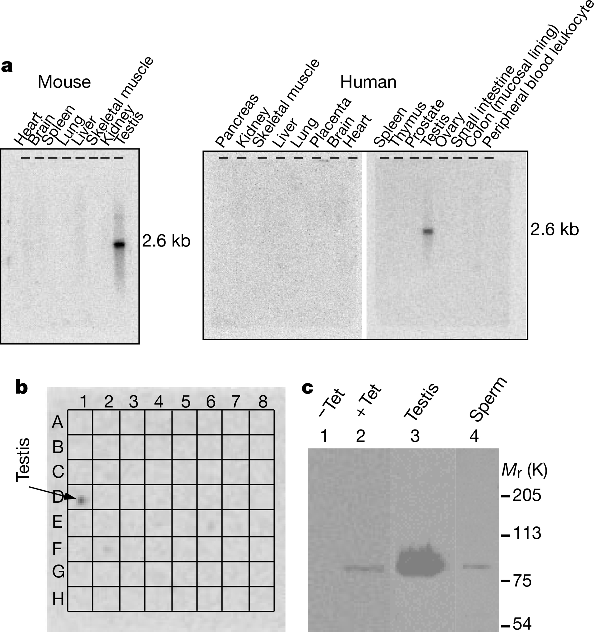
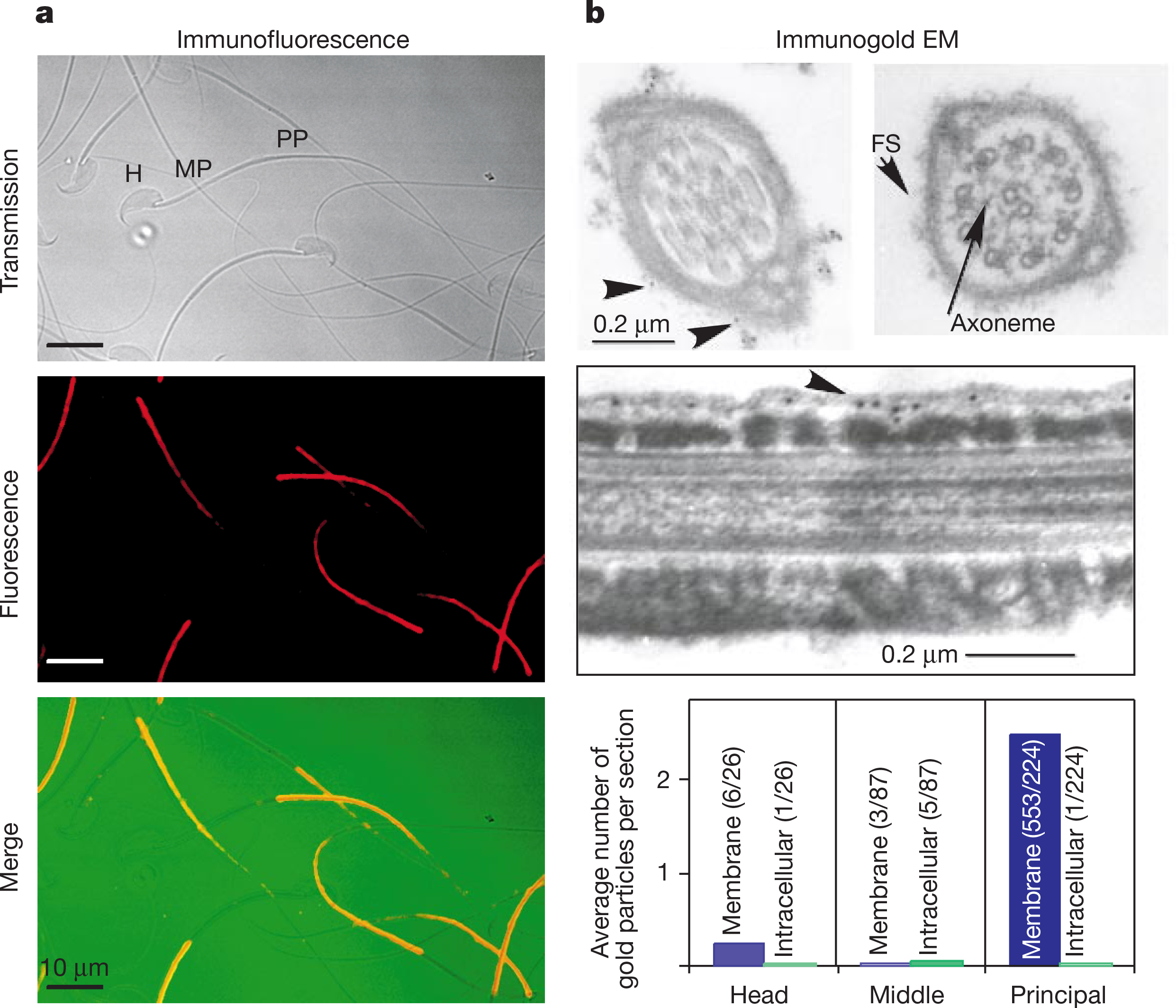
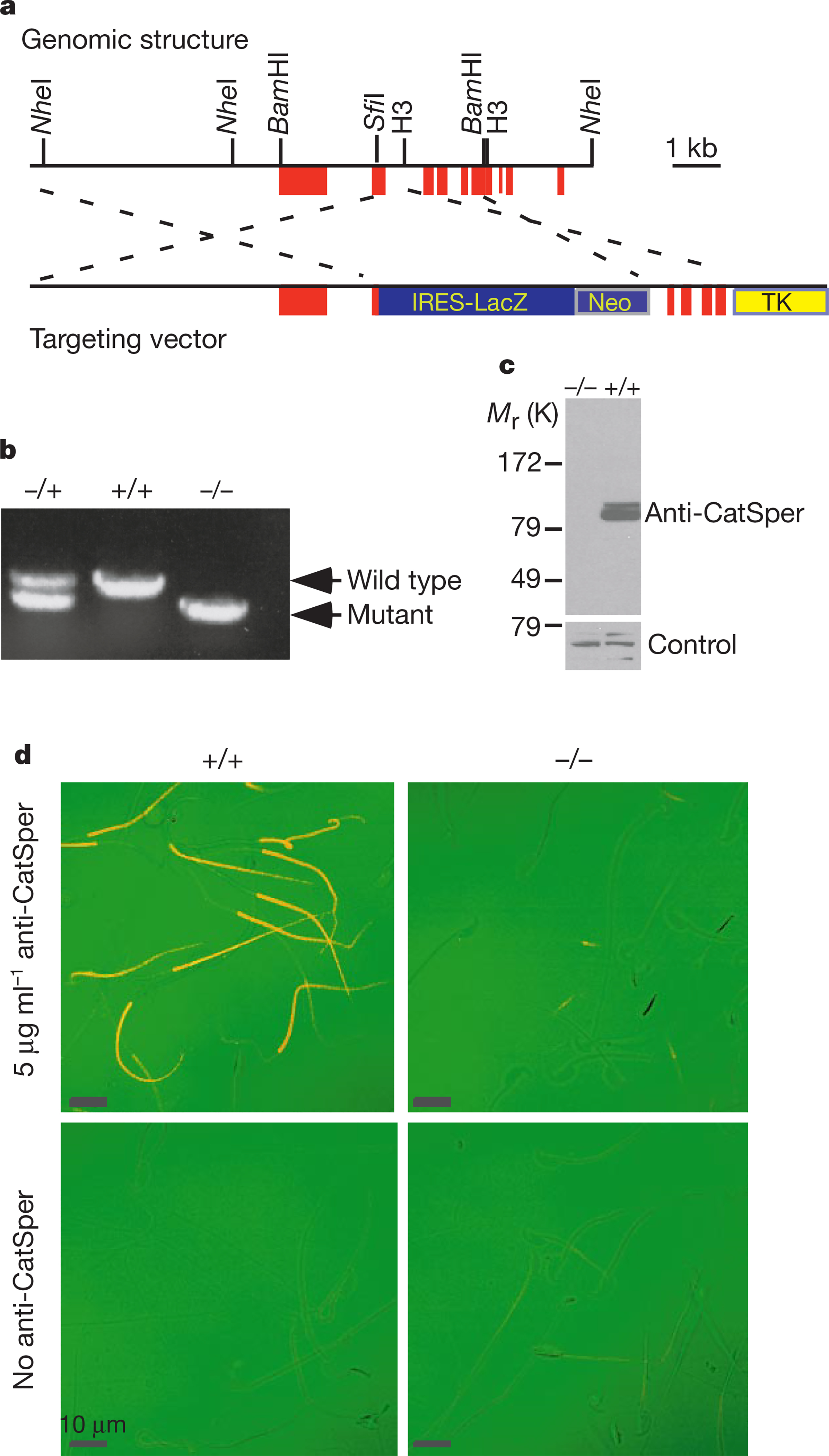
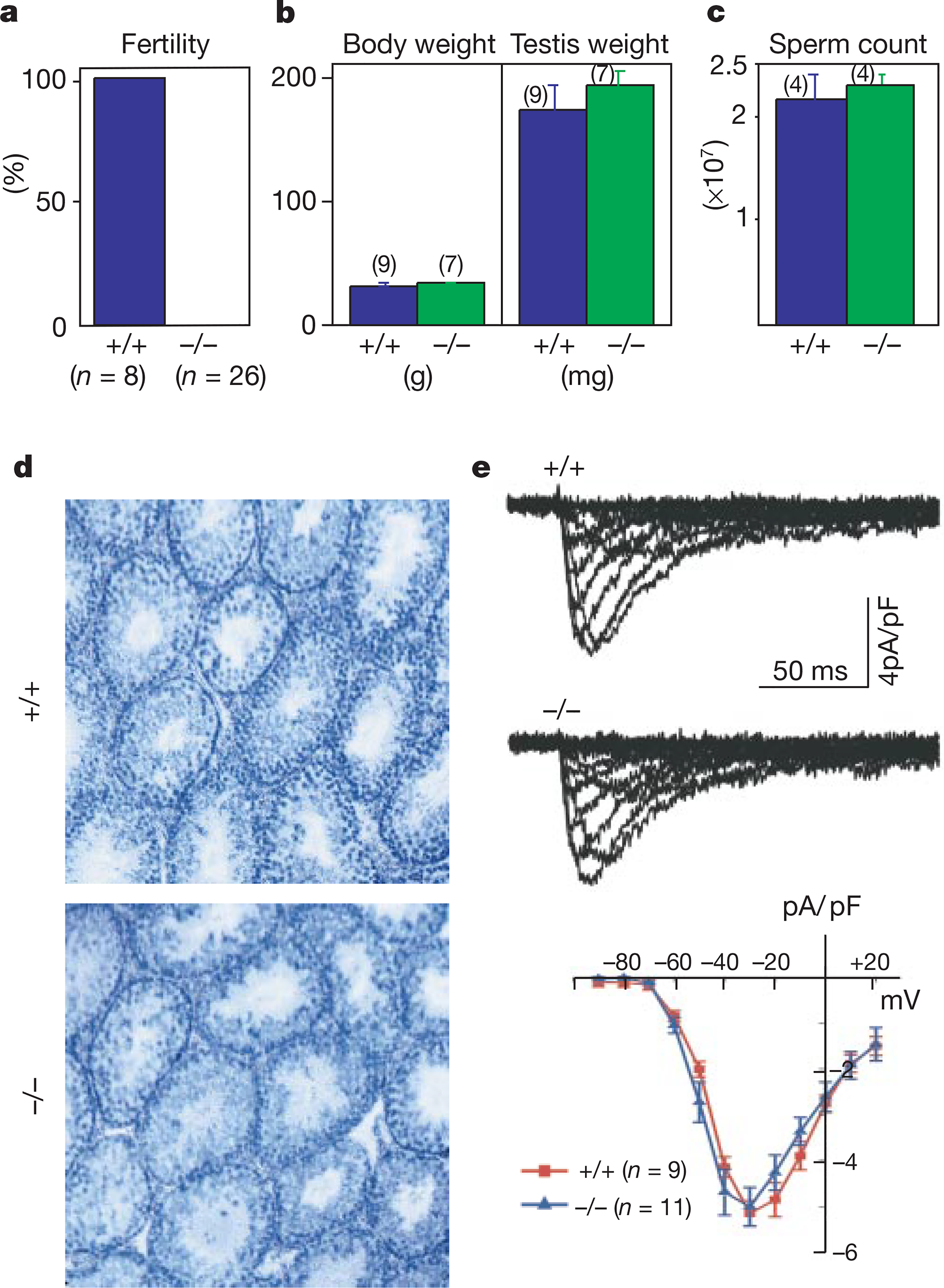
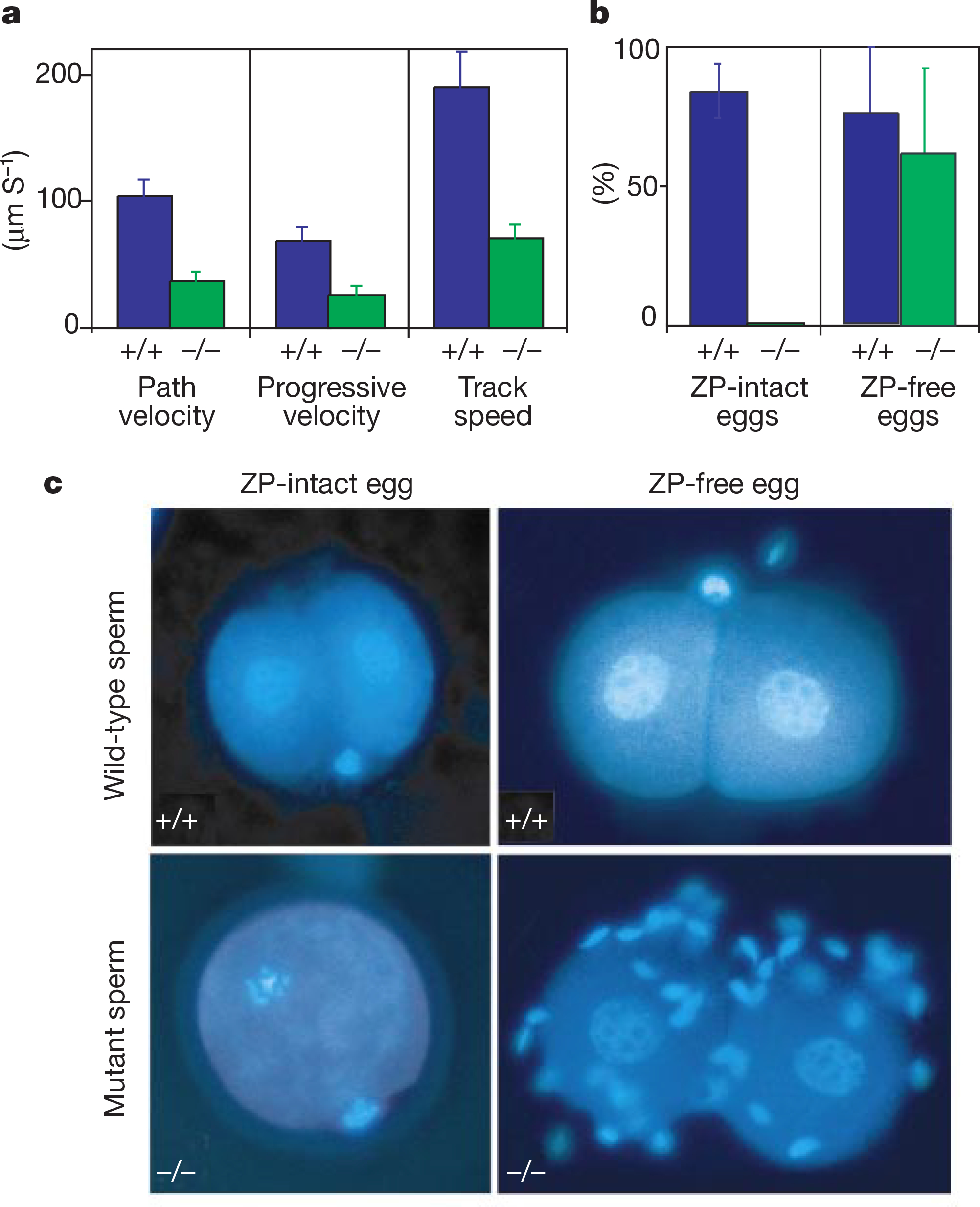
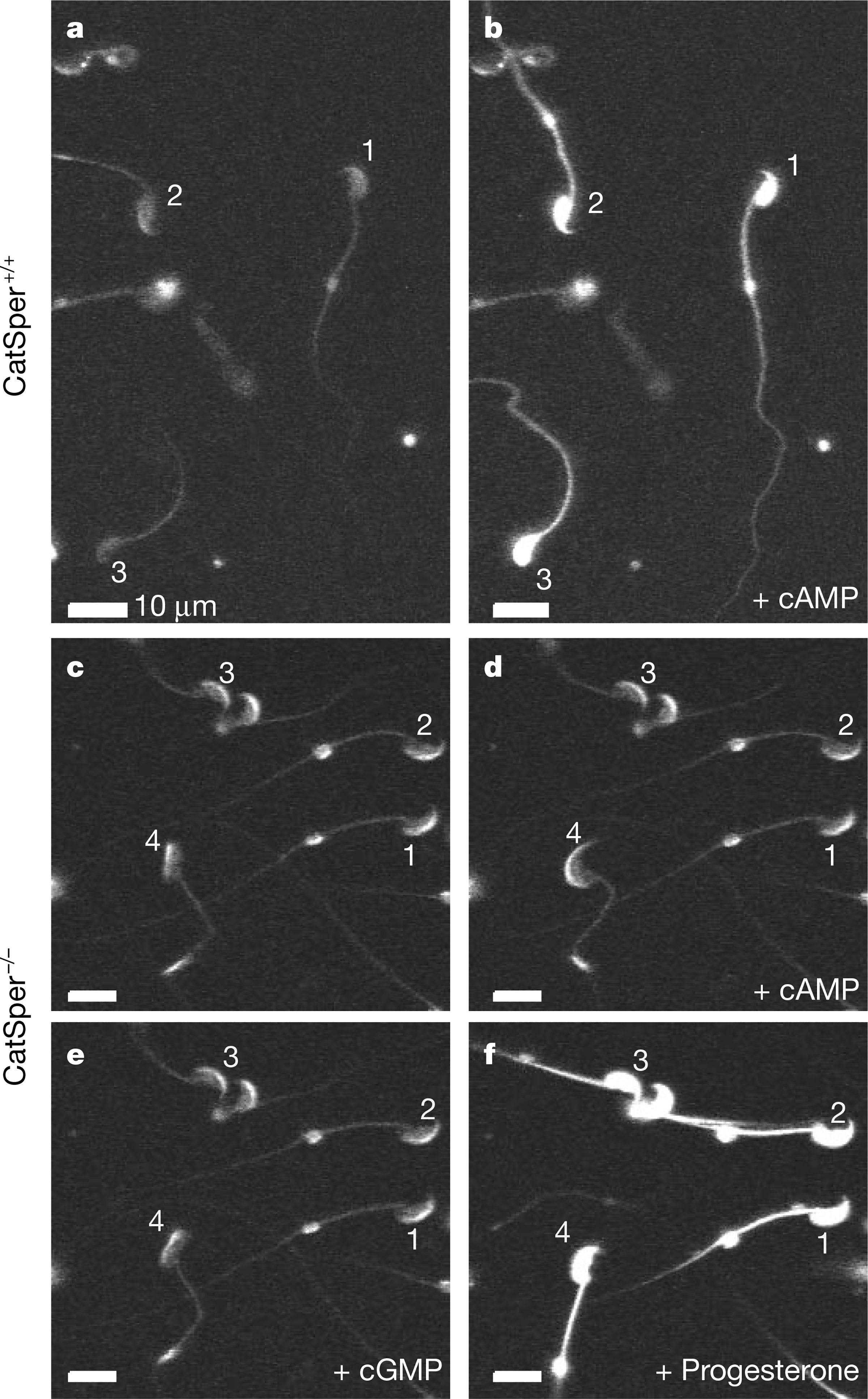
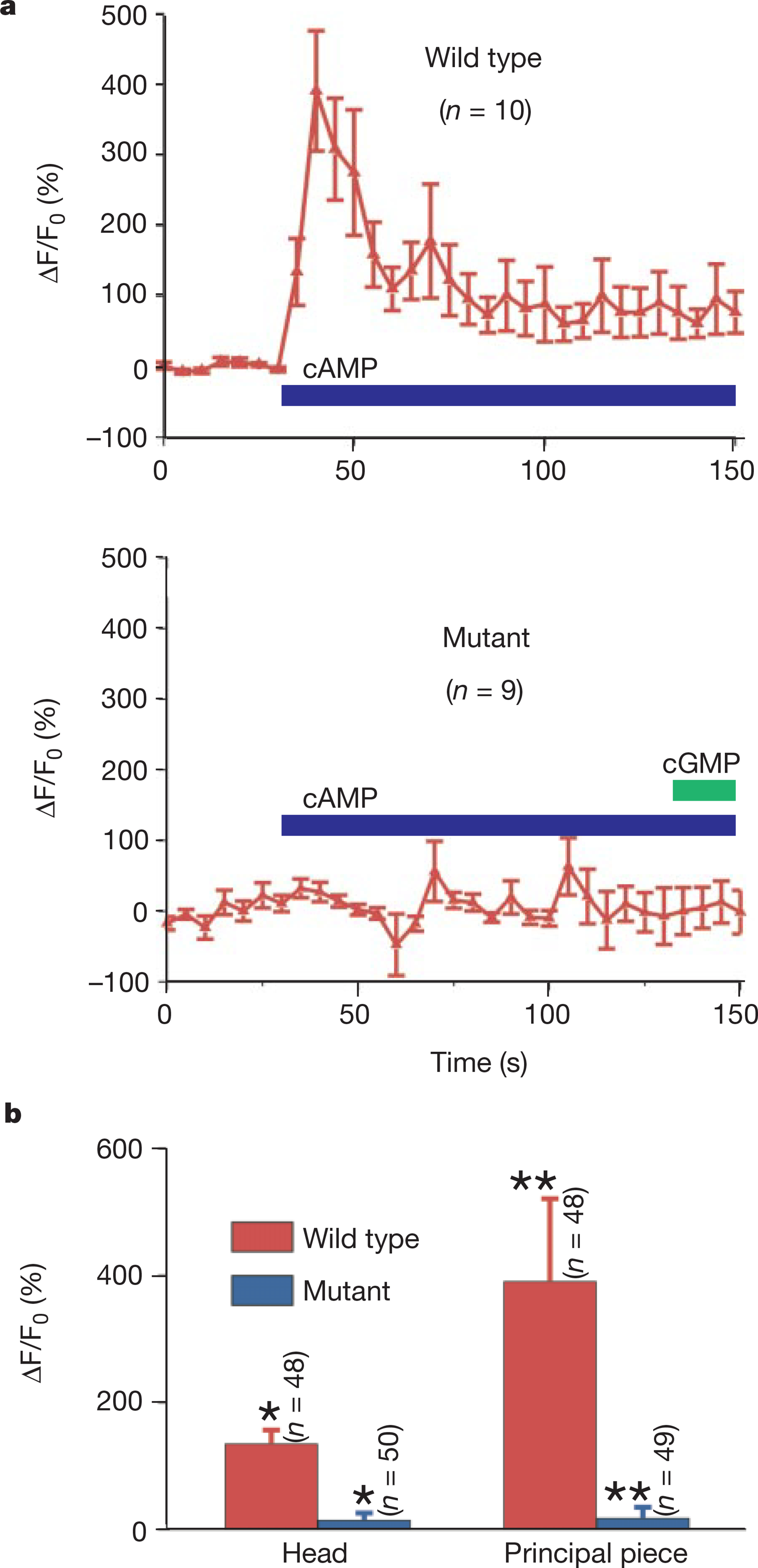
Calcium and cyclic nucleotides have crucial roles in mammalian fertilization, but the molecules comprising the Ca2+-permeation pathway in sperm motility are poorly understood. Here we describe a putative sperm cation channel, CatSper, whose amino-acid sequence most closely resembles a single, six-transmembrane-spanning repeat of the voltage-dependent Ca2+-channel four-repeat structure. CatSper is located specifically in the principal piece of the sperm tail. Targeted disruption of the gene results in male sterility in otherwise normal mice. Sperm motility is decreased markedly in CatSper-/- mice, and CatSper-/- sperm are unable to fertilize intact eggs. In addition, the cyclic-AMP-induced Ca2+ influx is abolished in the sperm of mutant mice. CatSper is thus vital to cAMP-mediated Ca2+ influx in sperm, sperm motility and fertilization. CatSper represents an excellent target for non-hormonal contraceptives for both men and women.


Figures

Figure 1
Figure 1
Primary structure of mouse CatSper. a, Amino-acid sequence; the six putative transmembrane domains (S1–S6) and the pore (P) region are boxed. The positively charged amino acids (K/R) in the S4 region are shown in red. Histidines in the first 250 amino acids are labelled in green. b, Hydropathy plot of CatSper predicts six transmembrane domains (1–6) and a P loop (P). Window size is 11. c, Alignment of the putative pore region of CatSper with that of the four domains (I, II, III, IV) from CaV1–3. The conserved residues in the T/S-x-E/D-x-W motif are shaded. GenBank accession numbers: X15539, CaV1.2; M94172, CaV2.2; 054898, CaV3.1; mouse CatSper, AF407332; human CatSper, AF407333.
Figure 2
Figure 2
The restricted expression pattern of CatSper in testis. a, Northern blot from mice (left) and human (right) organs. b, Dot blot of mRNAs from 50 human tissues (grid superimposed). The tissues in the blot are from left to right: row A, whole brain, amygdala, caudate nucleus, cerebellum, cerebral cortex, frontal lobe, hippocampus, medulla oblongata; row B, occipital lobe, putamen, substantia nigra, temporal lobe, thalamus, nucleus accumbens, spinal cord; row C, heart, aorta, skeletal muscle, colon, bladder, uterus, prostate, stomach; row D, testis, ovary, pancreas, pituitary gland, adrenal gland, thyroid gland, salivary gland, mammary gland; row E, kidney, liver, small intestine, spleen, thymus, peripheral leukocyte, lymph node, bone marrow; row F, appendix, lung, trachea, placenta; row G, fetal brain, fetal heart, fetal kidney, fetal liver, fetal spleen, fetal thymus, fetal lung; and row H, negative controls. c, Western blot of CatSper protein. Lanes 1 and 2 were loaded with total protein from HEK-293 cells stably expressing inducible CatSper without (−Tet) and with (+Tet) tetracycline induction. Lanes 3 and 4 were loaded with testis membrane protein and sperm total protein, respectively. Mr, relative molecular mass.
Figure 3
Figure 3
Localization of CatSper to the plasma membrane of the principal piece of sperm. a, Immunostaining of mouse sperm. Top, phase contrast. The head (H), midpiece (MP) and principal piece (PP) regions of the sperm are indicated. Middle, immunofluorescence. Bottom, merged signal where immunofluorescence is labelled red and transmission green. b, Sperm immunogold-labelled electron microscopy. Top left, cross-section through the principal piece; arrows indicate gold particles. Top right, fibrous sheath (FS). Middle, longitudinal section. The immunoreactive gold particles are at the cell membrane cytoplasmic face. Bottom, distribution of gold particles along the sperm head, midpiece and principal piece. Gold particles located outside the fibrous sheath were counted as being membrane localized, and inside the fibrous sheath as intracellular. The numbers of sections counted and particles detected are indicated in parentheses.
Figure 4
Figure 4
Targeted disruption of CatSper. a, Partial genomic structure of mouse CatSper and targeting vector. Filled red boxes, exons; thin lines, introns. b, PCR genotyping confirmed gene disruption. Tail genomic DNA was amplifed with primers specifc for CatSper+/+ and CatSper−/−. c, CatSper protein was absent in mutant mice by western blot. Roughly equal amounts of membrane protein from wild-type and mutant testes were blotted with anti-CatSper antibody. An unrelated antibody controlled for equal protein loading. d, Immunostaining of wild-type (+/+) and mutant (−/−) sperm. Primary antibody was omitted in the control. The transmission signal is green, immunofluorescence is red, and overlap is orange.
Figure 5
Figure 5
Male infertility caused by CatSper disruption. a, Fertility of CatSper+/+ and CatSper−/− males. b–d, Mice body and testis weight (b), sperm count (c), and testis histology (d) were not significantly different. Testes sections were stained with haematoxylin/eosin in d. e, Ca2+-channel currents were indistinguishable between wild-type (+/+) and mutant (−/−) spermatocytes in range of activation, amplitude and kinetics. Currents were elicited by test pulses from −90 to +20 mV (Δ10 mV). Bottom, averaged I/V relationships of inward currents. The holding potential was −100 mV; the average cell capacitances were 10.8 ± 1.4 pF (+/+; n = 9) and 12.9 ± 0.9 pF (−/−; n = 11).
Figure 6
Figure 6
CatSper−/− sperm motility and in vitro fertilization defects. a, Quantified sperm motility: path velocity, progressive velocity and track speed. b, In vitro fertilization (IVF) rates of mutant and the wild-type sperm with zona pellucida (ZP)-intact and ZP-free eggs (three pairs of mutant and wild-type mice each). c, Eggs with and without the ZP were incubated with CatSper+/+ or CatSper−/− sperm. After 24 h, cells were stained with Hoechst 33342 for chromatin analysis. Note the decondensed chromatin in the two nuclei of the individual blastomeres at the two-cell stage.
Figure 7
Figure 7
cAMP-induced calcium influx defect in CatSper−/− sperm. a, b, Fluo-4 fluorescence images of wild-type sperm before (a) and after (b) addition of 1 mM cell-permeant cAMP (8-Br-cAMP). c–f, Incubation in cell-permeant cAMP (1 mM) or cGMP (1 mM) for 10 s did not induce Ca2+ influx in mutant (CatSper−/−) sperm. Progesterone (~1 mM), which was used as a positive control for Ca2+ responses, initiated a large Ca2+ increase in both mutant (f) and control (not shown) sperm. Numbers identify individual spermatozoa.
Figure 8
Figure 8
Summary of cyclic-nucleotide-induced Ca2+ changes. a, Time courses of cAMP-induced Ca2+ changes in the principal pieces of wild-type (CatSper+/+) and mutant (CatSper−/−) sperm. b, Mean fluorescence change in sperm heads and principal pieces from CatSper+/+ and CatSper−/− mice. Fluorescence was normalized to the average of the first seven sampling points of the pre-stimulation trace. **P<0.005; *P<0.001; indicating statistical significance.

Comment in

Similar articles

Cited by

References

    1. Wassarman PM, Jovine L& Litscheer ES Aprofile of fertilization inmammals. Nature Cell Biol. 3, E59–E64 (2001). - PubMed
    1. Yanagimachi R in The Physiology of Reproduction (eds Knobill E & Neill JD ) 189–315 (Raven, New York, 1994).
    1. O’Toole CM, Arnoult C, Darszon A, Steinhardt RA & Florman HM Ca2+ entry through store-operated channels in mouse sperm is initiated by egg ZP3 and drives the acrosome reaction. Mol. Biol. Cell 11, 1571–1584 (2000). - PMC - PubMed
    1. Jungnickel MK,Marrero H, Birnbaumer L, Lemos JR & Florman HM Trp2 regulates entry of Ca2+ into mouse sperm triggered by egg ZP3. Nature Cell Biol. 3, 499–502 (2001). - PubMed
    1. Bedford JM Mammalian fertilization misread? Sperm penetration of the eutherian zona pellucida is unlikely to be a lytic event. Biol. Reprod. 59, 1275–1287 (1998). - PubMed

Publication types

MeSH terms

Associated data