Comparative Study
. 2001;2(10):RESEARCH0044.
doi: 10.1186/gb-2001-2-10-research0044. Epub 2001 Sep 19.

The ESAT-6 gene cluster of Mycobacterium tuberculosis and other high G+C Gram-positive bacteria

Affiliations
Comparative Study

The ESAT-6 gene cluster of Mycobacterium tuberculosis and other high G+C Gram-positive bacteria

N C Gey Van Pittius et al. Genome Biol. 2001.

Abstract

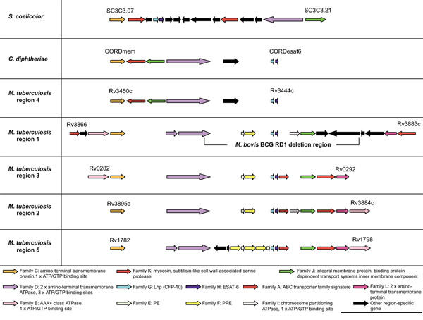
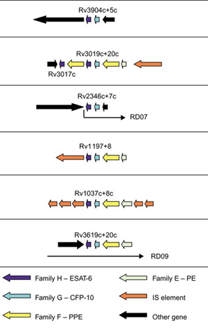
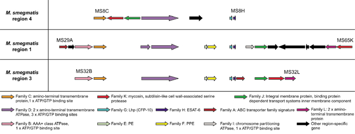
Background: The genome of Mycobacterium tuberculosis H37Rv has five copies of a cluster of genes known as the ESAT-6 loci. These clusters contain members of the CFP-10 (lhp) and ESAT-6 (esat-6) gene families (encoding secreted T-cell antigens that lack detectable secretion signals) as well as genes encoding secreted, cell-wall-associated subtilisin-like serine proteases, putative ABC transporters, ATP-binding proteins and other membrane-associated proteins. These membrane-associated and energy-providing proteins may function to secrete members of the ESAT-6 and CFP-10 protein families, and the proteases may be involved in processing the secreted peptide.

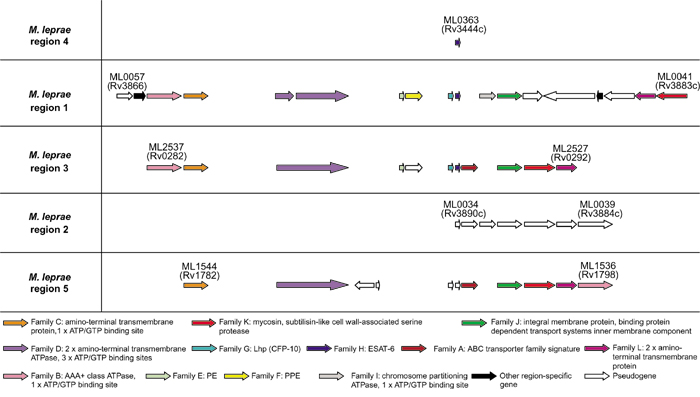
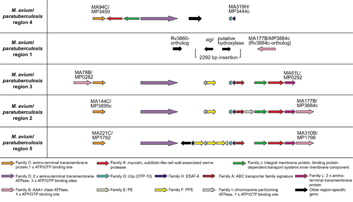
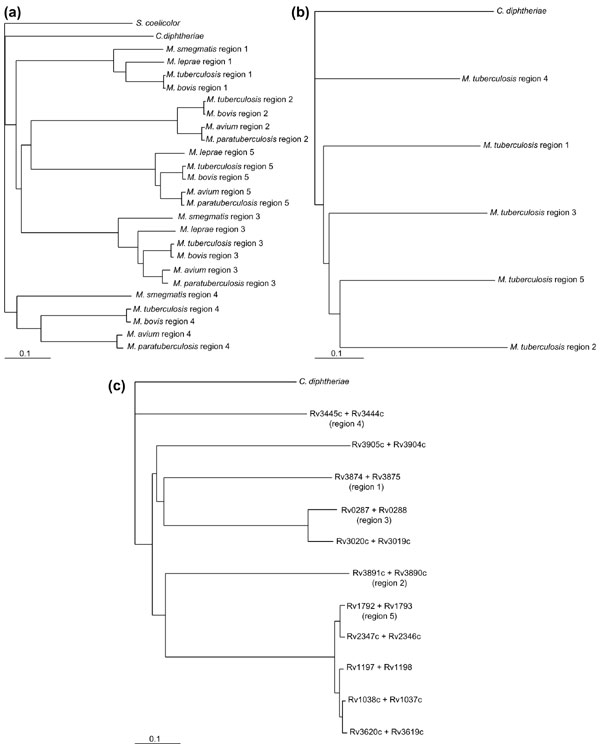
Results: Finished and unfinished genome sequencing data of 98 publicly available microbial genomes has been analyzed for the presence of orthologs of the ESAT-6 loci. The multiple duplicates of the ESAT-6 gene cluster found in the genome of M. tuberculosis H37Rv are also conserved in the genomes of other mycobacteria, for example M. tuberculosis CDC1551, M. tuberculosis 210, M. bovis, M. leprae, M. avium, and the avirulent strain M. smegmatis. Phylogenetic analyses of the resulting sequences have established the duplication order of the gene clusters and demonstrated that the gene cluster known as region 4 (Rv3444c-3450c) is ancestral. Region 4 is also the only region for which an ortholog could be found in the genomes of Corynebacterium diphtheriae and Streptomyces coelicolor.

Conclusions: Comparative genomic analysis revealed that the presence of the ESAT-6 gene cluster is a feature of some high-G+C Gram-positive bacteria. Multiple duplications of this cluster have occurred and are maintained only within the genomes of members of the genus Mycobacterium.


Figures

Figure 1
Figure 1
Schematic representation of the genomic organization of the genes present in the five ESAT-6 gene cluster regions of Mycobacterium tuberculosis H37Rv as well as the regions in C. diphtheriae and S. coelicolor. ORFs are represented as blocked arrows showing the direction of transcription, with the different colors reflecting the specific gene family and the length of the arrow reflecting the relative lengths of the genes. Annotations of M. tuberculosis H37Rv genes are according to Cole et al. [13]. Black arrows indicate unconserved genes present in these regions. Gaps between genes do not represent physical gaps between genes on the genome, but have been inserted to aid in indicating conservation among gene positions. Gene families were named arbitrarily according to their position in M. tuberculosis H37Rv region 1. The regions were named after the numbering system of Brown et al. [19] used arbitrarily for the five mycosin (subtilisin-like serine protease) genes identified from these regions (family K). M. tuberculosis regions are shown in order of suggested duplication events (see phylogenetic results) and not by numbering. The results of the analyses of the primary features of these genes and their corresponding proteins are included in a short summary at the bottom of the figure (see also Table 2).
Figure 2
Figure 2
Schematic representation of the six additional esat-6/lhp operon duplications and the regions that surround them in the genome of M. tuberculosis H37Rv. ORFs are represented by blocked arrows indicating direction of transcription, with the different colors reflecting the specific gene family and the length of the arrow reflecting the relative lengths of the genes as in Figure 1. The esat-6 and lhp genes deleted in M. bovis RD07 and RD09 deletion regions [7] are indicated.
Figure 3
Figure 3
Schematic representation of the genomic organization of the genes present in the five ESAT-6 gene cluster regions of Mycobacterium leprae. ORF's are represented as blocked arrows showing the direction of transcription, with the different colors reflecting the specific gene family and the length of the arrow reflecting the relative lengths of the genes as in Figure 1. Black arrows indicate unconserved genes present in these regions, while open arrows indicate pseudogenes. Annotations of M. leprae genes are according to Cole et al. [25].
Figure 4
Figure 4
Schematic representation of the genomic organization of the genes present in the four ESAT-6 gene cluster regions of Mycobacterium avium and Mycobacterium paratuberculosis, as well as the flanking genes of the region 1 deletion. ORFs are represented as blocked arrows showing the direction of transcription, with the different colors reflecting the specific gene family and the length of the arrow reflecting the relative lengths of the genes as in Figure 1. Black arrows indicate unconserved genes present in these regions. M. avium and M. paratuberculosis genes were arbitrarily annotated by the authors of this paper.
Figure 5
Figure 5
Schematic representation of the genomic organization of the genes present in the three ESAT-6 gene cluster regions of Mycobacterium smegmatis. ORFs are represented as blocked arrows showing the direction of transcription, with the different colors reflecting the specific gene family and the length of the arrow reflecting the relative lengths of the genes as in Figure 1. Black arrows indicate unconserved genes present in these regions. M. smegmatis genes were arbitrarily annotated by the authors of this paper.
Figure 6
Figure 6
Taxonomic position of the bacterial species that have the ESAT-6 gene clusters present in their genomes. This indicates that the ESAT-6 gene clusters seem to be a feature of only the high G+C Gram-positive bacteria (Actinobacteria) and that the presence of multiple copies of the gene clusters seems to be a characteristic only found in the mycobacteria. Phylogenetic relationships of members of the genus Mycobacterium indicated are based on 16S rRNA gene sequence information [56].
Figure 7
Figure 7
Phylogenetic trees showing the relationships between the five duplicated gene cluster regions. (a) Neighbor-joining phylogenetic tree of all available protein sequences of the ATP/GTP-binding protein family (family D in Table 2) with the protein ortholog of Streptomyces coelicolor as the outgroup. This tree is representative of all the trees drawn using the six most conserved proteins in these regions as well as using the protein ortholog of Corynebacterium diphtheriae as the outgroup. (b) Neighbor-joining phylogenetic tree of all six conserved proteins from the M. tuberculosis gene clusters combined into one protein per region. The combined protein of C. diphtheriae was used as the outgroup. (c) Neighbor-joining phylogenetic tree of the ESAT-6 and CFP-10 protein families combined (family G and H), using the combined protein of C. diphtheriae as the outgroup.

References

    1. Mahairas GG, Sabo PJ, Hickey MJ, Singh DC, Stover CK. Molecular analysis of genetic differences between Mycobacterium bovis BCG and virulent M. bovis. J Bacteriol. 1996;178:1274–1282. - PMC - PubMed
    1. Philipp WJ, Nair S, Guglielmi G, Lagranderie M, Gicquel B, Cole ST. Physical mapping of Mycobacterium bovis BCG pasteur reveals differences from the genome map of Mycobacterium tuberculosis H37Rv and from M. bovis. Microbiology. 1996;142:3135–3145. - PubMed
    1. Brosch R, Gordon SV, Billault A, Garnier T, Eiglmeier K, Soravito C, Barrell BG, Cole ST. Use of a Mycobacterium tuberculosis H37Rv bacterial artificial chromosome library for genome mapping, sequencing, and comparative genomics. Infect Immun. 1998;66:2221–2229. - PMC - PubMed
    1. Brosch R, Philipp WJ, Stavropoulos E, Colston MJ, Cole ST, Gordon SV. Genomic analysis reveals variation between Mycobacterium tuberculosis H37Rv and the attenuated M. tuberculosis H37Ra strain. Infect Immun. 1999;67:5768–5774. - PMC - PubMed
    1. Brosch R, Gordon SV, Buchrieser C, Pym AS, Garnier T, Cole ST. Comparative genomics uncovers large tandem chromosomal duplications in Mycobacterium bovis BCG Pasteur. Yeast. 2000;17:111–123. - PMC - PubMed

Publication types

MeSH terms

LinkOut - more resources