. 2001 Oct 12;89(8):732-9.
doi: 10.1161/hh2001.097872.

Vascular morphogenesis and remodeling in a human tumor xenograft: blood vessel formation and growth after ovariectomy and tumor implantation

Affiliations

Vascular morphogenesis and remodeling in a human tumor xenograft: blood vessel formation and growth after ovariectomy and tumor implantation

S Patan et al. Circ Res. .

Abstract

To determine mechanisms of blood vessel formation and growth in solid tumors, we used a model in which LS174T human colon adenocarcinomas are grown in the isolated ovarian pedicle of nude mice. Reconstruction of 3500 histological serial sections demonstrated that a new vascular network composed of venous-venous loops of varying sizes grows inside the tumor from the wall of the adjacent main vein. Loops elongate and remodel to establish complex loop systems. The mechanisms of loop formation and remodeling correspond to intussusceptive microvascular growth (IMG). In the tissue surrounding the tumor segmentation, another mechanism of IMG is prevalent in venous vessels. Comparison to vascular morphogenesis in the ovariectomized pedicle not only confirms the existence of corresponding mechanisms in both systems, but also reveals numerous sprouts that are superimposed onto loop systems and pathological deviations of loop formation, remodeling, and segmentation in the tumor. These pathological mechanisms interfere with vessel patency that likely cause heterogenous perfusion and hypoxia thus perpetuating angiogenesis. Blood vessel formation based on IMG was also detected in a large thrombus that completely occluded a part of an ovarian artery branch.


Figures
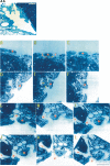
Figure 1
Figure 1
AA, Overview of the dilated main ovarian vein located close to the tumor margin (at the right) and near the ovarian artery (at the left). The venous lumen is divided by folds and ITSs, day 7. Bar=132 μm. A through L, Three elementary loop systems are illustrated: loop E1, A through C (serial sections 6168, 6174, 6178); loop E2, D through F (6136, 6142, 6146); and loop E3, G through L (6588, 6596, 6604, 6608, 6614, 6616). Loops E1 and E2 are single loops derived from the main vein and have each one central ITS (asterisks). A neighboring ITS, not yet entirely separated from the lateral venous wall, is about to split as indicated by two evaginations of the venous lumen within its center (D through F). These are filled with erythrocytes (arrows, B). The last system (E3) consists of three loops of different sizes (G through L). The large ITS remains connected indicating that the large loop is not patent yet (H). The small loop exhibits an ITS in the size range of a tissue pillar (I through L, arrow). Bar=80 μm.
Figure 2
Figure 2
A through U, Loop 1 (serial sections 6214, 6236, 6264, 6294, 6310, 6316, 6318, 6322, 6328, 6330, 6332, 6302, 6284, 6276, 6344, 6352, 6362, 6370, 6376, 6376, 6394) is illustrated (in red letters). Part of loop 2 (red asterisk) and part of loop 4 (yellow letters) are illustrated where these loops are in close spatial relationship to loop 1 and influence its structure. Segment (a, red) originates from the main vein (A through D); the red star indicates a segment of loop 2. (a) (red) connects to (b) (red) (G), and (b) (red) nearly connects to (b) (yellow), which is inhibited by the existence of an intraluminal fold (I through K). (b) (red) originates from the main vein (L through N). (a) (red) reconnects to the main vein via (c) (red) (O through U). Bar=60 μm.
Figure 2
Figure 2
A through U, Loop 1 (serial sections 6214, 6236, 6264, 6294, 6310, 6316, 6318, 6322, 6328, 6330, 6332, 6302, 6284, 6276, 6344, 6352, 6362, 6370, 6376, 6376, 6394) is illustrated (in red letters). Part of loop 2 (red asterisk) and part of loop 4 (yellow letters) are illustrated where these loops are in close spatial relationship to loop 1 and influence its structure. Segment (a, red) originates from the main vein (A through D); the red star indicates a segment of loop 2. (a) (red) connects to (b) (red) (G), and (b) (red) nearly connects to (b) (yellow), which is inhibited by the existence of an intraluminal fold (I through K). (b) (red) originates from the main vein (L through N). (a) (red) reconnects to the main vein via (c) (red) (O through U). Bar=60 μm.
Figure 3
Figure 3
A through D, Part of loop 4 (serial sections 6324, 6320, 6318, 6316) is illustrated (yellow letters). Patency between segments (a) and (b) is prevented by the presence of a single intraluminal cell (yellow arrow, B) (A through D). Bar=90 μm.
Figure 4
Figure 4
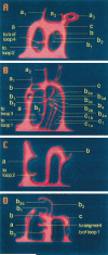
A through D, Reconstruction of loop systems. The vessel lumen is shown in red and the surrounding tissue in blue. Four loop systems (loops 1 through 4) originate and reconnect to the main vein (forming the bottom of the loop systems) and to each other (A through D). Individual segments are labeled with letters corresponding to Figures 2 and 3.
Figure 5
Figure 5
A through H, Mechanisms of loop formation and remodeling based on IMG. The vessel lumen is shown in red or yellow, respectively, and the surrounding tissue in blue. In situ elementary loop formation in the wall of the main vein based on lumen evagination around an intervening part of the vessel wall, which forms the ITS (A and B). Growth of the elementary loop is achieved by its elongation into the wall of the main vein and surrounding tissue and by growth of the central ITS (C). Loop remodeling causes “loop multiplication.” It occurs by splitting of the central ITS, which leads to the formation of a new segment that reconnects to the main vein (D, indicated by broken line). Loop remodeling can also occur by fold formation from the central ITS or from the lateral wall. The subsequent separation of the tip of the fold forms a free tissue pillar (E, indicated by broken line). Results of loop remodeling: loop remodeling results in the formation of compound loop systems, as double, triple, and quadruple loops (F). Pathological loop formation and remodeling: Sometimes loop patency is reached extremely late when the loop has reached a large size, based on the persistence of one intraluminal cell (see Figures 3 and 4D) (G). Loop remodeling by formation and insertion of intraluminal folds at the opposite vessel wall causes occlusion of the loop and loss of patency at a specific site (see Figures 2I through 2K) (H).

Comment in

References

    1. Fülleborn F. Beiträge zur Entwicklung der Allantois der Vögel (dissertation). G. Schade; Berlin, Germany: 1895.
    1. Clark ER, Clark EL. Microscopic observations on the growth of blood capillaries in the living mammal. Am J Anat. 1939;64:251–299.
    1. Maximow A. Experimentelle Untersuchungen über die entzündliche Neubildung von Bindegewebe. In: Ziegler E, editor. Beiträge zur pathologischen Anatomie und zur allgemeinen Pathologie (suppl Heft 5) G. Fischer; Jena, Germany: 1902.
    1. Wakui S, Furusato M, Matsumoto I, Takai K, Ushigome S, Ishikawa E. Two- and three-dimensional ultrastructural observation of angiogenesis at a capillary sprout with front-to-front growth in a human granuloma. Microcirculation. 1987;2:797–798.
    1. Schoefl G. Studies on inflammation, III: growing capillaries: their structure and permeability. Virchows Arch Pathol Anat. 1963;337:97–141. - PubMed

Publication types

MeSH terms