. 2001 Oct 15;20(20):5578-86.
doi: 10.1093/emboj/20.20.5578.

Link of the unique oncogenic properties of adenovirus type 9 E4-ORF1 to a select interaction with the candidate tumor suppressor protein ZO-2

Affiliations

Link of the unique oncogenic properties of adenovirus type 9 E4-ORF1 to a select interaction with the candidate tumor suppressor protein ZO-2

B A Glaunsinger et al. EMBO J. .

Abstract

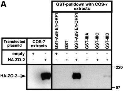
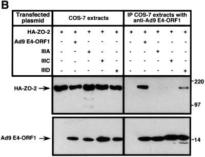
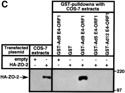
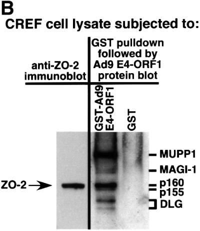
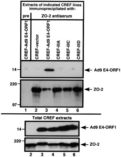
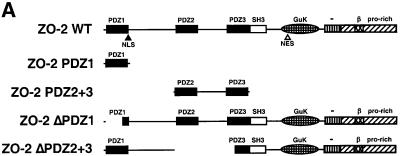
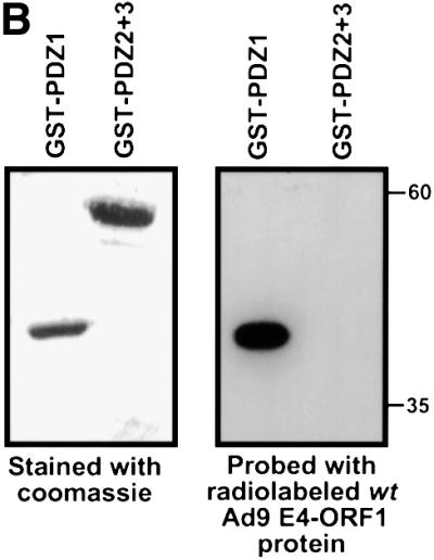
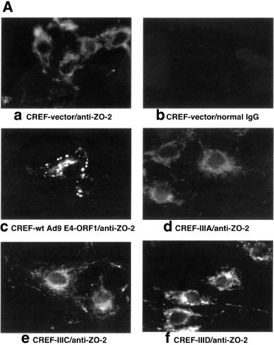
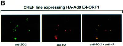
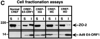
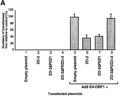
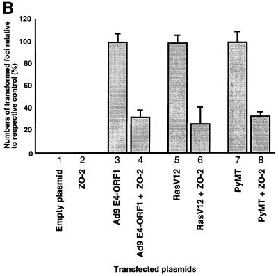
Adenovirus type 9 (Ad9) is distinct among human adenoviruses because it elicits solely mammary tumors in animals and its primary oncogenic determinant is the E4 region-encoded ORF1 (E4-ORF1) protein. We report here that the PDZ domain-containing protein ZO-2, which is a candidate tumor suppressor protein, is a cellular target for tumorigenic Ad9 E4-ORF1 but not for non-tumorigenic wild-type E4-ORF1 proteins encoded by adenovirus types 5 and 12. Complex formation was mediated by the C-terminal PDZ domain-binding motif of Ad9 E4- ORF1 and the first PDZ domain of ZO-2, and in cells this interaction resulted in aberrant sequestration of ZO-2 within the cytoplasm. Furthermore, transformation-defective Ad9 E4-ORF1 mutants exhibited impaired binding to and sequestration of ZO-2 in cells, and overexpression of wild-type ZO-2, but not mutant ZO-2 lacking the second and third PDZ domains, interfered with Ad9 E4-ORF1-induced focus formation. Our results suggest that the select capacity to complex with the candidate tumor suppressor protein ZO-2 is key to defining the unique transforming and tumorigenic properties of the Ad9 E4-ORF1 oncoprotein.


Figures

None
Fig. 1. Binding of wild-type but not mutant Ad9 E4-ORF1 to ZO-2 in GST pulldown assays. (A) ZO-2 binds to wild-type Ad9 E4-ORF1 in GST pulldown assays. Protein (100 µg) from RIPA buffer-lysed COS-7 cells transfected with 5 µg of either empty GW1 plasmid or GW1 plasmid expressing HA–ZO-2 was subjected to GST pulldown assays with the fusion protein indicated. Recovered proteins were separated by SDS–PAGE and immunoblotted with anti-HA antibodies. As a control, one-tenth the amount of protein used in GST pulldown assays was directly immunoblotted with the same antibodies. (B) Wild-type Ad9 E4-ORF1 co-immunoprecipitates with HA–ZO-2. Protein (150 µg) from RIPA buffer-lysed COS-7 cells co-transfected with 5 µg of GW1 plasmid expressing HA–ZO-2 and 5 µg of either empty GW1 plasmid or GW1 plasmid expressing wild-type or the indicated mutant Ad9 E4-ORF1 was immunoprecipitated with Ad9 E4-ORF1 antibodies. Recovered proteins were separated by SDS–PAGE and immunoblotted with either anti-HA or anti-Ad9 E4-ORF1 antibodies. As a control, one-tenth the amount of protein used in the immunoprecipitation reactions was directly immunoblotted with the same antibodies. (C) Binding of ZO-2 to Ad9 E4-ORF1 but not to Ad5 and Ad12 E4-ORF1 in GST pulldown assays. Experiments were performed as described above in (A).
None
Fig. 2. Co-migration of ZO-2 and Ad9 E4-ORF1-associated protein p160. (A) Gel mobilities of ZO-2 proteins expressed in various cell lines. Protein (100 µg) from the indicated RIPA buffer-lysed cells was separated by SDS–PAGE and immunoblotted with ZO-2 antibodies. As controls, 2.5 µg of protein from RIPA buffer-lysed COS-7 cells transfected with 4 µg of either empty GW1 plasmid or GW1 plasmid expressing wild-type ZO-2 was run on the same protein gel. (B) Ad9 E4-ORF1-associated cellular protein p160 co-migrates with endogenous ZO-2 of CREF cells. Protein (50 µg) from RIPA buffer-lysed normal CREF cells was separated by SDS–PAGE and then immunoblotted with ZO-2 antibodies (left panel). Alternatively, 4 mg of protein from RIPA buffer-lysed normal CREF cells was subjected to GST pulldown assays with the fusion proteins indicated. Recovered proteins were separated by SDS–PAGE and blotted with a radiolabeled Ad9 E4-ORF1 protein probe (right panel). Samples in both panels were run on the same gel to allow comparison of protein mobilities.
None
Fig. 3. Binding of wild-type but not mutant Ad9 E4-ORF1 to endogenous ZO-2 of CREF cells. One milligram of protein from RIPA buffer-lysed normal CREF cells or CREF cells stably expressing wild-type or mutant Ad9 E4-ORF1 were immunoprecipitated with ZO-2 antiserum or the matched pre-immune serum (pre). Recovered proteins were separated by SDS–PAGE and immunoblotted with either ZO-2 or Ad9 E4-ORF1 antiserum (top panel). As a control, 100 µg of protein from RIPA buffer-lysed normal CREF cells was also directly immunoblotted with the same antisera (bottom panel). Sample 1 of the top panel was not analyzed in the bottom panel.
None
Fig. 4. ZO-2 PDZ1 mediates binding of ZO-2 to Ad9 E4-ORF1. (A) Illustration of ZO-2 protein fragments and ZO-2 deletion mutants. (B) Specific binding of Ad9 E4-ORF1 to ZO-2 PDZ1 in protein blotting assays. Approximately 5 µg of the indicated ZO-2 GST fusion protein, separated by SDS–PAGE and immobilized on a membrane, was either stained with Coomassie Blue dye to verify the presence of equivalent amounts of protein (left panel) or probed with a radiolabeled wild-type Ad9 E4-ORF1 protein probe (right panel). (C) Requirement of ZO-2 PDZ1 for ZO-2 to co-immunoprecipitate with Ad9 E4-ORF1 from cell extracts. Protein (400 µg) from RIPA buffer-lysed COS-7 cells co-transfected with 5 µg of either empty GW1 plasmid or GW1 plasmid expressing Ad9 E4-ORF1 and 5 µg of GW1 plasmid expressing wild-type HA–ZO-2 or mutant HA–ZO-2 lacking either PDZ1 (HA–ZO-2ΔPDZ1) or both PDZ2 and PDZ3 (HA–ZO-2ΔPDZ2+3) was immunoprecipitated with Ad9 E4-ORF1 antibodies. Recovered proteins were separated by SDS–PAGE and immunoblotted with anti-HA or anti-Ad9 E4-ORF1 antibodies. As a control, one-fifteenth the amount of protein used in the immuno precipitation reactions was also directly immunoblotted with the same antibodies.
None
Fig. 5. Ad9 E4-ORF1 aberrantly sequesters ZO-2 in the cytoplasm of CREF cells. (A) Distribution of ZO-2 in normal CREF cells or CREF cells stably expressing either wild-type or the indicated mutant Ad9 E4-ORF1. IF assays were performed with either affinity-purified ZO-2 antibodies (a, c–f) or normal rabbit IgG (b) and visualized by fluorescence microscopy. (B) ZO-2 and Ad9 E4-ORF1 co-localize within cytoplasmic punctate bodies in CREF cells. Double-label IF assays were performed using both affinity-purified ZO-2 and anti-HA antibodies in CREF cells stably expressing HA–Ad9 E4-ORF1. Each of the three panels represents the same field of five cells stained for ZO-2 (left panel), HA–Ad9 E4-ORF1 (center panel) or the merged images (right panel). (C) Ad9 E4-ORF1 aberrantly sequesters ZO-2 within detergent-insoluble complexes in CREF cells. Normal CREF cells or CREF cells stably expressing either wild-type or the indicated mutant Ad9 E4-ORF1 were lysed in RIPA buffer, and extracts were centrifuged to produce RIPA buffer-soluble (S) supernatant and RIPA buffer-insoluble (I) pellet fractions. Protein (100 µg) from S fractions or an equivalent amount from I fractions (see Materials and methods) was separated by SDS–PAGE and immunoblotted with either ZO-2 or Ad9 E4-ORF1 antiserum.
None
Fig. 6. ZO-2 inhibits oncogene-induced focus formation in CREF cells. (A) ZO-2 interferes with Ad9 E4-ORF1-induced focus formation. CREF cells were transfected either alone with 8 µg of empty GW1 plasmid or GW1 plasmid expressing the indicated wild-type or mutant HA–ZO-2 protein (lanes 1–4) or together with 2 µg of pJ4Ω plasmid expressing wild-type Ad9 E4-ORF1 (lanes 5–8). At 3 weeks post-transfection, transformed foci were counted. Numbers of transformed foci are presented as percent relative to the Ad9 E4-ORF1 plasmid alone (control), which was normalized to 100%. Data are compiled from three independent assays, each performed in duplicate. (B) ZO-2 also blocks focus formation by the RasV12 and polyomavirus middle T oncoproteins. CREF cells were transfected either alone with 9 µg of empty GW1 plasmid or GW1 plasmid expressing wild-type HA–ZO-2 protein (lanes 1–2) or together with 3 µg of either pJ4Ω plasmid expressing wild-type Ad9 E4-ORF1 (lanes 3–4), GW1 plasmid expressing RasV12 (lanes 5–6) or pPyMT1 plasmid expressing polyomavirus middle T (PyMT) (lanes 7–8). Assays were scored as indicated above in (A). Numbers of transformed foci are presented as percent relative to the respective Ad9 E4-ORF1, RasV12 or PyMT plasmid alone (control), each of which was normalized to 100%. Data are compiled from two independent assays, each performed in duplicate.

Similar articles

Cited by

References

    1. Balda M.S. and Matter,K. (2000) The tight junction protein ZO-1 and an interacting transcription factor regulate ErbB-2 expression. EMBO J., 19, 2024–2033. - PMC - PubMed
    1. Beatch M., Jesaitis,L.A., Gallin,W.J., Goodenough,D.A. and Stevenson,B.R. (1996) The tight junction protein ZO-2 contains three PDZ (PSD-95/Discs-Large/ZO-1) domains and an alternatively spliced region. J. Biol. Chem., 271, 25723–25726. - PubMed
    1. Ben-Ze’ev A. (1997) Cytoskeletal and adhesion proteins as tumor suppressors. Curr. Opin. Cell Biol., 9, 99–108. - PubMed
    1. Bradford M.M. (1976) A rapid and sensitive method for the quantitation of microgram quantities of protein utilizing the principle of protein-dye binding. Anal. Biochem., 72, 248–254. - PubMed
    1. Chlenski A., Ketels,K.V., Engeriser,J.L., Talamonti,M.S., Tsao,M.S., Koutnikova,H., Oyasu,R. and Scarpelli,D.G. (1999a) ZO-2 gene alternative promoters in normal and neoplastic human pancreatic duct cells. Int. J. Cancer, 83, 349–358. - PubMed

Publication types

MeSH terms