. 2001 Nov;69(11):6625-32.
doi: 10.1128/IAI.69.11.6625-6632.2001.

Characterization of FasG segments required for 987P fimbria-mediated binding to piglet glycoprotein receptors

Affiliations

Characterization of FasG segments required for 987P fimbria-mediated binding to piglet glycoprotein receptors

B K Choi et al. Infect Immun. 2001 Nov.

Abstract

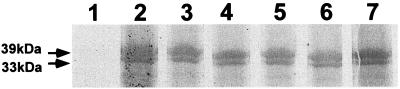
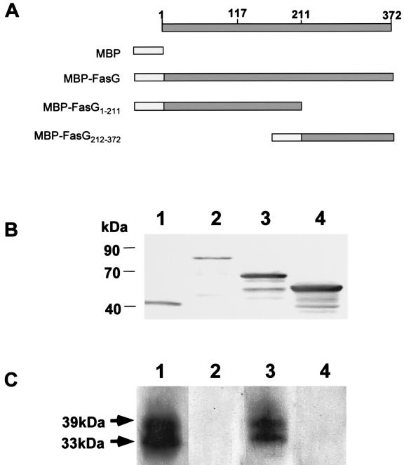
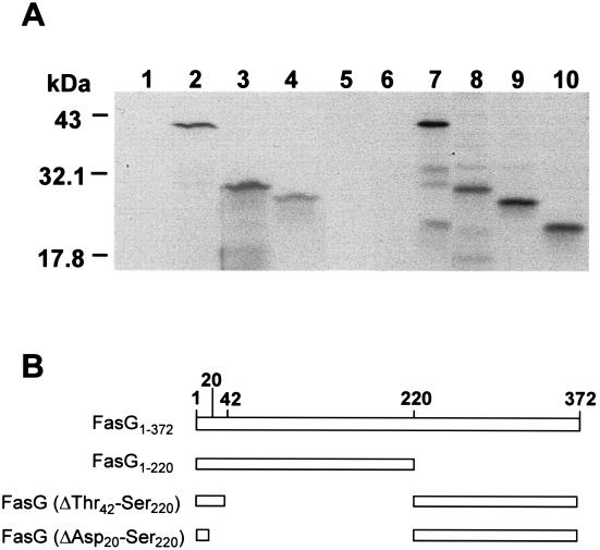
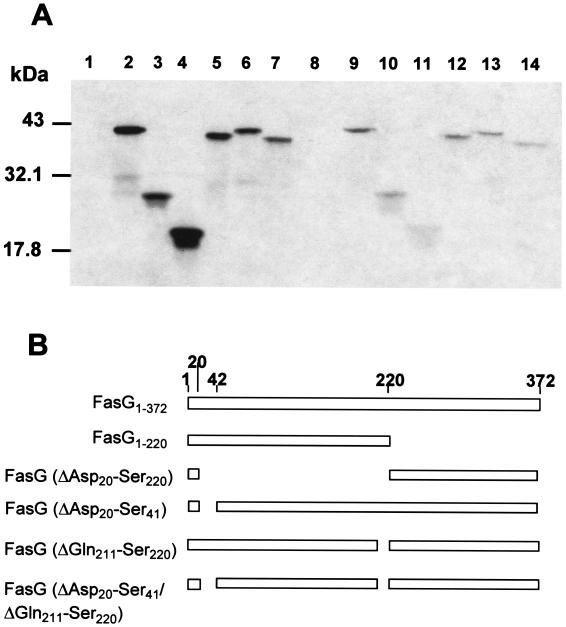
The 987P fimbriae of enterotoxigenic strains of Escherichia coli bind to both glycoprotein and glycolipid receptors on the brush borders of piglet enterocytes. A mutation in lysine residue 117 of the adhesive subunit FasG [fasG(K117A)] previously shown to abrogate 987P binding to the lipid receptor sulfatide did not affect the interaction with the glycoprotein receptors. Both the fimbriae and the FasG subunits of the wild type and the fasG(K117A) mutant bound to the glycoprotein receptors, confirming that lysine 117 was not required for binding to the glycoprotein receptors. Truncated FasG molecules were used to identify domains required for glycoprotein receptor recognition. At least two segments which did not include lysine117, namely, residues 211 (glutamine) to 220 (serine) and 20 (aspartic acid) to 41 (serine), were shown to be involved in the FasG-glycoprotein receptor interactions by ligand-blotting assays. Changing isoleucine 217 or leucine 215 of FasG to alanine abolished the property of a truncated FasG fusion protein to inhibit 987P recognition of its glycoprotein receptors. Thus, the K117 residue of FasG is required only for binding to the glycolipid receptor, whereas the newly identified hydrophobic residues of the FasG subunit are required specifically for the recognition of the glycoprotein receptor. Taken together, our data indicate that different residues of the FasG adhesin are important in 987P fimbrial binding to sulfatide and glycoprotein receptors, suggesting different mechanisms of interaction.


Figures

FIG. 1
FIG. 1
Direct binding of wild-type and mutated FasG fimbrial subunits to blotted BBV proteins. Each membrane strip was incubated with 35S-labeled periplasmic proteins isolated from a different strain. FasG binding was visualized by fluorogaphy. Periplasmic proteins were isolated from the following strains: control strain, E. coli SE5000(pGP1-2) (lane 1), SE5000(pGP1-2, pDMS127) expressing wild-type FasG (lane 2), SE5000(pGP1-2, pBKC-R17A) expressing FasG(R17A) (lane 3), SE5000(pGP1-2, pBKC-R116A) expressing FasG(R116A) (lane 4), SE5000(pGP1-2, pBKC-K117A) expressing FasG(K117A) (lane 5), SE5000(pGP1-2, pBKC-K118A) expressing FasG(K118A) (lane 6), and SE5000(pGP1-2, pBKC-R200A) expressing FasG(R200A) (lane 7).
FIG. 2
FIG. 2
Ligand-blotting inhibition assay. (A) Constructs of FasG and truncated FasG proteins fused to the MBP of E. coli. The residue numbers relate to mature FasG. (B) Western blot analysis of periplasmic fractions of bacteria expressing MBP or MBP-FasG fusion products detected with anti-MBP antibodies. Lane 1, MBP; lane 2, MBP-FasG; lane 3, MBP-FasG1–211; lane 4, MBP-FasG212–372. (C) Fimbrial ligand-blotting assays using MBP or MBP-FasG fusion proteins as inhibitors. Each membrane strip with SDS-PAGE-separated BBV proteins was incubated with the periplasmic fraction. The periplasmic fractions used were isolated from bacteria expressing the following proteins: MBP (lane 1), MBP-FasG (lane 2), MBP-FasG1–210 (lane 3), and MBP-FasG211–372 (lane 4).
FIG. 3
FIG. 3
Direct binding of FasG and its truncates to BBV. (A) Comparable amounts of all isolated 35S-labeled full-length or truncated FasG proteins were mixed with BBV. After 2 h of incubation, the BBV with bound FasG were washed by centrifugation, as described in Materials and Methods. BBV-associated FasG protein or truncates were separated by SDS–15% PAGE and detected by fluorography. Periplasmic FasG proteins were from the following strains: control strain, E. coli SE5000(pGP1-2) expressing no FasG (lanes 1 and 6), SE5000(pGP1-2, pDMS127) expressing full-length FasG (lanes 2 and 7), SE5000(pGP1-2, pBKC8) expressing FasG1–220 (lanes 3 and 8), SE5000(pGP1-2, pBKC10) expressing FasG(ΔThr42–Ser220) (lanes 4 and 9), and SE5000(pGP1-2, pBKC12) expressing FasG(ΔAsp20–Ser220) (lanes 5 and 10). Lanes 1 to 5, periplasmic FasG proteins which bound to the BBV; lanes 6 to 10, periplasmic FasG proteins before addition to the BBV. (B) DNA segments of the fasG gene included in each construct. The numbers correspond to the amino acid residues of mature FasG.
FIG. 4
FIG. 4
Direct binding of internally deleted FasG proteins to BBV. (A) Comparable amounts of isolated 35S-labeled full-length, truncated, or internally deleted FasG proteins were mixed with BBV. After 2 h of incubation, the BBV with bound FasG were washed by centrifugation, as described in Materials and Methods. BBV-associated FasG proteins were separated by SDS–15% PAGE and detected by fluorography. The periplasmic FasG proteins were from the following strains: control strain, E. coli SE5000(pGP1-2) expressing no FasG (lanes 1 and 8), SE5000(pGP1-2, pDMS127) expressing full-length FasG (lanes 2 and 9), SE5000(pGP1-2, pBKC8) expressing FasG1–220 (lanes 3 and 10), SE5000(pGP1-2, pBKC12) expressing FasG(ΔAsp20–Ser220) (lanes 4 and 11), SE5000(pGP1-2, pBKC18) expressing FasG(ΔAsp20–Ser41) (lanes 5 and 12), SE5000(pGP1-2, pBKC19) expressing FasG(ΔGln211–Ser220) (lanes 6 and 13), and SE5000(pGP1-2, pBKC20) expressing FasG(ΔAsp20–Ser41 ΔGln211–Ser220) (lanes 7 and 14). Lanes 1 to 7, periplasmic FasG proteins before addition to the BBV; lanes 8 to 14, periplasmic FasG proteins which bound to the BBV. (B) DNA segments of the fasG gene included in each construct. The numbers correspond to the amino acid residues of mature FasG.
FIG. 5
FIG. 5
Ligand-blotting inhibition assay. Periplasmic MBP-FasG fusion proteins were isolated from the following strains: SE5000(pGP1-2, pBKC-P9) expressing MBP-FasG1–220 (lanes 1, 6, and 11), SE5000(pGP1-2, pBKC12) expressing MBP-FasG221–372 (lanes 2, 7, and 12), SE5000[pGP1-2, pBKC-P9(L215A)] expressing MBP-FasG1–220 (L215A) (lanes 3, 8, and 13), SE5000[pGP1-2, pBKC-P9(I217A)] expressing MBP-FasG1–220 (I217A) (lanes 4, 9, and 14), and SE5000[pGP1-2, pBKC-P9(L215A I217A)] expressing MBP-FasG1–220 (L215AI217A) (lanes 5, 10, and 15). Three dilutions (lanes 1 to 5, undiluted; lanes 6 to 10, diluted 1/2; lanes 11 to 15, diluted 1/4) of normalized concentrations of MBP-FasG fusion proteins from periplasmic fractions were used to inhibit fimbrial binding to glycoprotein receptors on the membrane.

References

    1. Ben-Tal N, Honig B, Peitzsch R M, Denisov G, McLaughlin S. Binding of small basic peptides to membranes containing acidic lipids: theoretical models and experimental results. Biophys J. 1996;71:561–575. - PMC - PubMed
    1. Cao J, Khan A S, Bayer M E, Schifferli D M. Ordered translocation of 987P fimbrial subunits through the outer membrane of Escherichia coli. J Bacteriol. 1995;177:3704–3713. - PMC - PubMed
    1. Choi B-K, Schifferli D M. Lysine residue 117 of the FasG adhesin of enterotoxigenic Escherichia coli is essential for binding of 987P fimbriae to sulfatide. Infect Immun. 1999;67:5755–5761. - PMC - PubMed
    1. Choudhury D, Thompson A, Stojanoff V, Langermann S, Pinkner J, Hultgren S J, Knight S D. X-ray structure of the FimC-FimH chaperone-adhesin complex from uropathogenic Escherichia coli. Science. 1999;285:1061–1066. - PubMed
    1. Dean E A. Comparison of receptors for 987P pili of enterotoxigenic Escherichia coli in the small intestines of neonatal and older pigs. Infect Immun. 1990;58:4030–4035. - PMC - PubMed

Publication types

MeSH terms

LinkOut - more resources