. 2001 Nov;69(11):6633-42.
doi: 10.1128/IAI.69.11.6633-6642.2001.

Administration of superantigens protects mice from lethal Listeria monocytogenes infection by enhancing cytotoxic T cells

Affiliations

Administration of superantigens protects mice from lethal Listeria monocytogenes infection by enhancing cytotoxic T cells

S Okamoto et al. Infect Immun. 2001 Nov.

Abstract

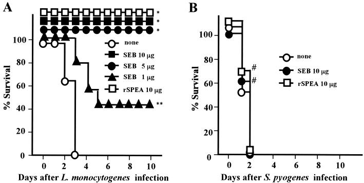
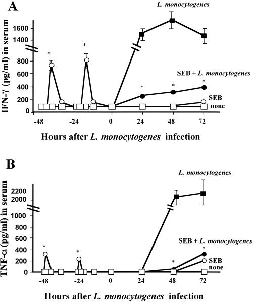
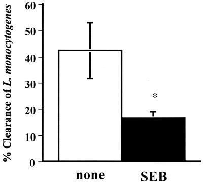
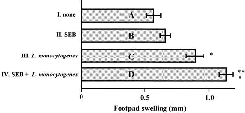
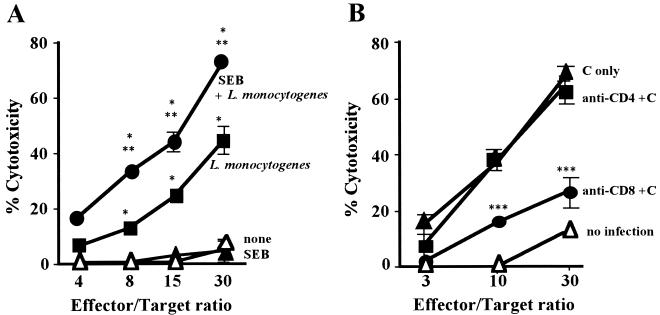
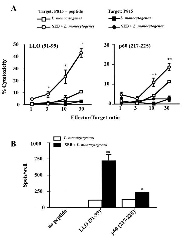
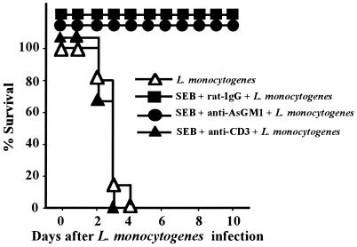
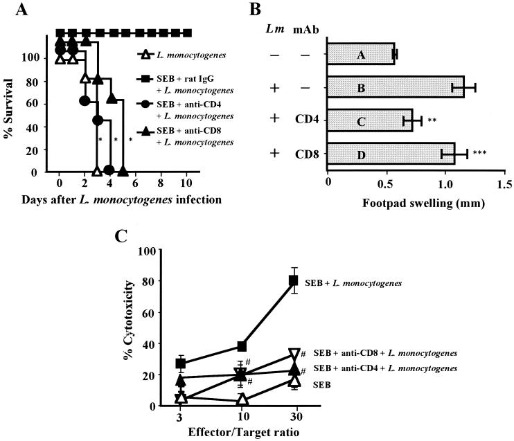
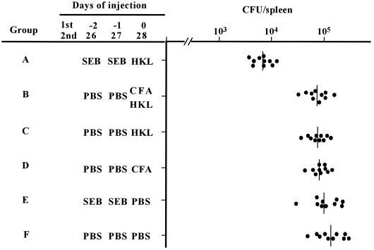
Superantigens stimulate T-cell-receptor Vbeta-selective T-cell proliferation accompanying the release of cytokines, which may eventually protect the host from microbial infections. We investigated here whether superantigens can rescue the host from lethal bacterial infection. Mice were pretreated with Staphylococcus aureus enterotoxin B (SEB) 1 and 2 days before bacterial infection, and the mortality of infected mice was assessed. SEB pretreatment protected mice from lethal infection with Listeria monocytogenes but not from lethal infection with Streptococcus pyogenes. This enhanced protection was also observed upon pretreatment with recombinant streptococcal pyrogenic exotoxin A. Furthermore, L. monocytogenes-specific delayed-type hypersensitivity (DTH) due to type 1 helper T (Th1) cells and the cytotoxicity of CD8(+) T cells were significantly enhanced after SEB administration and bacterial infection. Depletion of either CD4(+) T cells or CD8(+) T cells in SEB-pretreated mice completely abolished this protection. This phenomenon was ascribed to the elimination of L. monocytogenes-specific CD8(+) cytotoxic T lymphocytes (CTL). It was found that CD4(+) T cells contributed to the induction of the CTL populations. Furthermore, SEB pretreatment of heat-killed L. monocytogenes-immunized mice enhanced the protection from challenge of L. monocytogenes. Taken together, these results indicated that administrations of superantigens protected mice from infection with L. monocytogenes, which was dependent on the enhanced L. monocytogenes-specific CTL activity in the presence of CD4(+) T cells, and superantigens exhibited adjuvant activity in the immunization against intracellular pathogens.


Figures

FIG. 1
FIG. 1
Effect of SEB pretreatment on the infection with L. monocytogenes or S. pyogenes in mice. Groups of 15 BALB/c mice were pretreated i.p. with SEB or rSPEA twice 1 and 2 days before infection with 106 CFU of L. monocytogenes (A) or 106 CFU of S. pyogenes SSI-1 (B). The mortality of mice was assessed for 10 days after the infection. None, no SEB pretreatment. ∗, P < 0.0001; ∗∗, P = 0.0088 compared to none in panel A; #, P > 0.5 compared to none in panel B.
FIG. 2
FIG. 2
Effect of SEB pretreatment on secretion of IFN-γ and TNF-α in sera of mice before and after infection with L. monocytogenes. Mice were divided into four groups as follows: no treatment (□), pretreatment with SEB (10 μg) twice (○), infection with 104 CFU of L. monocytogenes (▪), and pretreatment with SEB (10 μg) twice and infection with 104 of CFU L. monocytogenes (●). Sera from five mice in groups □ and ○ were harvested at 48, 45, 42, 36, 24, 21, 18, and 12 h prior to infection. Additionally, sera from all mice in the four groups were harvested at 0, 24, 48, and 72 h after infection. The amounts of IFN-γ (A) and TNF-α (B) in sera were measured by ELISA. Values are presented as the mean picograms/milliliter ± the standard deviation. ∗, P < 0.01 compared to L. monocytogenes.
FIG. 3
FIG. 3
Effect of SEB pretreatment on macrophage phagocytosis of L. monocytogenes. Mice were divided into two groups (six mice per group). One group was not treated (none [open bar]), and the other was pretreated i.p. with SEB (10 μg) twice 1 and 2 days before being killed (SEB [closed bar]). Spleen macrophages were harvested, and phagocytosis of L. monocytogenes was assessed as described in Materials and Methods. Value are presented as the mean percentage of eliminated L. monocytogenes ± the standard deviation. ∗, P = 0.0095 compared to none.
FIG. 4
FIG. 4
Effect of SEB pretreatment on L. monocytogenes-specific Th cells in DTH response in mice. Twenty mice were divided into four groups (five mice per group): A to D. Suspensions of HKL and T cells from groups I to IV, as described in Materials and Methods, were injected s.c. into the left hind footpad of animals in groups A to D, respectively. In the right hind footpad, PBS (25 μl) was injected s.c. as a control. The thicknesses of the left and right footpads were measured 24 h after injection. Values are presented as the mean difference of thickness between the footpads ± the standard deviation. ∗, P < 0.01 compared to none; ∗ (P = 0.0095) and ∗∗ (P = 0.0065) compared to bar A; #, P = 0.039 compared to bar C.
FIG. 5
FIG. 5
Effect of SEB pretreatment on L. monocytogenes-specific cytotoxicity of CD8+ T cells. (A) Twenty mice were divided into four groups as follows: no treatment (▵), pretreatment with SEB (10 μg) twice (▴), infection with 104 CFU of L. monocytogenes (▪), and pretreatment with SEB (10 μg) twice and infection with 104 CFU of L. monocytogenes (●). Spleen cells were harvested 5 days after infection and effector T cells were established as described in Materials and Methods. The cytotoxicity of the effector T cells for the L. monocytogenes-infected J774A.1 cells was assayed. (B) Groups of five mice pretreated with SEB (10 μg) twice were infected i.v. with 104 CFU of L. monocytogenes. Effector T cells were established as described above and treated with complement (C) (▴), anti-CD4 MAb plus C (▪), or anti-CD8 MAb plus C (●). Effector T cells from untreated mice were established as described above and treated with C (▵). These T cells were used for a cytotoxic assay for L. monocytogenes infected J774A.1 cells. ∗, P < 0.01 compared to none in panel A; ∗∗, P < 0.01 compared to L. monocytogenes in panel A; ∗∗∗, P < 0.01 compared to C only in panel B.
FIG. 6
FIG. 6
Effect of SEB pretreatment on Listeria epitope-specific cytotoxicity. (A) Twenty mice were divided into four groups (five mice per group). Two groups of mice were infected i.v. with 104 CFU of L. monocytogenes (□ and ▪), and the other two were pretreated i.p.with SEB (10 μg) twice and then infected i.v. with L. monocytogenes (○ and ●). Spleen cells were harvested 5 days after infection and effector T cells were established as described in Materials and Methods. The cytotoxicity of these T cells for Listeria epitope-labeled P815 cells (104 cells/well; □ and ▪) or unlabeled P815 cells (104 cells/well; ○ and ●) was assayed. (B) Groups of five mice were infected with 104 CFU of L. monocytogenes (open bar) or pretreated with SEB (10 μg) twice and then infected with 104 CFU of L. monocytogenes (closed bar). Effector T cells were established as described above. Listeria epitope-labeled P815 cells and effector cells (105 cells/well) were poured into Multiscreen 96-well HA plates precoated with anti-IFN-γ MAb, and an ELISPOT assay was performed. ∗, P < 0.01 compared to P815 plus peptide-L. monocytogenes in LLO (91-99); ∗∗, P < 0.05 compared to P815 plus peptide-L. monocytogenes in the p60 (217-225) panel; #, P = 0.018 compared to L. monocytogenes in the p60 (217-225) panel; ##, P = 0.002 compared to L. monocytogenes in the LLO (91-99) section.
FIG. 7
FIG. 7
Protection of SEB-pretreated mice from L. monocytogenes infection by depletion of T cells. Groups of 15 mice were pretreated i.p. with SEB (10 μg) twice and infected i.v. with 106 CFU of L. monocytogenes. Purified anti-AsGM1 antibody (50 μl), anti-CD3 MAb (600 μg), or rat IgG (600 μg) was administered i.v. to mice 18 h before the infection. Mortality was assessed for 10 days after the infection (P < 0.0001 for SEB plus rat IgG plus L. monocytogenes versus SEB plus anti-CD3 plus L. monocytogenes; P = 0.4331 for L. monocytogenes versus SEB plus anti-CD3 plus L. monocytogenes).
FIG. 8
FIG. 8
Role of CD4+ T cells in protection from lethal L. monocytogenes infection due to CTLs. (A) Groups of 15 mice pretreated i.p. with SEB (10 μg) twice were infected with 106 CFU of L. monocytogenes. Anti-CD4 MAb (600 μg) or anti-CD8 MAb (600 μg) was injected i.v. into mice 18 h before the infection. Mortality was assessed for 10 days after the infection. (B) Groups of seven mice pretreated with SEB and MAb as described above were infected i.v. with 103 CFU of L. monocytogenes (Lm), and spleen T cells were harvested 7 days after the infection. T cells (5 × 10⁶ cells) and HKL (5 × 10⁷ CFU) were injected into the footpads of uninfected littermates. Swelling of the footpads was measured 24 h after the injection. (C) Groups of five mice pretreated as described above were infected i.v. with 104 CFU of L. monocytogenes. Spleen cells were harvested 5 days after the infection, and effector T cells were established. The cytotoxicity of these effector T cells for L. monocytogenes-infected J774A.1 cells was assayed. ∗, P < 0.0001 compared to SEB plus rat IgG plus L. monocytogenes in panel A; P = 0.0095 (∗∗) and 0.3402 (∗∗∗) compared to bar B in panel B; #, P < 0.01 compared to SEB plus L. monocytogenes in panel C.
FIG. 9
FIG. 9
Protection against L. monocytogenes challenge in HKL-immunized mice and effect of SEB. Groups of 10 mice were injected with SEB (10 μg) or CFA (100 μl) and immunized s.c. with HKL (5 × 10⁷ CFU) twice as indicated. These mice were infected with 104 CFU of L. monocytogenes/mouse 63 days after the initial immunization. Spleen was harvested 4 days after the infection, and the number of L. monocytogenes colonies in the spleen was determined. P = 0.0095, 0.0065, 0.0020, 0.0057, and 0.0012 for group A compared to groups B to F, respectively.

Similar articles

Cited by

References

    1. Ashman R B, Müllbacher A. A T helper cell for anti-viral cytotoxic T-cell responses. J Exp Med. 1979;150:1277–1282. - PMC - PubMed
    1. Bancroft G J, Sheehan K C F, Schreiber R D, Unanue E R. Tumor necrosis factor is involved in the T cell-independent pathway of macrophage activation in scid mice. J Immunol. 1989;143:127–130. - PubMed
    1. Blackman M A, Woodland D L. In vivo effects of superantigens. Life Sci. 1995;57:1717–1735. - PubMed
    1. Bouwer H G A, Hinrichs D J. Cytotoxic-T-lymphocyte responses to epitopes of listeriolysin O and p60 following infection with Listeria monocytogenes. Infect Immun. 1996;64:2515–2522. - PMC - PubMed
    1. Buller R M L, Holmes K L, Hügin A, Frederickson T N, Morse H C., III Induction of cytotoxic T-cell responses in vivo in the absence of CD4 helper cells. Nature. 1987;328:77–79. - PubMed

Publication types

MeSH terms