. 2001 Nov;69(11):6785-95.
doi: 10.1128/IAI.69.11.6785-6795.2001.

Type III secretion-dependent cell cycle block caused in HeLa cells by enteropathogenic Escherichia coli O103

Affiliations

Type III secretion-dependent cell cycle block caused in HeLa cells by enteropathogenic Escherichia coli O103

J P Nougayrède et al. Infect Immun. 2001 Nov.

Abstract

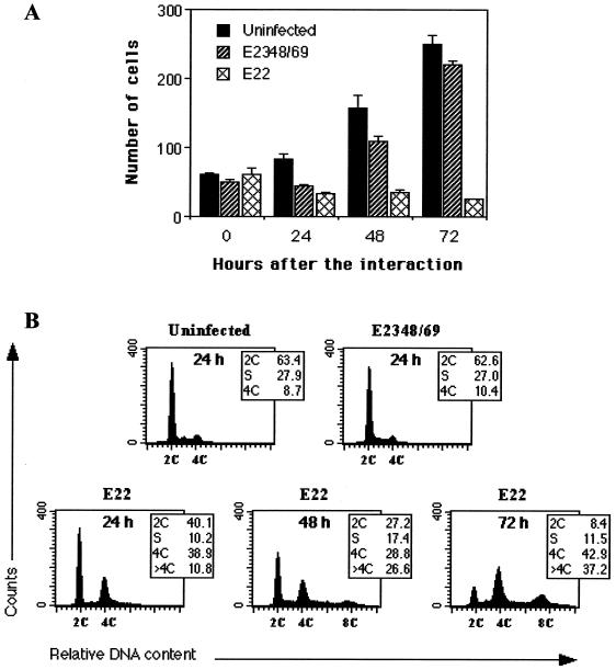
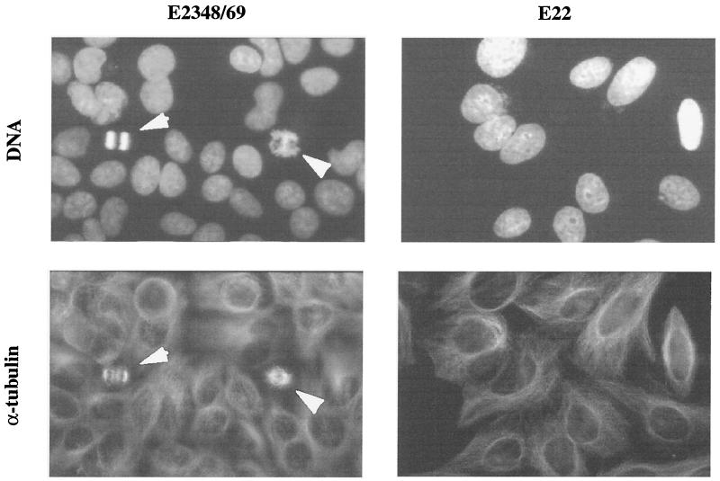
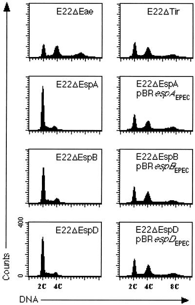
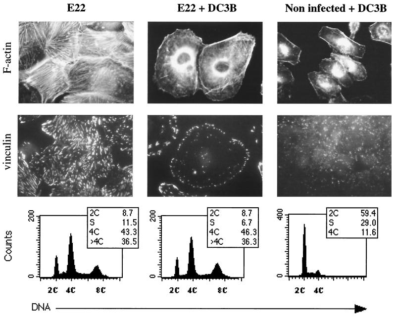
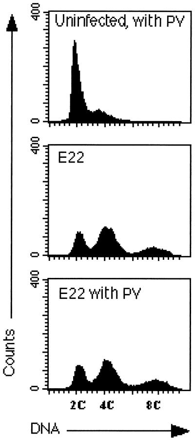
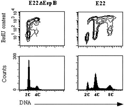
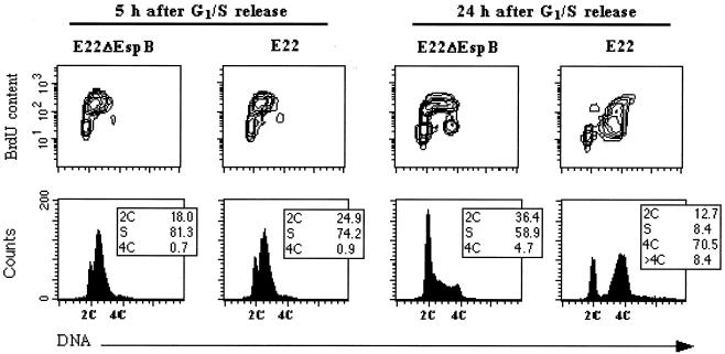
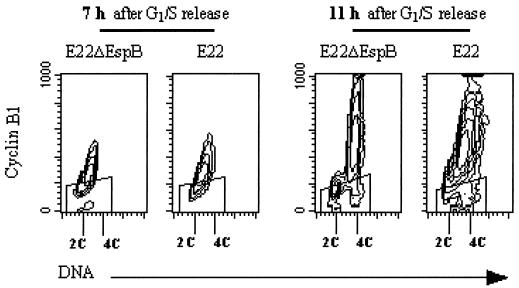
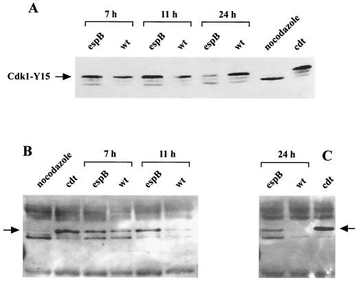
Rabbit enteropathogenic Escherichia coli (EPEC) O103 induces in HeLa cells an irreversible cytopathic effect characterized by the recruitment of focal adhesions, formation of stress fibers, and inhibition of cell proliferation. We have characterized the modalities of the proliferation arrest and investigated its underlying mechanisms. We found that HeLa cells that were exposed to the rabbit EPEC O103 strain E22 progressively accumulated at 4C DNA content and did not enter mitosis. A significant proportion of the cells were able to reinitiate DNA synthesis without division, leading to 8C DNA content. This cell cycle inhibition by E22 was abrogated in mutants lacking EspA, -B, and -D and was restored by transcomplementation. In contrast, intimin and Tir mutants retained the antiproliferative effect. The cell cycle arrest was not a direct consequence of the formation of stress fibers, since their disruption by toxins during exposure to E22 did not reverse the cell cycle inhibition. Likewise, the cell cycle arrest was not dependent on the early tyrosine dephosphorylation events triggered by E22 in the cells. Two key partner effectors controlling entry into mitosis were also investigated: cyclin B1 and the associated cyclin-dependent kinase 1 (Cdk1). Whereas cyclin B1 was not detectably affected in E22-exposed cells, Cdk1 was maintained in a tyrosine-phosphorylated inactive state and lost its affinity for p13(suc1)-agarose beads. This shows that Cdk1 is implicated in the G2/M arrest caused by EPEC strain E22.


Figures

FIG. 1
FIG. 1
Demonstration of the irreversible arrest in HeLa cell proliferation (A) and cell cycle perturbation (B) triggered by E22. Cells were exposed for 4 h to E2348/69 or E22 or left uninfected, and they were further cultivated for the indicated times. (A) Cell proliferation after interaction. Cultures were fixed and stained with Giemsa, and cells within random microscope fields were counted (objective, ×20). Each point is the mean of four independent measures. (B) Cell distribution according to DNA content, analyzed by flow cytometry after staining of DNA with propidium iodide. The percentages of cell populations are shown in each case.
FIG. 2
FIG. 2
Absence of mitotic figures in E22-exposed cells. Cells were exposed for 4 h to E2348/69 or E22 and further cultivated for 24 h. DNA was stained with diaminophenylindole, and α-tubulin was labeled by FITC indirect immunofluorescence. The same fields were photographed with a ×100 objective. Note the increased nuclear size in E22-exposed cells, compared to E2348/69-exposed cells, where figures of mitosis are visible (arrows).
FIG. 3
FIG. 3
Cell cycle patterns of HeLa cells 72 h after interaction with E22 mutant strains. The cell cycle arrest triggered by E22 required EspA, -B, and -D but neither intimin nor Tir. Each esp mutant was fully complemented by the corresponding esp gene cloned from E2348/69.
FIG. 4
FIG. 4
Cytoskeletal rearrangement and cell distribution according to DNA content of HeLa cells 72 h after exposition to E22 in the presence or absence of the Rho inhibitor DC3B. HeLa cells were pretreated for 2 h with DC3B, and then E22 was added and the interaction was continued for 4 h. Control cells were treated with DC3B but left uninfected. After several washes, the cells were incubated for 72 h without bacteria and toxin. F-actin was stained with rhodamine-phalloidin, and vinculin was labeled by FITC indirect immunofluorescence. Corresponding cell distribution according to DNA content was determined by flow cytometry.
FIG. 5
FIG. 5
Distribution of HeLa cells according to DNA content 72 h after the interaction in the presence or absence of the tyrosine phosphatase inhibitor PV. Cells were infected with E22 for 2 h, PV was added, and the interaction was continued for 2 h. After several washes, the cells were incubated for 72 h without bacteria and PV, and then the cell distribution according to DNA content was determined by flow cytometry.
FIG. 6
FIG. 6
Determination of BrdU incorporation in E22ΔEspB- and E22-exposed cells by bivariate flow cytometry. Seventy-two hours after exposition to bacteria, cells were treated for 6 h with BrdU (5 μg/ml). Incorporated BrdU was labeled by FITC indirect immunofluorescence, and DNA was labeled with propidium iodide. Contour maps of DNA red fluorescence versus FITC fluorescence are shown on the upper row, and corresponding DNA frequency distributions are displayed below.
FIG. 7
FIG. 7
Determination of BrdU incorporation in synchronized cells exposed to E22ΔEspB or E22. HeLa cells were synchronized at the G1/S border and exposed for 4 h to bacteria at the time of release. Cells were treated for 30 min with BrdU (5 μg/ml) 5 or 24 h after release. Incorporated BrdU was labeled by FITC indirect immunofluorescence, and DNA was labeled by propidium iodide. Contour maps of DNA versus BrdU contents are on the upper row, and corresponding DNA frequency distributions are displayed below.
FIG. 8
FIG. 8
Evolution of cyclin B1 content in synchronized cells exposed to E22 or E22ΔEspB. HeLa cells were synchronized at the G1/S border and exposed for 4 h to bacteria starting at the time of release. The cells were harvested 7 and 11 h after release and processed for bivariate flow cytometry analysis. Cyclin B1 was labeled by FITC indirect immunofluorescence, and DNA was labeled by propidium iodide. Contour maps of cyclin B1 as a function of DNA content are shown. The “windows” represent the level of nonspecific fluorescence, i.e., fluorescence of the cells that were treated with irrelevant isotypic IgG rather than antibodies against cyclin.
FIG. 9
FIG. 9
Demonstration of Cdk1 in E22- and E22ΔEspB-exposed cells. HeLa cells synchronized at the G1/S border (A and B) or left unsynchronized (C) were exposed to bacteria for 4 h and were harvested after 7, 11, and 24 h. (A) Cell lysates (40 μg of proteins from 5 × 10⁵ cells) were resolved by SDS-PAGE, and the three isoforms of Cdk1 were revealed by Western blotting. (B and C) Cdk1 was affinity-purified from cell lysates (200 μg of proteins from 2 × 10⁶ cells) using p13suc1-agarose beads. After SDS-PAGE, the three isoforms of Cdk1 were revealed by Western blotting. Cells blocked in prometaphase by nocodazole (dephosphorylated Cdk1) and blocked in G2/M by CDT-I (hyperphosphorylated Cdk1) were used as controls. The blots were stripped and reprobed with anti-Cdk1-Tyr15 or anti-phosphotyrosine antibodies to confirm that the slow-migrating isoform (noted by an arrow) was tyrosine phosphorylated (data not shown).

Similar articles

Cited by

References

    1. Albert M J, Faruque S M, Faruque A S, Bettelheim K A, Neogi P K, Bhuiyan N A, Kaper J B. Controlled study of cytolethal distending toxin-producing Escherichia coli infections in Bangladeshi children. J Clin Microbiol. 1996;34:717–719. - PMC - PubMed
    1. Assoian R K, Zhu X. Cell anchorage and the cytoskeleton as partners in growth factor dependent cell cycle progression. Curr Opin Cell Biol. 1997;9:93–98. - PubMed
    1. Aullo P, Giry M, Olsnes S, Popoff M R, Kocks C, Boquet P. A chimeric toxin to study the role of the 21 kDa GTP binding protein rho in the control of actin microfilament assembly. EMBO J. 1993;12:921–931. - PMC - PubMed
    1. Barth H, Olenik C, Sehr P, Schmidt G, Aktories K, Meyer D K. Neosynthesis and activation of rho by Escherichia coli cytotoxic necrotizing factor (CNF1) reverse cytopathic effects of ADP-ribosylated Rho. J Biol Chem. 1999;274:27407–27414. - PubMed
    1. Brizuela L, Draetta G, Beach D. p13suc1 acts in the fission yeast cell division cycle as a component of the p34cdc2 protein kinase. EMBO J. 1987;6:3507–3514. - PMC - PubMed

Publication types

LinkOut - more resources