CD4(+) T lymphocytes from calves immunized with Anaplasma marginale major surface protein 1 (MSP1), a heteromeric complex of MSP1a and MSP1b, preferentially recognize the MSP1a carboxyl terminus that is conserved among strains
- PMID: 11598059
- PMCID: PMC100064
- DOI: 10.1128/IAI.69.11.6853-6862.2001
CD4(+) T lymphocytes from calves immunized with Anaplasma marginale major surface protein 1 (MSP1), a heteromeric complex of MSP1a and MSP1b, preferentially recognize the MSP1a carboxyl terminus that is conserved among strains
Abstract
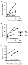
Native major surface protein 1 (MSP1) of the ehrlichial pathogen Anaplasma marginale induces protective immunity in calves challenged with homologous and heterologous strains. MSP1 is a heteromeric complex of a single MSP1a protein covalently associated with MSP1b polypeptides, of which at least two (designated MSP1F1 and MSP1F3) in the Florida strain are expressed. Immunization with recombinant MSP1a and MSP1b alone or in combination fails to provide protection. The protective immunity in calves immunized with native MSP1 is associated with the development of opsonizing and neutralizing antibodies, but CD4(+) T-lymphocyte responses have not been evaluated. CD4(+) T lymphocytes participate in protective immunity to ehrlichial pathogens through production of gamma interferon (IFN-gamma), which promotes switching to high-affinity immunoglobulin G (IgG) and activation of phagocytic cells to produce nitric oxide. Thus, an effective vaccine for A. marginale and related organisms should contain both T- and B-lymphocyte epitopes that induce a strong memory response that can be recalled upon challenge with homologous and heterologous strains. This study was designed to determine the relative contributions of MSP1a and MSP1b proteins, which contain both variant and conserved amino acid sequences, in stimulating memory CD4(+) T-lymphocyte responses in calves immunized with native MSP1. Peripheral blood mononuclear cells and CD4(+) T-cell lines from MSP1-immunized calves proliferated vigorously in response to the immunizing strain (Florida) and heterologous strains of A. marginale. The conserved MSP1-specific response was preferentially directed to the carboxyl-terminal region of MSP1a, which stimulated high levels of IFN-gamma production by CD4(+) T cells. In contrast, there was either weak or no recognition of MSP1b proteins. Paradoxically, all calves developed high titers of IgG antibodies to both MSP1a and MSP1b polypeptides. These findings suggest that in calves immunized with MSP1 heteromeric complex, MSP1a-specific T lymphocytes may provide help to MSP1b-specific B lymphocytes. The data provide a basis for determining whether selected MSP1a CD4(+) T-lymphocyte epitopes and selected MSP1a and MSP1b B-lymphocyte epitopes presented on the same molecule can stimulate a protective immune response.
Figures


Similar articles
-
Major histocompatibility complex class II DR-restricted memory CD4(+) T lymphocytes recognize conserved immunodominant epitopes of Anaplasma marginale major surface protein 1a.Infect Immun. 2002 Oct;70(10):5521-32. doi: 10.1128/IAI.70.10.5521-5532.2002. Infect Immun. 2002. PMID: 12228278 Free PMC article.
-
CD4(+) T-lymphocyte and immunoglobulin G2 responses in calves immunized with Anaplasma marginale outer membranes and protected against homologous challenge.Infect Immun. 1998 Nov;66(11):5406-13. doi: 10.1128/IAI.66.11.5406-5413.1998. Infect Immun. 1998. PMID: 9784551 Free PMC article.
-
Antibodies to Anaplasma marginale major surface proteins 1a and 1b inhibit infectivity for cultured tick cells.Vet Parasitol. 2003 Feb 13;111(2-3):247-60. doi: 10.1016/s0304-4017(02)00378-3. Vet Parasitol. 2003. PMID: 12531299
-
Mapping of B-cell epitopes in the N-terminal repeated peptides of Anaplasma marginale major surface protein 1a and characterization of the humoral immune response of cattle immunized with recombinant and whole organism antigens.Vet Immunol Immunopathol. 2004 Apr;98(3-4):137-51. doi: 10.1016/j.vetimm.2003.11.003. Vet Immunol Immunopathol. 2004. PMID: 15010223
-
Adaptations of the tick-borne pathogen, Anaplasma marginale, for survival in cattle and ticks.Exp Appl Acarol. 2002;28(1-4):9-25. doi: 10.1023/a:1025329728269. Exp Appl Acarol. 2002. PMID: 14570114 Review.
Cited by
-
Complete genome sequencing of Anaplasma marginale reveals that the surface is skewed to two superfamilies of outer membrane proteins.Proc Natl Acad Sci U S A. 2005 Jan 18;102(3):844-9. doi: 10.1073/pnas.0406656102. Epub 2004 Dec 23. Proc Natl Acad Sci U S A. 2005. PMID: 15618402 Free PMC article.
-
Functional and immunological relevance of Anaplasma marginale major surface protein 1a sequence and structural analysis.PLoS One. 2013 Jun 11;8(6):e65243. doi: 10.1371/journal.pone.0065243. Print 2013. PLoS One. 2013. PMID: 23776456 Free PMC article.
-
Development of a subcutaneous ear implant to deliver an anaplasmosis vaccine to dairy steers.J Anim Sci. 2020 Jun 1;98(6):skz392. doi: 10.1093/jas/skz392. J Anim Sci. 2020. PMID: 31889177 Free PMC article.
-
Targeting the tick/pathogen interface for developing new anaplasmosis vaccine strategies.Vet Res Commun. 2007 Aug;31 Suppl 1:91-6. doi: 10.1007/s11259-007-0070-z. Vet Res Commun. 2007. PMID: 17682853
-
Immunogenicity of Anaplasma marginale type IV secretion system proteins in a protective outer membrane vaccine.Infect Immun. 2007 May;75(5):2333-42. doi: 10.1128/IAI.00061-07. Epub 2007 Mar 5. Infect Immun. 2007. PMID: 17339347 Free PMC article.
References
-
- Adler H, Peterhans E, Nicolet J, Jungi T W. Inducible l-arginine-dependent nitric oxide synthase activity in bone marrow-derived macrophages. Biochem Biophys Res Commun. 1994;198:510–515. - PubMed
Publication types
MeSH terms
Substances
Grants and funding
LinkOut - more resources
Full Text Sources
Other Literature Sources
Research Materials

