. 2001 Nov;69(11):7029-38.
doi: 10.1128/IAI.69.11.7029-7038.2001.

Generation and surface localization of intact M protein in Streptococcus pyogenes are dependent on sagA

Affiliations

Generation and surface localization of intact M protein in Streptococcus pyogenes are dependent on sagA

I Biswas et al. Infect Immun. 2001 Nov.

Abstract

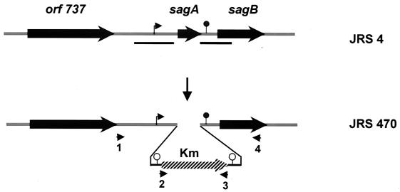
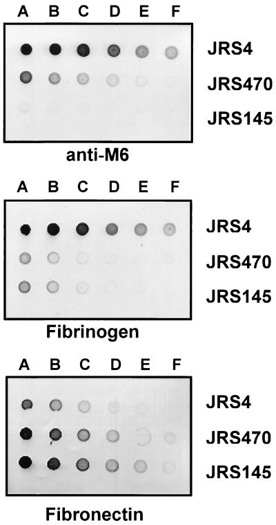
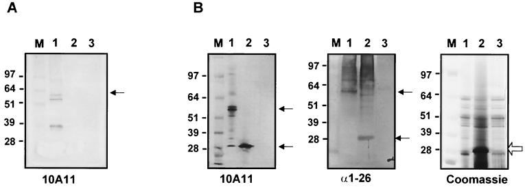
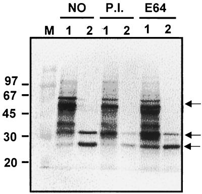
The M protein is an important surface-located virulence factor of Streptococcus pyogenes, the group A streptococcus (GAS). Expression of M protein is primarily controlled by Mga, a transcriptional activator protein. A recent report suggested that the sag locus, which includes nine genes necessary and sufficient for production of streptolysin S, another GAS virulence factor, is also needed for transcription of emm, encoding the M protein (Z. Li, D. D. Sledjeski, B. Kreikemeyer, A. Podbielski, and M. D. Boyle, J. Bacteriol. 181:6019-6027, 1999). To investigate this in more detail, we constructed an insertion-deletion mutation in sagA, the first gene in the sag locus, in the M6 strain JRS4. The resulting strain, JRS470, produced no detectable streptolysin S and showed a drastic reduction in cell surface-associated M protein, as measured by cell aggregation and Western blot analysis. However, transcription of the emm gene was unaffected by the sagA mutation. Detailed analysis with monoclonal antibodies and an antipeptide antibody showed that the M protein in the sagA mutant strain was truncated so that it lacks the C-repeat region and the C-terminal domain required for anchoring it to the cell surface. This truncated M protein was largely found, as expected, in the culture supernatant. Lack of surface-located M protein made the sagA mutant strain susceptible to phagocytosis. Thus, although sagA does not affect transcription of the M6 protein gene, it is needed for the surface localization of this important virulence factor.


Figures

FIG. 1
FIG. 1
Schematic diagram of sagA insertion-deletion mutant derivative of JRS4. The omega-Km2 cassette (hatched box) was used to replace the sagA locus to generate JRS470. Thick arrows indicate ORFs. The chromosomal segments used as the homologous region for gene replacement are shown by black lines below the diagram of JRS4. The putative promoter of the sag operon is indicated by a bent arrow and the putative transcriptional terminator is shown by a filled circle. Transcriptional terminators present in the omega-Km2 cassette are shown by open circles. Arrowheads represent primers (1 to 4) used to confirm the gene replacement event.
FIG. 2
FIG. 2
Expression of surface proteins in the sagA mutant. Twofold serial dilutions starting with 2.5 cell units/ml (A to F) from overnight cultures of the wild type (JRS4), the sagA mutant (JRS470), and the M deletion mutant strain (JRS145) were spotted onto a nitrocellulose membrane and probed with anti-M6 antibody (MAb 10A11), DIG-labeled fibrinogen, and DIG-labeled fibronectin as indicated.
FIG. 3
FIG. 3
Transcription of the emm gene in the wild-type and sagA mutant strains at different stages of growth. (A) Growth curve indicating the times of RNA isolation at early and mid-log phases of growth and at the transition to the stationary phase of growth (arrows). (B) Hybridization of specific DNA probes internal to the genes indicated to RNA isolated at the stages of growth indicated in panel A. On each filter, 2.0 and 0.5 μg of RNA was applied in vertically arranged duplicates.
FIG. 4
FIG. 4
Analysis of the M protein of the wild type and the sagA mutant. (A) Schematic diagram of the structure of the M6 protein indicating the location of the epitopes recognized by the antibodies used. Repeat regions are shown by capital letters and the cell-associated region and cell wall anchoring signals are indicated. (B and C) Western blot analysis of whole-cell extracts separated by SDS–10% (B) or 4 to 12% (C) PAGE. The antibodies used are indicated below the gels. Lanes contain extracts from JRS4 (lanes 1), JRS470 (lanes 2), and JRS145 (lanes 3). M, Rainbow (B; Amersham) or See Blue (C; Invitrogen) molecular mass marker. Bands that reacted with anti-M antibody are indicated by arrows.
FIG. 5
FIG. 5
Localization of M6 protein in the mutant and wild-type strains. Analysis of cell wall fractions (A) or concentrated culture supernatants (B) from various GAS strains separated on SDS–4 to 12% polyacrylamide gels and probed with MAb 10A11 or anti-amino acids 1 to 26 (α1-26) or stained with Coomassie blue, as indicated below each panel. Lanes contain samples from JRS4 (lanes 1), JRS470 (lanes 2), and JRS145 (lanes 3). M, See Blue (Invitrogen) molecular mass markers whose sizes are indicated on the left of each panel. Bands that reacted with anti-M antibody are indicated by arrows.
FIG. 6
FIG. 6
M proteins in cell extracts grown in the presence or absence of protease inhibitors. The wild-type (lane 1) and sagA mutant (lane 2) strains were grown in the absence of protease inhibitor (NO), the presence of complete protease inhibitor (P.I.), or with cysteine protease inhibitor E64. Whole-cell extracts were separated on SDS–10% polyacrylamide gels and reacted with MAb 10A11. M, molecular mass marker (Rainbow).

References

    1. Akesson P, Sjoholm A G, Bjorck L. Protein SIC, a novel extracellular protein of Streptococcus pyogenes interfering with complement function. J Biol Chem. 1996;271:1081–1088. - PubMed
    1. Ashbaugh C D, Warren H B, Carey V J, Wessels M R. Molecular analysis of the role of the group A streptococcal cysteine protease, hyaluronic acid capsule, and M protein in a murine model of human invasive soft-tissue infection. J Clin Investig. 1998;102:550–560. - PMC - PubMed
    1. Beckert S, Kreikemeyer B, Podbielski A. Group A streptococcal rofA gene is involved in the control of several virulence genes and eukaryotic cell attachment and internalization. Infect Immun. 2001;69:534–537. - PMC - PubMed
    1. Berge A, Bjorck L. Streptococcal cysteine proteinase releases biologically active fragments of streptococcal surface proteins. J Biol Chem. 1995;270:9862–9867. - PubMed
    1. Betschel S D, Borgia S M, Barg N L, Low D E, De Azavedo J C. Reduced virulence of group A streptococcal Tn916 mutants that do not produce streptolysin S. Infect Immun. 1998;66:1671–1679. - PMC - PubMed

Publication types

MeSH terms

LinkOut - more resources