. 2001 Oct;12(10):2907-20.
doi: 10.1091/mbc.12.10.2907.

Adaptins: the final recount

Affiliations
Free PMC article

Adaptins: the final recount

M Boehm et al. Mol Biol Cell. 2001 Oct.
Free PMC article

Abstract

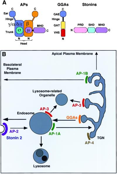
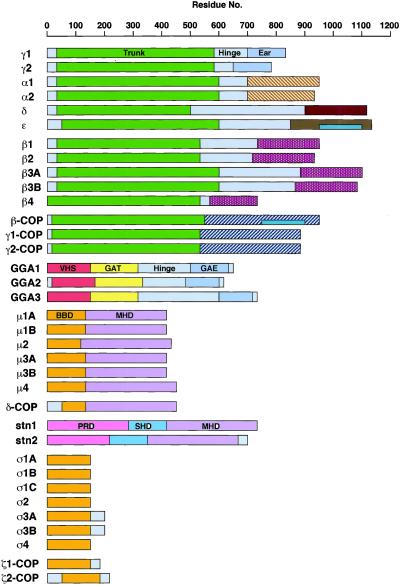
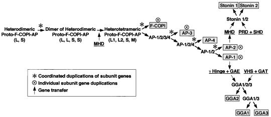
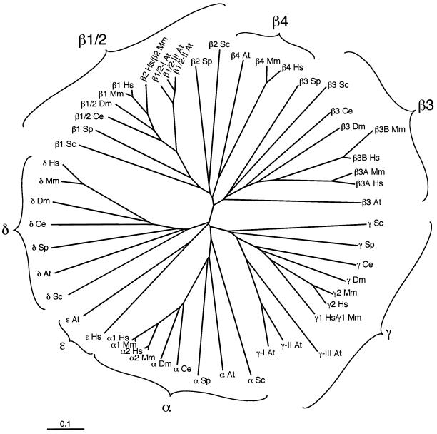
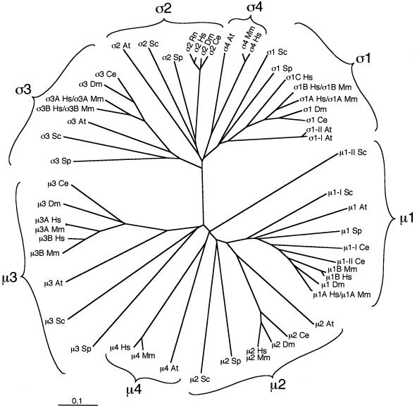
Adaptins are subunits of adaptor protein (AP) complexes involved in the formation of intracellular transport vesicles and in the selection of cargo for incorporation into the vesicles. In this article, we report the results of a survey for adaptins from sequenced genomes including those of man, mouse, the fruit fly Drosophila melanogaster, the nematode Caenorhabditis elegans, the plant Arabidopsis thaliana, and the yeasts, Saccharomyces cerevisiae and Schizosaccharomyces pombe. We find that humans, mice, and Arabidopsis thaliana have four AP complexes (AP-1, AP-2, AP-3, and AP-4), whereas D. melanogaster, C. elegans, S. cerevisiae, and S. pombe have only three (AP-1, AP-2, and AP-3). Additional diversification of AP complexes arises from the existence of adaptin isoforms encoded by distinct genes or resulting from alternative splicing of mRNAs. We complete the assignment of adaptins to AP complexes and provide information on the chromosomal localization, exon-intron structure, and pseudogenes for the different adaptins. In addition, we discuss the structural and evolutionary relationships of the adaptins and the genetic analyses of their function. Finally, we extend our survey to adaptin-related proteins such as the GGAs and stonins, which contain domains homologous to the adaptins.


Figures

Figure 1
Figure 1
Schematic representation of adaptins and related proteins (A) and of the trafficking pathways in which they might be involved (B). The scheme in A depicts the structure of a generic AP complex, a GGA, and a stonin. N and C indicate the amino- and carboxy-termini of the proteins, respectively. GAE, γ-adaptin ear-homology domain; GAT, GGA, and TOM1-homology domain; MHD, μ-homology domain; PRD, proline-rich domain; SHD, stonin-homology domain; VHS, Vps27p/HRS/STAM-homology domain. B is a scheme of postGolgi trafficking pathways, showing the putative localization and role AP complexes, GGAs, and stonins within the cell. Of these, only the localization and role of AP-2 have been established with certainty. All the others should be considered tentative.
Figure 2
Figure 2
Bar diagram showing the structural relationships between the different adaptins and adaptin-related proteins. Regions in the proteins as defined by homology, function, or tertiary structural information, as well as protein family (Pfam) domains are depicted as colored boxes.
Figure 2
Figure 2
Bar diagram showing the structural relationships between the different adaptins and adaptin-related proteins. Regions in the proteins as defined by homology, function, or tertiary structural information, as well as protein family (Pfam) domains are depicted as colored boxes.
Figure 3
Figure 3
Possible evolution of the COPI-AP coat components. The order of the appearance of distinct complexes was deduced from the phylogenetic analysis of all adaptins present in A. thaliana, C. elegans, D. melanogaster, H. sapiens, M. musculus, and S. cerevisiae. The GGAs and stonins are hypothesized to have evolved after the γ and μ2 adaptins were differentiated, respectively, based on their higher degree of homology to domains from these adaptins. Asterisks indicate coordinated rounds of gene duplication, while dashed arrows show gene transfer. See text for more details.
Figure 4
Figure 4
An unrooted phylogenetic tree displaying the evolutionary relationship between the large adaptins from A. thaliana (At), C. elegans (Ce), D. melanogaster (Dm), H. sapiens (Hs), M. musculus (Mm), S. cerevisiae (Sc), and S. pombe (Sp). The γ/α/δ/ε and β1–4 subunits from these organisms were aligned, and a phylogenetic tree was calculated using the EMBL European Bioinformatics Institute ClustalW algorithm (http://www2.ebi.ac.uk/clustalw) and displayed using the TreeviewPPC program. The evolutionary distance is indicated by substitutions per site.
Figure 5
Figure 5
Analysis of the evolutionary relationship between the small and medium adaptins calculated as described for Figure 4. Note that for ς2, the R. norvegicus (Rn) sequence was used, as the M. musculus ς2 sequence is not yet available.

References

    1. Adams MD, et al. The genome sequence of Drosophila melanogaster. Science. 2000;287:2185–2195. - PubMed
    1. Aguilar RC, Boehm M, Gorshkova I, Crouch RJ, Tomita K, Saito T, Ohno H, Bonifacino JS. Signal-binding specificity of the μ4 subunit of the adaptor protein complex AP-4. J Biol Chem. 2001;276:13145–13152. - PubMed
    1. Aguilar RC, Ohno H, Roche KW, Bonifacino JS. Functional domain mapping of the clathrin-associated adaptor medium chains μ1 and μ2. J Biol Chem. 1997;272:27160–27166. - PubMed
    1. Andrews J, Smith M, Merakovsky J, Coulson M, Hannan F, Kelly LE. The stoned locus of Drosophila melanogasterproduces a dicistronic transcript and encodes two distinct polypeptides. Genetics. 1996;143:1699–1711. - PMC - PubMed
    1. Ball CL, Hunt SP, Robinson MS. Expression and localization of α-adaptin isoforms. J Cell Sci. 1995;108:2865–2875. - PubMed

Publication types

MeSH terms

Substances