. 2001 Oct;12(10):2921-33.
doi: 10.1091/mbc.12.10.2921.

Cytoplasmic dynein light intermediate chain is required for discrete aspects of mitosis in Caenorhabditis elegans

Affiliations
Free PMC article

Cytoplasmic dynein light intermediate chain is required for discrete aspects of mitosis in Caenorhabditis elegans

J H Yoder et al. Mol Biol Cell. 2001 Oct.
Free PMC article

Abstract

We describe phenotypic characterization of dli-1, the Caenorhabditis elegans homolog of cytoplasmic dynein light intermediate chain (LIC), a subunit of the cytoplasmic dynein motor complex. Animals homozygous for loss-of-function mutations in dli-1 exhibit stochastic failed divisions in late larval cell lineages, resulting in zygotic sterility. dli-1 is required for dynein function during mitosis. Depletion of the dli-1 gene product through RNA-mediated gene interference (RNAi) reveals an early embryonic requirement. One-cell dli-1(RNAi) embryos exhibit failed cell division attempts, resulting from a variety of mitotic defects. Specifically, pronuclear migration, centrosome separation, and centrosome association with the male pronuclear envelope are defective in dli-1(RNAi) embryos. Meiotic spindle formation, however, is not affected in these embryos. DLI-1, like its vertebrate homologs, contains a putative nucleotide-binding domain similar to those found in the ATP-binding cassette transporter family of ATPases as well as other nucleotide-binding and -hydrolyzing proteins. Amino acid substitutions in a conserved lysine residue, known to be required for nucleotide binding, confers complete rescue in a dli-1 mutant background, indicating this is not an essential domain for DLI-1 function.


Figures

Figure 1
Figure 1
Map position and genetic structure of dli-1. (A) Top line shows a portion of the genetic map from linkage group IV with relative positions of relevant genetic markers. Below are the three cosmids injected as a pool that confer rescue in the dli-1 alleles. The cosmid C39E9 confers rescue alone as does the large XhoI deletion and the subclone pJHY10. (B) Structure of the dli-1 gene showing locations of lesions within the three alleles and the two splice variants as determined by cDNA sequence comparison.
Figure 2
Figure 2
Alignment between DLI-1, rat LIC1, and human LIC1. Dark shaded boxes show identical residues in two or more of the three proteins shown. Light gray boxes highlight similar residues. There is 33% sequence identity throughout the full length of the proteins and 51% similarity. The underlined residues constitute the putative P-loop sequence and the asterisk highlights the conserved lysine residue that is mutated in our rescuing constructs to either alanine or asparginine. Sequences were aligned with the use of Clustal W.
Figure 3
Figure 3
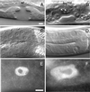
dli-1 alleles undergo stochastic failed cell divisions in many postembryonic lineages. (A and C) Wild-type C. elegans at late L4, the last larval stage before the adult molt. (B and D) Similarly staged ku266 animals. (A) Wild-type late L4 vulva. Only 12 of 22 cells are visible in this plane of focus. (B) ku266 late L4 vulva. The enlarged nuclei are the result of failed divisions. Several of these cells contain multiple nucleoli (arrowheads) indicative of failed chromosome segregation. (C) Gonad of a wild-type animal composed of many syncitial germline nuclei. (D) Germline nuclei in ku266 animals also have abnormal morphology and are probably polyploid as a result of failed mitotic division in the distal end of the gonad arm. (E) Wild-type somatic sheath cell nucleus visualized by staining with anti-CEH-18 antibodies. (F) Sheath cell nucleus of a ku266 animal. Like all other late postembryonic lineages observed, the somatic sheath cells also appear to undergo failed division attempts, resulting in too few cells (average of 6 per gonad arm compared with 10 in wild-type) with abnormal nuclear morphology. Bar, ∼10 μm.
Figure 4
Figure 4
Comparison of pronuclear migration in wild-type and dli-1(RNAi) embryos. All images are oriented with anterior to the left. (A–D) Wild-type embryo; female (anterior) and male (posterior) pronuclei begin migration before regression of the pseudocleavage furrow. The two pronuclei meet and fuse before nuclear envelope breakdown. (E–H) In the majority (16/19) of dli-1(RNAi) embryos harvested from injected wild-type mothers (shown) and 100% of embryos harvested from injected dli-1 heterozygous mothers (n = 19), pronuclear migration does not occur. (F) At the completion of pseudocleavage furrow regression, neither pronucleus has initiated migration. (G) At a time point equivalent to that in B in which the wild-type embryo is in metaphase, no pronuclear migration is apparent, and the male pronucleus has undergone NEB before female pronuclear NEB. (H) Multiple nuclei were formed in all dli-1(RNAi) embryos observed (arrowheads). Timestamps are relative to completion of anterior cytoplasmic contractions. In a wild-type embryo almost immediately after this, the female pronucleus initiates posteriorward migration. Bar, ∼10 μm.
Figure 5
Figure 5
Meiotic spindles are formed in dli-1(RNAi) embryos, but not in all dhc-1(RNAi) embryos observed. All embryos are oriented with anterior to the left. (A, D, and G) Anti-tubulin staining to reveal microtubules. (B, E, and H) DAPI staining to reveal DNA. (A–C) Wild-type embryo at meiosis II. A barrel-shaped spindle surrounds the female meiotic chromosomes. One polar body has already been extruded (B and C, arrow). The male meiotic chromosomes are tightly condensed at the posterior end of the embryo. (D–F) dli-1(RNAi) embryo slightly later in meiosis II. A barrel-shaped meiotic spindle has also formed around the condensed meiotic chromosomes. The spindle has rotated to the anterior/posterior axis to complete the meiosis II division. A single polar body has also already been extruded (E and F, arrow). (G–I) dhc-1(RNAi) embryo. No meiotic spindle is observable and the female meiotic chromosomes are scattered throughout the embryo. Bar, ∼10 μm.
Figure 6
Figure 6
DLI-1 is required for centrosome separation in the one-cell C. elegans embryo. Cytological analysis of metaphase embryos stained for the centrosomal component ZYG-9, tubulin, or DNA as indicated. All images are oriented with anterior to the left. (A–C) Wild-type embryo at metaphase. (A) Anti-ZYG-9 stains the two centrosomes that have rotated and aligned along an anterior-posterior axis. (B) Centrosomes nucleate microtubules forming the bipolar spindle visualized by anti-tubulin staining. (C) DAPI staining shows chromosomes condensed to the metaphase plate. (D–F) dli-1(RNAi) embryo in which centrosomes did separate. (D) Centrosomes have separated but have failed to centrate. This panel represents the most successful degree of rotation and spindle alignment observed. (E and F) Centrosomes do nucleate microtubules and chromosomes are condensed but have not yet congressed to a metaphase plate. (G–I) dli-1(RNAi) embryo exhibiting the most severe centrosome separation phenotype. (G) Some embryos (10/17) from wild-type injections failed to separate centrosomes and therefore formed no bipolar spindle. (H) Microtubules are nucleated and chromosomes have condensed and congressed to a metaphase plate (I). Embryos from heterozygous injections (7/8) were phenotypically identical to the embryo in G–I. Bar, ∼10 μm.
Figure 7
Figure 7
Centrosomes are dissociated from the male pronucleus in 32% of dli-1(RNAi). In 5/17 embryos from wild-type–injected mothers and 3/8 embryos from dli-1 heterozygous injected mothers, centrosomes (arrows) were dissociated from the male pronucleus (arrowheads). (A–D) Embryo from a dli-1 heterozygous-injected mother showing centrosomes as visualized by anti-ZYG-9 staining (A), microtubules visualized by anti-tubulin staining (B), DAPI staining to reveal DNA (C), and merged (D). Bar, ∼10 μm.
Figure 8
Figure 8
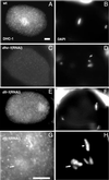
DHC-1 localizes correctly in dli-1(RNAi) embryos. (A) DHC-1 localization to the mitotic spindle in a wild-type metaphase embryo. (B) The same wild-type embryo stained with DAPI to reveal metaphase chromosomes. (C and D) dli-1(RNAi) embryo also at metaphase. DHC-1 localizes to either side of the condensed metaphase chromosomes and the mitotic spindle. Centrosomes have apparently separated enough in this embryo for a mitotic spindle to form. (E and F) dhc-1(RNAi) embryo also at metaphase. No DHC-1 staining is detectable. (G and H) Posterior end of a dli-1(RNAi) embryo as chromosomes are condensing at metaphase. When centrosomes fail to separate and no spindle is formed, DHC-1 staining is still observed on metaphase (arrowhead) and prometaphase (arrow) chromosomes. Bars, ∼10 μm.

Similar articles

Cited by

References

    1. Albertson DG. C. elegans II. In: Riddle DL, Blumenthal T, Meyer BJ, Priess JR, editors. C. elegans II. Cold Spring Harbor, NY: Cold Spring Harbor Laboratory Press; 1997. pp. 47–78.
    1. Azzaria M, Schurr E, Gros P. Discrete mutations introduced in the predicted nucleotide-binding sites of the mdr1 gene abolish its ability to confer multidrug resistance. Mol Cell Biol. 1989;9:5289–5297. - PMC - PubMed
    1. Barstead RJ, Kleiman L, Waterston RH. Cloning, sequencing, and mapping of an alpha-actinin gene from the nematode Caenorhabditis elegans. Cell Motil Cytoskeleton. 1991;20:69–78. - PubMed
    1. Beckwith SM, Roghi CH, Liu B, Ronald Morris N. The “8-kD” cytoplasmic dynein light chain is required for nuclear migration and for dynein heavy chain localization in Aspergillus nidulans. J Cell Biol. 1998;143:1239–1247. - PMC - PubMed
    1. Bielli A, Thornqvist PO, Hendrick AG, Finn R, Fitzgerald K, McCaffrey MW. The small GTPase Rab4A interacts with the central region of cytoplasmic dynein light intermediate chain-1. Biochem Biophys Res Commun. 2001;281:1141–1153. - PubMed

Publication types

MeSH terms

LinkOut - more resources