. 2001 Oct;12(10):3004-15.
doi: 10.1091/mbc.12.10.3004.

Characterization of a di-leucine-based signal in the cytoplasmic tail of the nucleotide-pyrophosphatase NPP1 that mediates basolateral targeting but not endocytosis

Affiliations
Free PMC article

Characterization of a di-leucine-based signal in the cytoplasmic tail of the nucleotide-pyrophosphatase NPP1 that mediates basolateral targeting but not endocytosis

V Bello et al. Mol Biol Cell. 2001 Oct.
Free PMC article

Abstract

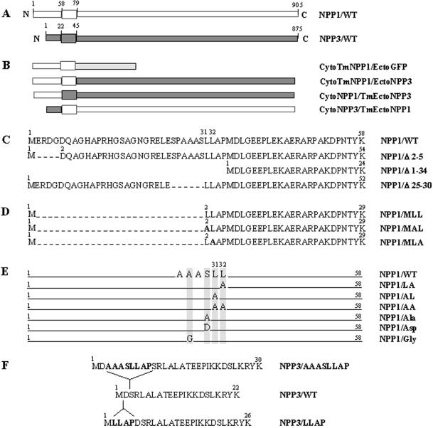
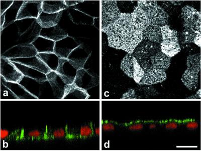
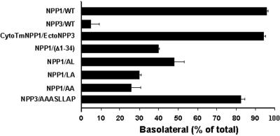
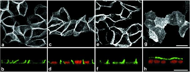
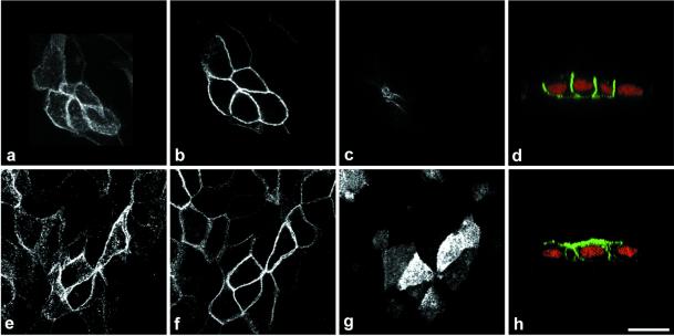
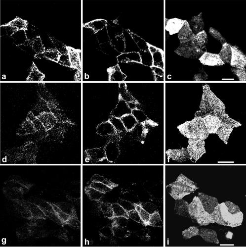
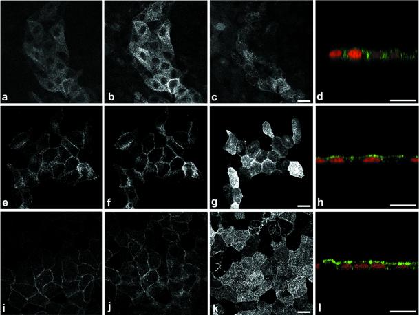
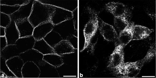
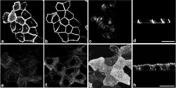
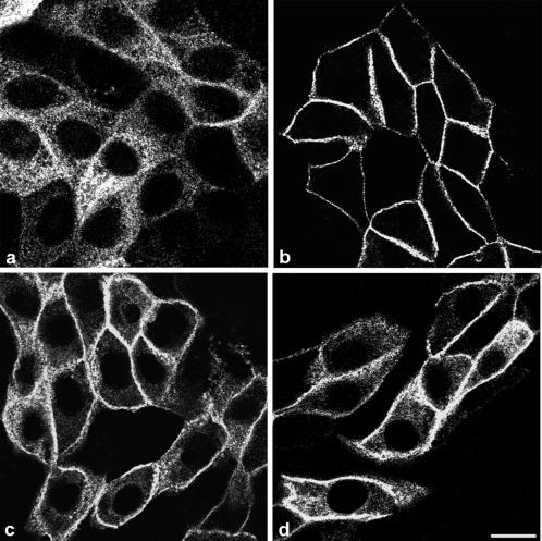
Enzymes of the nucleotide pyrophosphatase/phosphodiesterase (NPPase) family are expressed at opposite surfaces in polarized epithelial cells. We investigated the targeting signal of NPP1, which is exclusively expressed at the basolateral surface. Full-length NPP1 and different constructs and mutants were transfected into the polarized MDCK cell line. Expression of the proteins was analyzed by confocal microscopy and surface biotinylation. The basolateral signal of NPP1 was identified as a di-leucine motif located in the cytoplasmic tail. Mutation of either or both leucines largely redirected NPP1 to the apical surface. Furthermore, addition of the conserved sequence AAASLLAP redirected the apical nucleotide pyrophosphatase/phosphodiesterase NPP3 to the basolateral surface. Full-length NPP1 was not significantly internalized. However, when the cytoplasmic tail was deleted upstream the di-leucine motif or when the six upstream flanking amino acids were deleted, the protein was mainly found intracellularly. Endocytosis experiments indicated that these mutants were endocytosed from the basolateral surface. These results identify the basolateral signal of NPP1 as a short sequence including a di-leucine motif that is dominant over apical determinants and point to the importance of surrounding amino acids in determining whether the signal will function as a basolateral signal only or as an endocytotic signal as well.


Figures

Figure 1
Figure 1
Schematic representation of wild-type proteins and constructs. (A) NPP1 and NPP3 wild-types. Numbers indicate the limits between the cytoplasmic, transmembrane and ectodomains. (B) Chimeras. In the CytoTmNPP1/EctoGFP and CytoTmNPP1/EctoNPP3 chimeras the ectodomain of NPP1 is replaced either by GFP or by the ectodomain of NPP3. The CytoNPP1/TmEctoNPP3 chimera is made of the cytoplasmic domain of NPP1 and the transmembrane and ectodomain of NPP3. The CytoNPP3/TmEctoNPP1 chimera combines the cytoplasmic domain of NPP3 and the transmembrane and ectodomain of NPP1. (C) Amino acid sequence of the cytoplasmic domain of NPP1/WT, NPP1/Δ2-5 deleted of residues 2–5, NPP1/Δ1-34 deleted of the first 34 residues and NPP1/Δ25-30 deleted of residues 25–30. (D) Amino acid sequence of the cytoplasmic domain of NPP1/MLL that is deleted from amino acids 2–30 and the mutants NPP1/MAL and NPP1/MLA. Alanine substitutions are in boldface. (E) Point and double mutations of leu31-leu32 (NPP1/LA, NPP1/AL, NPP1/AA) and point mutations of Ser30 (NPP1/Ala, NPP1/Asp), and Ala28 (NPP1/Gly) in full-length NPP1. (F) Amino acid sequence of the cytoplasmic domain of NPP3/WT and of the NPP3/AAASLLAP and NPP3/LLAP chimeras which respectively contain the additional amino acid stretches AAASLLAP and LLAP indicated in bold.
Figure 2
Figure 2
Confocal microscopic examination of the expression of NPP1 and NPP3 wild-types in polarized MDCK cells. MDCK cells stably transfected with NPP1 or NPP3 wild-types were grown on Transwell filters units, fixed, permeabilized and stained with mAb IR518 anti-mouse NPP1 (a and b) or with mAB B10 anti-rat NPP3 (c and d) and secondary FITC-conjugated antibody as described in MATERIALS AND METHODS. NPP1/WT is mainly restricted to the basolateral surface (a and b), whereas NPP3/WT is expressed at the apical surface (c and d). (a and c) Projections of all 0.5-μm xy focal sections taken throughout the height of the cells. (b and d) xz focal sections perpendicular to the xy focal sections; nuclei are stained in red with propidium iodide. Bar, 20 μm.
Figure 3
Figure 3
Quantification of the steady-state distribution of NPP1 and NPP3 wild-types, and different constructs. MDCK cells expressing NPP1/WT, NPP3/WT, CytoTmNPP1/EctoNPP3, NPP1/Δ1-34, NPP1/LA, NPP1/AL, NPP1/AA, and NPP3/AAASLLAP were grown to confluence on Transwell filters, labeled for 16 h with [35S]methionine/cysteine, and biotinylated either from the apical or from the basolateral sides. Proteins were immunoprecipitated, then the biotinylated proteins were recovered by streptavidin precipitation and analyzed by electrophoresis and fluorography. Results are means ± SD of three to five determinations.
Figure 4
Figure 4
The cytoplasmic domain of NPP1/WT is necessary and sufficient for basolateral sorting. Cells were processed for indirect immunofluorescence microscopy as described in the legend of Figure 2 and analyzed by projections of all 0.5-μm confocal xy sections taken through the height of the cells (a, c, e, and g), and xz sections (b, d, f, and h) are represented for each chimera. CytoTmNPP1/EctoGFP (a and b), CytoTmNPP1/EctoNPP3 (c and d), CytoNPP1/TmEctoNPP3 (e and f), and CytoNPP3/TmEctoNPP1 (g and h). Bars, 20 μm.
Figure 5
Figure 5
The basolateral signal is localized within amino acids 6–34. Localization of truncated protein NPP1/Δ2-5 (a–d) and NPP1/Δ1-34 (e–h) was analyzed by confocal laser scanning microscopy. Each picture is a sum of three 0.5-μm confocal microscopy xy sections from the bottom (a and e), middle (b and f), and top (c and g) of the cells. (d and h) xz sections. Bar, 20 μm.
Figure 6
Figure 6
Basolateral sorting requires a di-leucine motif. Expression of point mutants NPP1/AL (a–c), NPP1/LA (d–f), and NPP1/AA (g–i) in MDCK cells. Each pictures is a sum of three 0.5-μm confocal microscopy xy sections from the bottom (a, d, and g), middle (b, e, and h), and top (c, f, and i) of the cells. Bars, 20 μm.
Figure 7
Figure 7
Truncation of the tail upstream the di-leucine motif unmasks an endocytosis signal. Expression of NPP1/MLL (a–d), NPP1/MAL (e–h), and NPP1/MLA (i–l). Each panel is a sum of three 0.5-μm confocal microscopy xy sections from the bottom (a, e, and i), middle (b, f, and j), and top (c, g, and k) of the cells. (d, h, and l) xz sections. Bars, 20 μm.
Figure 8
Figure 8
Morphological analysis of NPP1/WT and NPP1/MLL endocytosis. Filter-grown cells stably transfected with NPP1/WT (a) or NPP1/MLL (b) were incubated for 1 h at 37°C with mAb IR 518 anti-mouse NPP1 added to the basolateral medium, as described in MATERIALS AND METHODS. After washing, fixation, and permeabilization, the antibody was visualized with a secondary FITC-conjugated antibody. (a) and (b) Single 0.5-μm xy sections taken from the middle of the cells. Bars, 20 μm.
Figure 9
Figure 9
The sequence AAASLLAP but not the sequence LLAP is able to redirect NPP3 to the basolateral surface. MDCK cells stably transfected with NPP3/AAASLLAP or with NPP3/LLAP were studied by confocal microscopy with the use of a mAb anti-NPP3 as described in the legend of Figure 2. Each picture is a sum of three 0.5-μm confocal microscopy xy focal sections taken at the bottom (a), middle (b), and top (c) of the cells. (d) An xz section. Bar, 20 μm.
Figure 10
Figure 10
Flanking upstream amino acids are required for specific basolateral targeting of NPP1. Expression of deletion mutant NPP1/Δ25-30 (a) and point mutants NPP1/Gly (b), NPP1/Asp (c), and NPP1/Ala (d) were studied by confocal microscopy. Each panel is a single xy section taken from the middle of the cells. Bars, 20 μm.

References

    1. Ali SA, Steinkasserer A. PCR-ligation-PCR mutagenesis: a protocol for creating gene fusions and mutations. Biotechniques. 1995;18:746–750. - PubMed
    1. Aroeti B, Okhrimenko H, Reich V, Orzech E. Polarized trafficking of plasma membrane proteins: emerging roles for coats, SNAREs, GTPases and their link to the cytoskeleton. Biochim Biophys Acta. 1998;1376:57–90. - PubMed
    1. Belli SI, Goding JW. Biochemical characterization of human PC-1, an enzyme possessing alkaline phosphodiesterase I and nucleotide pyrophosphatase activities. Eur J Biochem. 1994;226:433–443. - PubMed
    1. Bollen M, Gijsbers R, Ceulemans H, Stalmans W, Stefan C. Nucleotide pyrophosphatases/phosphodiesterases on the move. Crit Rev Biochem Mol Biol. 2000;35:393–432. - PubMed
    1. Bonifacino JS, Dell'Angelica EC. Molecular bases for the recognition of tyrosine-based sorting signals. J Cell Biol. 1999;145:923–926. - PMC - PubMed

Publication types

MeSH terms

Substances

LinkOut - more resources