. 2001 Oct;12(10):3074-86.
doi: 10.1091/mbc.12.10.3074.

Dual role of H-Ras in regulation of lymphocyte function antigen-1 activity by stromal cell-derived factor-1alpha: implications for leukocyte transmigration

Affiliations
Free PMC article

Dual role of H-Ras in regulation of lymphocyte function antigen-1 activity by stromal cell-derived factor-1alpha: implications for leukocyte transmigration

K S Weber et al. Mol Biol Cell. 2001 Oct.
Free PMC article

Abstract

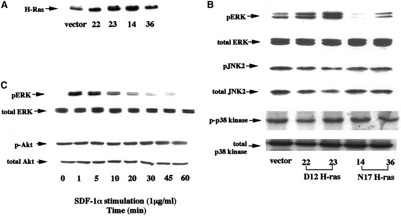
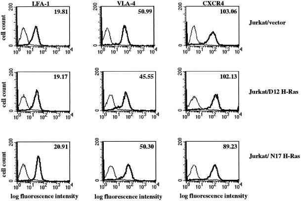
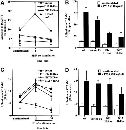
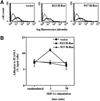
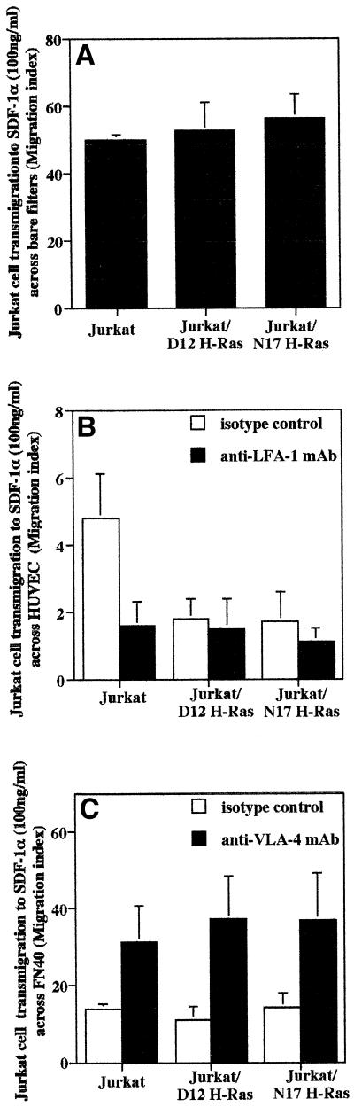
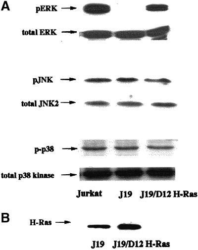
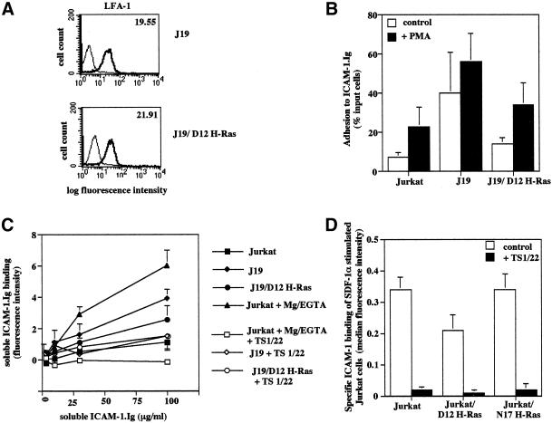
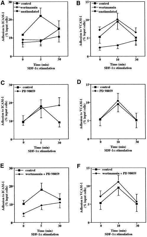
We investigated the role of H-Ras in chemokine-induced integrin regulation in leukocytes. Stimulation of Jurkat T cells with the CXC chemokine stromal cell-derived factor-1alpha (SDF-1alpha) resulted in a rapid increase in the phosphorylation, i.e., activation of extracellular signal receptor-activated kinase (ERK) but not c-Jun NH(2)-terminal kinase or p38 kinase, and phosphorylation of Akt, reflecting phosphatidylinositol 3-kinase (PI3-K) activation. Phosphorylation of ERK in Jurkat cells was enhanced and attenuated by expression of dominant active (D12) or inactive (N17) forms of H-Ras, respectively, while N17 H-Ras abrogated SDF-1alpha-induced Akt phosphorylation. SDF-1alpha triggered a transient regulation of adhesion to intercellular adhesion molecule-1 (ICAM-1) and vascular cell adhesion molecule-1 mediated by lymphocyte function antigen-1 (LFA-1) and very late antigen-4 (VLA-4), respectively, and a rapid increase in LFA-1 binding to soluble ICAM-1.Ig, which was inhibited by D12 but not N17 H-Ras. Both D12 and N17 H-Ras abrogated the regulation of LFA-1 but not VLA-4 avidity, and impaired LFA-1-mediated transendothelial chemotaxis but not VLA-4-dependent transmigration induced by SDF-1alpha. Analysis of the mutant Jurkat J19 clone revealed LFA-1 with constitutively high affinity and reduced ERK phosphorylation, which were partially restored by expression of active H-Ras. Inhibition of PI3-K blocked the up-regulation of Jurkat cell adhesion to ICAM-1 by SDF-1alpha, whereas inhibition of mitogen-activated protein kinase kinase impaired the subsequent down-regulation and blocking both pathways abrogated LFA-1 regulation. Our data suggest that inhibition of initial PI3-K activation by inactive H-Ras or sustained activation of an inhibitory ERK pathway by active H-Ras prevail to abolish LFA-1 regulation and transendothelial migration induced by SDF-1alpha in leukocytes, establishing a complex and bimodal involvement of H-Ras.


Figures

Figure 1
Figure 1
SDF-1α induces substantial phosphorylation of ERK and Akt but not JNK or p38. Jurkat cells were stimulated with SDF-1α (1 μg/ml) for indicated periods (A, B, and D) or with SDF-1α at indicated concentrations (C). Cell lysates were separated by 10% SDS-PAGE and specific mAbs were used to detect phosphorylated and total ERK (A–C), JNK, and p38 kinase (A and B) or Akt (D). Shown are representative blots from at least three separate experiments or quantitative densitometry (B) expressed as phosphorylated relative to total protein (percentage of control) of a representative experiment.
Figure 2
Figure 2
Expression of dominant active (D12) or inactive (N17) forms of H-Ras results in a selective regulation of phosphorylated ERK. Jurkat cells were stably transfected with vector alone, active (D12) or inactive (N17) H-Ras. Cell lysates from selected clones were separated by 10% SDS-PAGE and specific mAbs were used to detect total H-Ras (A), and phosphorylated and total ERK, JNK, and p38 kinase (B). Jurkat/N17 H-Ras transfectants were stimulated with SDF-1α (1 μg/ml) for indicated times, cell lysates were separated by 10% SDS-PAGE, and specific mAbs were used to detect phosphorylated and total ERK and Akt (C). Shown are representative blots from at least three separate experiments.
Figure 3
Figure 3
Expression of LFA-1, VLA-4, and CXCR4 on Jurkat cell transfectants. Jurkat cells transfected with vector alone, active (D12) or inactive (N17) H-Ras were stained with specific mAbs to LFA-1, VLA-4, and CXCR4 (bold lines) and the corresponding isotype controls (thin lines). Surface expression was measured by FACScan with appropriate gates. Shown are representative histograms with the respective specific mean fluorescence intensities.
Figure 4
Figure 4
Adhesion of Jurkat/D12 and Jurkat/N17 transfectants to ICAM-1 and VCAM-1. BCECF-AM–labeled Jurkat cell transfectants were stimulated with SDF-1α (1 μg/ml) (A and C) or PMA (100 ng/ml) (B and D) and allowed to adhere to ICAM-1 (A and B) or VCAM-1 (C and D) for indicated times. Nonadherent cells were removed by a flick wash. For mAb inhibition, vector-transfected cells were preincubated with the anti-LFA-1 mAb (TS1/22) or the anti-VLA-4 mAb (HP1/2) or the respective isotype control for 30 min on ice before being added to the assay. Fluorescence of input and adherent cells was measured in a fluorescence plate reader. Data represent the mean ± SD of at least four separate experiments performed in triplicate.
Figure 5
Figure 5
Adhesion of Jurkat cells transiently transfected with H-Ras mutants on ICAM-1. (A) Cells were transiently cotransfected with vector, or D12 or N17 H-Ras together with a cDNA-encoding GFP. Expression of GFP was measured by flow cytometry. (B) Adhesion of vector, D12 and N17 H-Ras Jurkat transfectants to ICAM-1. Transfectants labeled with BCECF-AM were stimulated with SDF-1α (1 μg/ml) and allowed to adhere to ICAM-1 for indicated times. Nonadherent cells were removed by a flick wash. Fluorescence of input and adherent cells was measured in a fluorescence plate reader. Data represent the mean ± SD of three experiments.
Figure 6
Figure 6
Transmigration of Jurkat cell transfectants to SDF-1α. Jurkat cell transfectants were allowed to transmigrate across bare filters (A), filters coated with endothelial cells (B) or the 40-kDa fragment (FN40) of fibronectin (C) to an SDF-1α gradient for 3 h (A and C) or 4 h (B). For mAb inhibition, cells were pretreated with LFA-1 mAb TS1/22, VLA-4 mAb HP1/2, or isotype control (all 10 μg/ml) for 30 min on ice which were kept present during the assays (B). Migrated cells and input were counted by flow cytometry with the use of standard beads. Data are expressed as migration index and shown are the mean ± SD of at least three separate experiments performed in duplicate.
Figure 7
Figure 7
Transfection of active H-Ras in the mutant J19 cells restores expression of phosphorylated ERK. J19 cells were transfected with dominant active D12 H-Ras. (A) Cell lysates from wild-type Jurkat, J19/vector, and J19/D12 H-Ras were separated by 10% SDS-PAGE and specific mAbs were used to detect phosphorylated and total ERK, JNK, and p38 kinase (A) and total H-Ras (B). Shown are representative blots from three separate experiments.
Figure 8
Figure 8
Transfection of active H-Ras restores low constitutive affinity of LFA-1 in the mutant J19 cells and impairs ICAM-1 binding upon SDF-1α stimulation. (A) Expression of LFA-1 on J19 cell transfectants. J19 and J19/D12 H-Ras cell transfectants were stained with specific mAb to LFA-1 (bold lines) and the corresponding isotype controls (thin lines). Surface expression was measured by FACScan with appropriate gates. Data shown are representative histograms and specific mean fluorescence intensities. (B) Jurkat, J19/vector and J19/D12 H-Ras cells were stimulated with PMA or left untreated and allowed to adhere to immobilized ICAM-1. Nonadherent cells were removed by a flick wash. Fluorescence of input and adherent cells was measured in a fluorescence plate reader. Data represent the mean ± SD of four separate experiments performed in triplicate. (C) Jurkat, J19/vector, and J19/D12 H-Ras cells were incubated with increasing concentrations of soluble ICAM-1. Ig in TBS/2 mM Mg2+/1 mM Ca2+/0.5% BSA at 37°C for 1 h, washed, and stained with FITC-conjugated anti-human IgG. Jurkat cells were also incubated in TBS/10 mM Mg2+/1 mM EGTA/0.5% BSA to induce the maximal binding to soluble ICAM-1.Ig. Binding was confirmed to be specific for LFA-1 by preincubation with the blocking LFA-1 mAb TS1/22. Shown is the mean fluorescence intensity. Data represent mean ± SD of three separate experiments. (D) Jurkat cells and transfectants were incubated with soluble ICAM-1.Ig (100 μg/ml) and FITC-conjugated antihuman Ig for 30 min, stimulated with SDF-1α (1 μg/ml) for 1 min, fixed, and analyzed in a FACScan. Induction of ICAM-1 binding was expressed as specific median fluorescence intensity stimulated by SDF-1α. The presence of LFA-1 mAb TS1/22 inhibited induction of ICAM-1 binding, revealing that it was mediated by LFA-1. Data represent mean ± SD of three separate experiments.
Figure 9
Figure 9
Involvement of PI3-K and the Raf-1/ERK kinase pathway in chemokine-induced integrin regulation. Jurkat cells were stimulated with SDF-1α (1 μg/ml) or left unstimulated and allowed to adhere for indicated times to immobilized ICAM-1 (A, C, and E) and VCAM-1 (B, D, and F). For inhibition of PI3-K and MEK kinase, cells were preincubated with wortmannin (A and B) and PD 98059 (C and D), respectively, or both (E and F) for 15 min at 37°C. Nonadherent cells were removed and fluorescence of input and adherent cells was measured in a fluorescence plate reader. Data represent the mean ± SD of at least four separate experiments performed in triplicate.

References

    1. Baggiolini M. Chemokines and leukocyte traffic. Nature. 1998;392:565–568. - PubMed
    1. Campbell JJ, Hedrick J, Zlotnik A, Siani MA, Thompson DA, Butcher EC. Chemokines and the arrest of lymphocytes rolling under flow conditions. Science. 1998;279:381–384. - PubMed
    1. Capodici C, Hanft S, Feoktistov M, Pillinger MH. Phosphatidylinositol 3-kinase mediates chemoattractant-stimulated, CD11b/CD18-dependent cell-cell adhesion of human neutrophils: evidence for an ERK-independent pathway. J Immunol. 1998;160:1901–1909. - PubMed
    1. Chaudhary A, King WG, Mattaliano MD, Frost JA, Diaz B, Morrison DK, Cobb MH, Marshall MS, Brugge JS. Phosphatidylinositol 3-kinase regulates Raf1 through Pak phosphorylation of serine 338. Curr Biol. 2000;10:551–554. - PubMed
    1. Clark EA, Brugge JS. Integrins and signal transduction pathways: the road taken. Science. 1995;268:233–238. - PubMed

Publication types

MeSH terms

Substances