. 2001 Oct;12(10):3214-25.
doi: 10.1091/mbc.12.10.3214.

The cytoplasmic domain of the integrin alpha9 subunit requires the adaptor protein paxillin to inhibit cell spreading but promotes cell migration in a paxillin-independent manner

Affiliations
Free PMC article

The cytoplasmic domain of the integrin alpha9 subunit requires the adaptor protein paxillin to inhibit cell spreading but promotes cell migration in a paxillin-independent manner

B A Young et al. Mol Biol Cell. 2001 Oct.
Free PMC article

Abstract

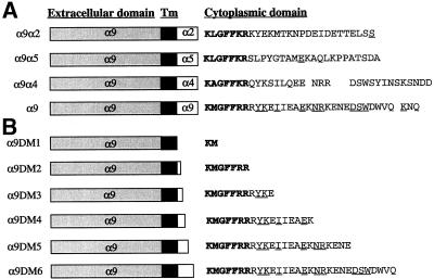
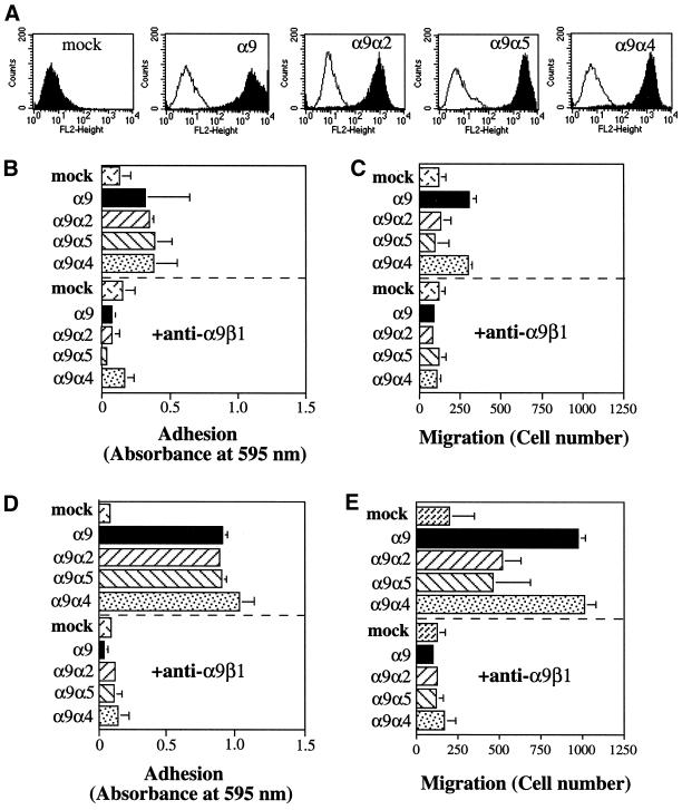
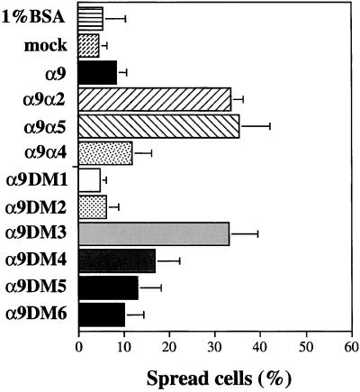
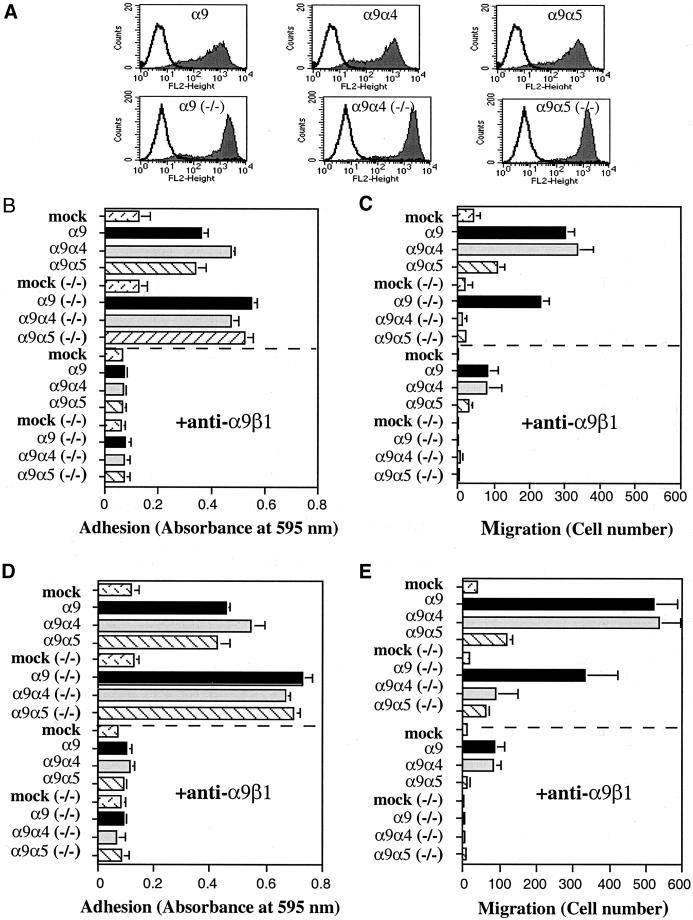
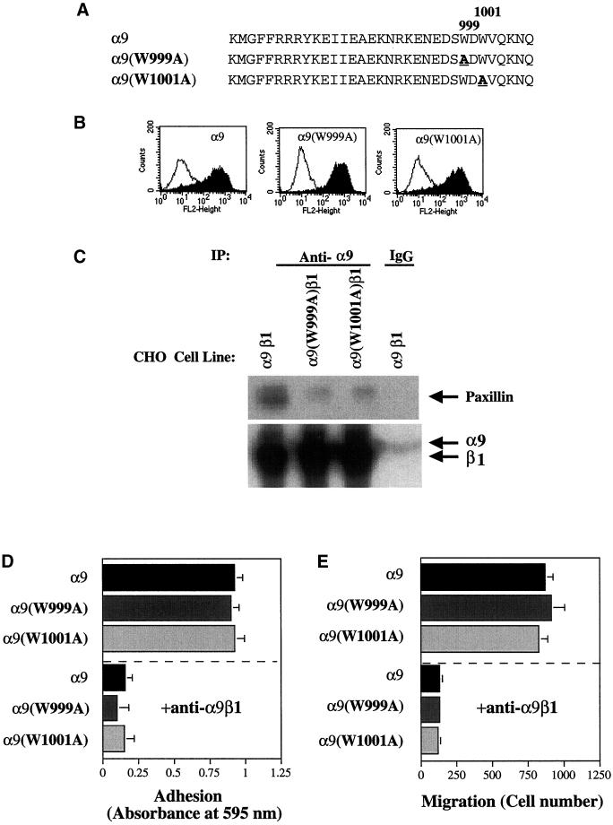
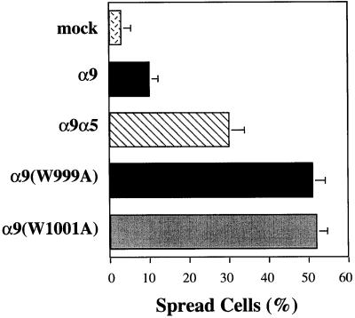
The integrin alpha9 subunit forms a single heterodimer, alpha9beta1. The alpha9 subunit is most closely related to the alpha4 subunit, and like alpha4 integrins, alpha9beta1 plays an important role in leukocyte migration. The alpha4 cytoplasmic domain preferentially enhances cell migration and inhibits cell spreading, effects that depend on interaction with the adaptor protein, paxillin. To determine whether the alpha9 cytoplasmic domain has similar effects, a series of chimeric and deleted alpha9 constructs were expressed in Chinese hamster ovary cells and tested for their effects on migration and spreading on an alpha9beta1-specific ligand. Like alpha4, the alpha9 cytoplasmic domain enhanced cell migration and inhibited cell spreading. Paxillin also specifically bound the alpha9 cytoplasmic domain and to a similar level as alpha4. In paxillin(-/-) cells, alpha9 failed to inhibit cell spreading as expected but surprisingly still enhanced cell migration. Further, mutations that abolished the alpha9-paxillin interaction prevented alpha9 from inhibiting cell spreading but had no effect on alpha9-dependent cell migration. These findings suggest that the mechanisms by which the cytoplasmic domains of integrin alpha subunits enhance migration and inhibit cell spreading are distinct and that the alpha9 and alpha4 cytoplasmic domains, despite sequence and functional similarities, enhance cell migration by different intracellular signaling pathways.


Figures

Figure 1
Figure 1
Schematic diagram of α9, α9 chimeras, and α9DMs. The cytoplasmic amino acid sequences of α9, α9 chimeras (A), and α9DMs (B) are shown. Amino acids indicated in bold type represent areas of homology between all constructs and the underlined amino acids represent areas of homology with the α4 cytoplasmic domain.
Figure 2
Figure 2
Adhesion and migration of α9-expressing CHO cells. (A) Flow cytometric evaluation of cell surface expression of the α9β1 integrins on α9-, α9 chimera-, and mock-expressing CHO cells. Open peaks represent fluorescence (FL) of unstained CHO cells, and shaded peaks represent fluorescence of CHO cells stained with the anti-α9β1 antibody, Y9A2. (B and D) α9-, α9 chimera-, or mock-expressing CHO cells were added to 96-well plates coated with either 3 μg/ml TNfn3RAA (B) or 10 μg/ml TNfn3RAA (D) after incubation with (below the dashed line) or without (above the dashed line) the anti-α9β1 mAb, Y9A2. Cells were allowed to attach for 60 min, and nonadherent cells were removed by centrifugation. Adherent cells were stained with crystal violet and quantified by measurement of absorbance at 595 nm. (C and E) α9-, α9 chimera-, and mock-expressing CHO cells suspended in serum-free medium were seeded onto membranes coated with 3 μg/ml TNfn3RAA (C) or 10 μg/ml TNfn3RAA (E) in the upper well of 24-well plates after preincubation with (below the dashed line) or without (above the dashed line) the anti-α9β1 mAb, Y9A2. After a 2-h incubation in the presence of 1% FCS in the bottom well, nonmigrated cells on the top side of the membrane were removed, and migrated cells on the bottom side of the membrane were fixed, stained, and counted with the use of a phase-contrast microscope in 10 HPF and expressed as number of migrated cells. Data (B–E) represent the means (±SEM) of triplicate experiments.
Figure 3
Figure 3
Adhesion and migration of α9- and α9DM-expressing CHO cells. (A) Flow cytometric evaluation of cell surface expression of the α9β1 integrin from α9- and α9DM-expressing CHO cells as described in Figure 2. (B and D) Adhesion of α9- and α9DM-expressing CHO cells on 3 μg/ml TNfn3RAA (B) or 10 μg/ml TNfn3RAA (D) with or without preincubation with the anti-α9β1 antibody, Y9A2, as described in Figure 2. (C and E) Migration of α9- and α9DM-expressing CHO cells on 3 μg/ml TNfn3RAA (C) or 10 μg/ml TNfn3RAA (E) with or without preincubation with the anti-α9β1 antibody, Y9A2, as described in Figure 2. Data (B–E) represent the means (±SEM) of triplicate experiments.
Figure 4
Figure 4
Spreading of α9-, α9 chimera-, and α9DM-expressing CHO cells. Cells were seeded onto sterile coverslips coated with TNfn3RAA (10 μg/ml) and allowed to spread for 6 h at 37°C. Percentage of spreading was determined by phase-contrast microscopy. Data represent the means (±SEM) of triplicate experiments.
Figure 5
Figure 5
Direct association of paxillin with α9. (A) HA-tagged recombinant GST-paxillin was added to Ni2+-charged resins loaded with the α9 or αIIb cytoplasmic domains. Bound fractions were collected and separated on 4–20% SDS-PAGE under reducing conditions, transferred to a nitrocellulose membrane, and stained with antibody specific for the HA-tag, 12CA5. S.M., starting material. Depicted are results of one of three experiments performed with similar results. (B) HA-tagged recombinant GST-paxillin was added to Ni2+-charged resins loaded with the α9, α4, or the αIIb cytoplasmic domains as described above. Depicted are results of one of three experiments performed with similar results. ●, α9; ▴, α4; ▪, αIIb.(C) CHO cells stably expressing α9β1, α9α2β1, or α9α4β1 integrins were surface labeled with biotin and subjected to immunoprecipitation with the anti-α9β1 antibody, Y9A2, or an irrelevant mouse IgG. The precipitates were separated on 4–20% SDS-PAGE and transferred to a nitrocellulose membrane. Paxillin coimmunoprecipitation was detected with an anti-paxillin antibody (top), and precipitated surface proteins were detected with streptavidin peroxidase followed by ECL (bottom). Depicted are results of one of three experiments performed with similar results.
Figure 6
Figure 6
Association of paxillin with α9 deletion mutants. (A) Binding of recombinant paxillin to α9, α9DM3, α9DM4, α9DM5, or the αIIb cytoplasmic domains as described in Figure 5. Depicted are results of one of two experiments performed with similar results. ♦, α9DM3; ▪, α9DM4; ●, α9DM5; ▴, α9; ▾, αIIb.
Figure 7
Figure 7
Adhesion and migration of α9-expressing MEF and paxillin−/− MEF cells. (A) Flow cytometric evaluation of cell surface expression of the α9β1 integrins from α9- and α9 chimera-expressing MEF cells (top row) and α9- and α9 chimera-expressing paxillin−/− MEF cells (−/−; bottom row) as described in Figure 2. (B and D) Adhesion of α9- and α9 chimera-expressing MEF and paxillin−/− MEF cells (−/−) on 3 μg/ml TNfn3RAA (B) or 10 μg/ml TNfn3RAA (D) with or without preincubation with the anti-α9β1 antibody, Y9A2, as described in Figure 2. Note: twice the amount of the anti-α9β1 antibody, Y9A2, was used to block adhesion of paxillin−/− MEF cells (−/−) on 10 μg/ml TNfn3RAA. (C and E) Migration of α9- and α9 chimera-expressing MEF and paxillin−/− MEF cells (−/−) on 3 μg/ml TNfn3RAA (C) or 10 μg/ml TNfn3RAA (E) for 3 h with or without preincubation with the anti-α9β1 antibody, Y9A2, as described in Figure 2. Data (B–E) represent the means (±SEM) of triplicate experiments.
Figure 8
Figure 8
Spreading of α9- and α9 chimera-expressing MEF and paxillin−/− MEF cells. α9- and α9 chimera-expressing MEF cells and α9- and α9 chimera-expressing paxillin−/− MEF cells (−/−) were seeded onto sterile coverslips coated with TNfn3RAA (10 μg/ml) and allowed to spread for 3 h at 37°C. Percentage of spreading was determined by phase-contrast microscopy. Data represent the means (±SEM) of triplicate experiments.
Figure 9
Figure 9
Adhesion and migration of CHO cells expressing α9 cytoplasmic domain mutations that abolish paxillin binding. (A) The cytoplasmic amino acid sequences of α9, α9(W999A), and α9(W1001A) are shown. Amino acids depicted in bold type represent sites of mutagenesis. (B) Flow cytometric evaluation of cell surface expression of the α9β1 integrins from α9-, α9(W999A)-, and α9(W1001A)-expressing CHO as described in Figure 2. (C) CHO cells stably expressing α9β1 or the α9(W999A)β1- or α9(W1001A)β1-chimeric integrins were surface labeled with biotin and subjected to immunoprecipitation (IP) with the anti-α9β1 antibody, Y9A2, or an irrelevant mouse IgG as described in Figure 5. Depicted are results of one of three experiments performed with similar results. (D) Adhesion of α9-, α9(W999A)-, and α9(W1001A)-expressing CHO cells on 10 μg/ml TNfn3RAA with or without preincubation with the anti-α9β1 antibody, Y9A2, as described in Figure 2. (E) Migration of α9-, α9(W999A)-, and α9(W1001A)-expressing CHO cells on 10 μg/ml TNfn3RAA with or without preincubation with the anti-α9β1 antibody, Y9A2, as described in Figure 2. Data (D–E) represent the means (±SEM) of triplicate experiments.
Figure 10
Figure 10
Spreading of α9-, α9(W999A)-, and α9(W1001A)-expressing CHO cells. α9-, α9(W999A)-, and α9(W1001A)-expressing CHO cells were seeded onto sterile coverslips coated with TNfn3RAA (10 μg/ml) and allowed to spread for 6 h at 37°C. Percentage of spreading was determined by phase-contrast microscopy. Data represent the means (±SEM) of triplicate experiments.

References

    1. Chan BM, Kassner PD, Schiro JA, Byers HR, Kupper TS, Hemler ME. Distinct cellular functions mediated by different VLA integrin alpha subunit cytoplasmic domains. Cell. 1992;68:1051–1060. - PubMed
    1. Clark EA, Brugge JS. Integrins and signal transduction pathways: the road taken. Science. 1995;268:233–239. - PubMed
    1. Eto K, Puzon-McLaughlin W, Sheppard D, Sehara-Fujisawa A, Zhang XP, Takada Y. RGD-independent binding of integrin alpha9beta1 to the ADAM-12 and -15 disintegrin domains mediates cell-cell interaction. J Biol Chem. 2000;275:34922–34930. - PubMed
    1. Gao JX, Issekutz AC. The beta 1 integrin, very late activation antigen-4 on human neutrophils can contribute to neutrophil migration through connective tissue fibroblast barriers. Immunology. 1997;90:448–454. - PMC - PubMed
    1. Horwitz AR, Parsons JT. Cell migration: movin' on [comment] Science. 1999;286:1102–1103. - PubMed

Publication types

MeSH terms