

doi: 10.1128/AAC.45.11.3195-3197.2001.
New model of oropharyngeal candidiasis in mice
Affiliations
- PMID: 11600377
- PMCID: PMC90803
- DOI: 10.1128/AAC.45.11.3195-3197.2001
Item in Clipboard
New model of oropharyngeal candidiasis in mice
Antimicrob Agents Chemother.
2001 Nov.
Abstract
We established a straightforward murine model of oropharyngeal candidiasis. Mice were immunosuppressed with cortisone acetate, anesthetized, and then inoculated by placing cotton wool balls saturated with Candida albicans sublingually for 2 h. A prolonged, reproducible infection was induced. This model may be useful for antifungal screening or pathogenesis studies.
Figures
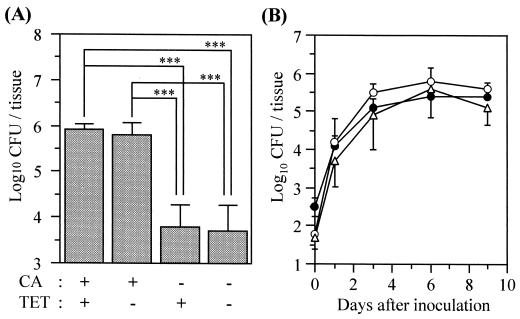
Development of OPC in mice. (A) Effect of cortisone acetate and tetracycline on the numbers of viable C. albicans cells in the oral tissue on day 6. Data are the mean plus standard deviation for five mice per group. ∗∗∗, P < 0.001 by the Tukey test. CA, cortisone acetate; TET, tetracycline. (B) Time course of OPC in mice treated with cortisone acetate and tetracycline. Data are the mean ± standard deviation for five mice per group. Open circles, tongue; closed circles, nonlingual oral tissue; open triangles, esophagus.
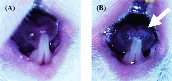
White patches on the tongue of a mouse inoculated with C. albicans. The tongues of an uninfected (A) and infected (B) mouse were photographed on day 3. The arrow indicates the white patches on the tip of the tongue.
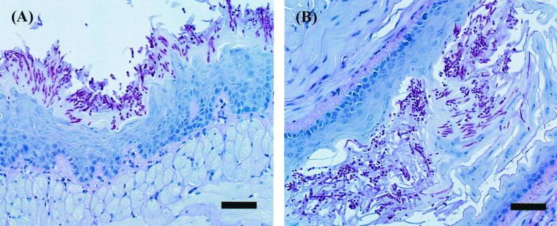
Histopathological analysis of the tongue (A) and the esophagus (B) tissue of mice infected with C. albicans for 4 days (PAS stain; bar = 50 μm).
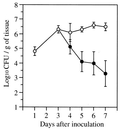
The number of viable C. albicans cells in the oral tissue of mice that were treated with daily FLC at 10 mg/kg/dose starting on day 3. Results are the mean ± standard deviation for six mice per time point. Open circles, control mice; closed circles, mice treated with FLC.
Similar articles
-
Mouse model of oropharyngeal candidiasis.Nat Protoc. 2012 Mar 8;7(4):637-42. doi: 10.1038/nprot.2012.011. Nat Protoc. 2012. PMID: 22402633 Free PMC article.
-
[Experimental oral candidiasis in healthy and immunocompromised BALB/c mice].Mikrobiyol Bul. 2011 Apr;45(2):336-43. Mikrobiyol Bul. 2011. PMID: 21644077 Turkish.
-
Antifungal activities of two new azasordarins, GW471552 and GW471558, in experimental models of oral and vulvovaginal candidiasis in immunosuppressed rats.Antimicrob Agents Chemother. 2001 Dec;45(12):3304-9. doi: 10.1128/AAC.45.12.3304-3309.2001. Antimicrob Agents Chemother. 2001. PMID: 11709301 Free PMC article.
-
Models hosts for the study of oral candidiasis.Adv Exp Med Biol. 2012;710:95-105. doi: 10.1007/978-1-4419-5638-5_10. Adv Exp Med Biol. 2012. PMID: 22127889 Review.
-
The role of yeasts other than Candida albicans in oropharyngeal candidiasis.Curr Opin Infect Dis. 2001 Dec;14(6):673-7. doi: 10.1097/00001432-200112000-00002. Curr Opin Infect Dis. 2001. PMID: 11964883 Review.
Cited by
-
Neutrophils Do Not Express IL-17A in the Context of Acute Oropharyngeal Candidiasis.Pathogens. 2015 Jul 24;4(3):559-72. doi: 10.3390/pathogens4030559. Pathogens. 2015. PMID: 26213975 Free PMC article.
-
Filamentation in Candida auris, an emerging fungal pathogen of humans: passage through the mammalian body induces a heritable phenotypic switch.Emerg Microbes Infect. 2018 Nov 28;7(1):188. doi: 10.1038/s41426-018-0187-x. Emerg Microbes Infect. 2018. PMID: 30482894 Free PMC article.
-
Host cell invasion by medically important fungi.Cold Spring Harb Perspect Med. 2014 Nov 3;5(1):a019687. doi: 10.1101/cshperspect.a019687. Cold Spring Harb Perspect Med. 2014. PMID: 25367974 Free PMC article. Review.
-
Development of Anti-Virulence Approaches for Candidiasis via a Novel Series of Small-Molecule Inhibitors of Candida albicans Filamentation.mBio. 2017 Dec 5;8(6):e01991-17. doi: 10.1128/mBio.01991-17. mBio. 2017. PMID: 29208749 Free PMC article.
-
Mucosal biofilms of Candida albicans.Curr Opin Microbiol. 2011 Aug;14(4):380-5. doi: 10.1016/j.mib.2011.06.001. Epub 2011 Jul 7. Curr Opin Microbiol. 2011. PMID: 21741878 Free PMC article.
References
-
- Gillum A M, Tsay E Y H, Kirsch D R. Isolation of the Candida albicans gene for orotidine-5′-phosphate decarboxylase by complementation of S. cerevisiae ura3 and E. coli pyrF mutations. Mol Gen Genet. 1984;198:179–182. - PubMed
-
- Klein R S, Carol A H, Small C B, Moll B, Lesser M, Friedland G H. Oral candidiasis in high-risk patients as the initial manifestation of the acquired immunodeficiency syndrome. N Engl J Med. 1984;311:354–358. - PubMed
-
- National Committee for Clinical Laboratory Standards. Reference method for broth dilution antifungal susceptibility testing of yeasts: approved standard. NCCLS document M27-A. Wayne, Pa: National Committee for Clinical Laboratory Standards; 1997.
Publication types
MeSH terms
Substances
Grants and funding
LinkOut - more resources
Full Text Sources
Other Literature Sources
Medical
